The
temporomandibular joint is the joint of the
jawThe jaw is any opposable articulated structure at the entrance of the mouth, typically used for grasping and manipulating food. The term jaws is also broadly applied to the whole of the structures constituting the vault of the mouth and serving to open and close it and is part of the body plan of...
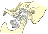
and is frequently referred to as
TMJ. There are two TMJs, one on either side, working in unison. The name is derived from the two bones which form the
jointA joint is the location at which two or more bones make contact. They are constructed to allow movement and provide mechanical support, and are classified structurally and functionally.-Classification:...
: the upper
temporal boneThe temporal bones are situated at the sides and base of the skull, and lateral to the temporal lobes of the cerebrum.The temporal bone supports that part of the face known as the temple.-Parts:The temporal bone consists of four parts:* Squama temporalis...
which is part of the
craniumThe human skull is a bony structure, skeleton, that is in the human head and which supports the structures of the face and forms a cavity for the brain.In humans, the adult skull is normally made up of 22 bones...
(
skullThe human skull is a bony structure, skeleton, that is in the human head and which supports the structures of the face and forms a cavity for the brain.In humans, the adult skull is normally made up of 22 bones...
), and the lower jaw bone called the
mandible. The unique feature of the TMJs is the
articular disc. The disc is composed of fibrocartilagenous tissue (like the firm and flexible
elastic cartilageElastic cartilage or yellow cartilage is a type of cartilage present in the outer ear, larynx, and epiglottis. It contains elastic fiber networks and collagen fibers. The principal protein is elastin....
of the ear) which is positioned between the two bones that form the joint. The TMJs are one of the only synovial joints in the human body with an articular disc, another being the sternoclavicular joint. The disc divides each joint into two. The lower joint compartment formed by the mandible and the articular disc is involved in
rotational movement—this is the initial movement of the jaw when the mouth opens. The upper joint compartment formed by the articular disk and the temporal bone is involved in
translational movement—this is the secondary gliding motion of the jaw as it is opened widely. The part of the mandible which mates to the under-surface of the disc is the
condyle and the part of the temporal bone which mates to the upper surface of the disk is the glenoid (or mandibular)
fossaThe mandibular fossa is the depression in the temporal bone that articulates with the mandibular condyle. In the temporal bone, the mandibular fossa is bounded, in front, by the articular tubercle; behind, by the tympanic part of the bone, which separates it from the external acoustic meatus; it is...
.
Pain or dysfunction of the temporomandibular joint is commonly referred to as "TMJ", when in fact, TMJ is really the name of the joint, and
Temporomandibular joint disorderTemporomandibular joint disorder , or TMJ syndrome, is an umbrella term covering acute or chronic inflammation of the temporomandibular joint, which connects the mandible to the skull. The disorder and resultant dysfunction can result in significant pain and impairment...
(or
dysfunction) is abbreviated TMD. This term is used to refer to a group of problems involving the TMJs and the muscles, tendons, ligaments, blood vessels, and other tissues associated with them. Some practitioners might include the neck, the back and even the whole body in describing problems with the TMJs.
Development
Formation of the TMJ occurs at around 12 weeks
in uteroIn utero is a Latin term literally meaning "in the womb". In biology, the phrase describes the state of an embryo or fetus. In legal contexts, the phrase is used to refer to unborn children. Under common law, unborn children are still considered to exist for property transfer purposes.-See also:*...
when the joint spaces and the articular disc develop.
Articulation
The TMJ is a
ginglymoarthrodial joint, referring to its dual compartment structure and function (
ginglymo-A hinge joint is a bone joint in which the articular surfaces are molded to each other in such a manner as to permit motion only in one plane—backward and forward—the extent of motion at the same time being considerable....
and
arthrodialA plane joint is a synovial joint which, under physiological conditions, allows only gliding movement....
).
The condyle articulates with the
temporal boneThe temporal bones are situated at the sides and base of the skull, and lateral to the temporal lobes of the cerebrum.The temporal bone supports that part of the face known as the temple.-Parts:The temporal bone consists of four parts:* Squama temporalis...
in the
mandibular fossaThe mandibular fossa is the depression in the temporal bone that articulates with the mandibular condyle. In the temporal bone, the mandibular fossa is bounded, in front, by the articular tubercle; behind, by the tympanic part of the bone, which separates it from the external acoustic meatus; it is...
. The mandibular fossa is a concave depression in the squamous portion of the temporal bone.
These two bones are actually separated by an articular disc, which divides the TMJ into two distinct compartments. The
inferior compartment allows for
rotation of the condylar head around an instantaneous axis of rotation, corresponding to the first 20 mm or so of the opening of the mouth. After the mouth is open to this extent, the mouth can no longer open without the
superior compartment of the TMJ becoming active.
At this point, if the mouth continues to open, not only is the condylar head rotating within the lower compartment of the TMJ, but the entire apparatus (condylar head and articular disc)
translates. Although this had traditionally been explained as a
forward and downward sliding motion, on the anterior concave surface of the glenoid fossa and the posterior convex surface of the articular eminence, this
translation actually amounts to a rotation around another axis. This effectively produces an
evoluteIn the differential geometry of curves, the evolute of a curve is the locus of all its centers of curvature. Equivalently, it is the envelope of the normals to a curve....
which can be termed the
resultant axis of mandibular rotation, which lies in the vicinity of the mandibular foramen, allowing for a low-tension environment for the vasculature and innervation of the mandible.
The necessity of translation to produce further opening past that which can be accomplished with sole rotation of the condyle can be demonstrated by placing a resistant fist against the chin and trying to open the mouth more than 20 or so mm.
Components
There are six main components of the TMJ.
- Mandibular condyles
- Articular surface of the temporal bone
- Capsule
- Articular disc
- Ligaments
- Lateral pterygoid
Capsule and articular disc
The capsule is a fibrous membrane that surrounds the joint and incorporates the articular eminence. It attaches to the articular eminence, the articular disc and the neck of the mandibular condyle.
The articular disc is a fibrous extension of the capsule in between the two bones of the joint. The disc functions as articular surfaces against both the temporal bone and the condyles and divides the joint into two sections, as described in more detail below. It is biconcave in structure and attaches to the condyle medially and laterally. The anterior portion of the disc splits in the vertical dimension, coincident with the insertion of the superior head of the lateral pterygoid. The posterior portion also splits in the vertical dimension, and the area between the split continues posteriorly and is referred to as the
retrodiscal tissue. Unlike the disc itself, this piece of connective tissue is vascular and innervated, and in some cases of
anterior disc displacement, the pain felt during movement of the mandible is due to the condyle compressing this area against the articular surface of the temporal bone.
Ligaments
There are three ligaments associated with the TMJ: one major and two minor ligaments.
- The major ligament, the temporomandibular ligament
The temporomandibular ligament consists of two short, narrow fasciculi, one in front of the other, attached, above, to the lateral surface of the zygomatic arch and to the tubercle on its lower border; below, to the lateral surface and posterior border of the neck of the mandible.It is broader...
, is actually the thickened lateral portion of the capsule, and it has two parts: an outer oblique portion (OOP) and an inner horizontal portion (IHP).
- The two minor ligaments, the stylomandibular and sphenomandibular ligaments are accessory and are not directly attached to any part of the joint.
- The stylomandibular ligament
The stylomandibular ligament is a specialized band of the cervical fascia, which extends from near the apex of the styloid process of the temporal bone to the angle and posterior border of the angle of the mandible, between the Masseter and Pterygoideus internus.This ligament separates the parotid...
separates the infratemporal region (anterior) from the parotid region (posterior), and runs from the styloid processThe styloid process is a pointed piece of bone that extends down from the human skull, just below the ear.-Structure:The styloid process is a slender pointed piece of bone just below the ear...
to the angle of the mandibleAt the junction of the lower border of the ramus of the mandible with the posterior border is the angle of the mandible, which may be either inverted or everted and is marked by rough, oblique ridges on each side, for the attachment of the Masseter laterally, and the Pterygoideus internus medially;...
.
- The sphenomandibular ligament
The sphenomandibular ligament is a flat, thin band which is attached above to the spina angularis of the sphenoid bone, and, becoming broader as it descends, is fixed to the lingula of the mandibular foramen...
runs from the spine of the sphenoid bone to the lingula of mandibleThe margin of the mandibular foramen is irregular; it presents in front a prominent ridge, surmounted by a sharp spine, the lingula of the mandible which gives attachment to the sphenomandibular ligament; at its lower and back part is a notch from which the mylohyoid groove runs obliquely downward...
.
These ligaments are important in that they define the border movements, or in other words, the farthest extents of movements, of the mandible. Movements of the mandible made past the extents functionally allowed by the muscular attachments will result in painful stimuli, and thus, movements past these more limited borders are rarely achieved in normal function.
Other ligaments, called "oto-mandibular ligaments", connect
middle earThe middle ear is the portion of the ear internal to the eardrum, and external to the oval window of the cochlea. The mammalian middle ear contains three ossicles, which couple vibration of the eardrum into waves in the fluid and membranes of the inner ear. The hollow space of the middle ear has...
(
malleusThe malleus or hammer is a hammer-shaped small bone or ossicle of the middle ear which connects with the incus and is attached to the inner surface of the eardrum...
) with temporomandibular joint:
- discomallear (or disco-malleolar) ligament,
- malleomandibular (or malleolar-mandibular) ligament.
Innervation and vascularization
Sensory innervation of the temporomandibular joint is derived from the auriculotemporal and
massetericMasseteric can refer to:* Masseteric artery* Masseteric nerve* Masseter muscle...
branches of
V3The mandibular nerve is the largest of the three branches of the trigeminal nerve.-Roots:It is made up of two roots:* a large sensory root proceeding from the inferior angle of the trigeminal ganglion....
(otherwise known as the mandibular branch of the
trigeminal nerveThe trigeminal nerve contains both sensory and motor fibres. It is responsible for sensation in the face and certain motor functions such as biting, chewing, and swallowing. Sensory information from the face and body is processed by parallel pathways in the central nervous system...
). These are only sensory innervation. Recall that motor is to the muscles.
Its arterial blood supply is provided by branches of the
external carotid arteryIn human anatomy, the external carotid artery is a major artery of the head and neck. It arises from the common carotid artery when it bifurcates into the external and internal carotid artery.-Course:...
, predominately the superficial temporal branch. Other branches of the external carotid artery namely: the
deep auricular arteryThe deep auricular artery often arises in common with the anterior tympanic artery.It ascends in the substance of the parotid gland, behind the temporomandibular articulation, pierces the cartilaginous or bony wall of the external acoustic meatus, and supplies its cuticular lining and the outer...
,
anterior tympanic arteryThe anterior tympanic artery usually arises as a branch of the mandibular part of the maxillary artery. It passes upward behind the temporomandibular articulation, enters the tympanic cavity through the petrotympanic fissure, and ramifies upon the tympanic membrane, forming a vascular circle...
,
ascending pharyngeal arteryThe ascending pharyngeal artery, the smallest branch of the external carotid, is a long, slender vessel, deeply seated in the neck, beneath the other branches of the external carotid and under the Stylopharyngeus...
, and maxillary artery- may also contribute to the arterial blood supply of the joint.
The specific mechanics of proprioception in the temporomandibular joint involve four receptors.
Ruffini endingThe Bulbous corpuscle or Ruffini ending or Ruffini corpuscle is a class of slowly adapting mechanoreceptor thought to exist only in the glabrous dermis and subcutaneous tissue of humans...
s function as static
mechanoreceptorA mechanoreceptor is a sensory receptor that responds to mechanical pressure or distortion. There are four main types in the glabrous skin of humans: Pacinian corpuscles, Meissner's corpuscles, Merkel's discs, and Ruffini corpuscles...
s which position the mandible.
Pacinian corpuscleLamellar corpuscles or Pacinian corpuscles are one of the four major types of mechanoreceptor. They are nerve endings in the skin, responsible for sensitivity to vibration and pressure. Vibrational role may be used to detect surface, e.g., rough vs...
s are dynamic mechanoreceptors which accelerate movement during reflexes.
Golgi tendon organThe Golgi organ , is a proprioceptive sensory receptor organ that is located at the insertion of skeletal muscle fibers into the tendons of skeletal muscle...
s function as static mechanoreceptors for protection of ligaments around the temporomandibular joint.
Free nerve endingA free nerve ending is an unspecialized, afferent nerve ending, meaning it brings information from the body's periphery toward the brain. They function as cutaneous receptors and are essentially used by vertebrates to detect pain.-Structure:...
s are the pain receptors for protection of the temporomandibular joint itself.
In order to work properly, there is neither innervation nor vascularization within the central portion of the articular disc. Had there been any nerve fibers or blood vessels, people would bleed whenever they moved their jaws, and movement itself would be too painful.
Jaw movements
During
jawThe jaw is any opposable articulated structure at the entrance of the mouth, typically used for grasping and manipulating food. The term jaws is also broadly applied to the whole of the structures constituting the vault of the mouth and serving to open and close it and is part of the body plan of...
movements, only the mandible moves.
Normal movements of the mandible during function, such as
masticationMastication or chewing is the process by which food is crushed and ground by teeth. It is the first step of digestion and it increases the surface area of foods to allow more efficient break down by enzymes. During the mastication process, the food is positioned between the teeth for grinding by...
, or chewing, are known as excursions. There are two lateral excursions (left and right) and the forward excursion, known as protrusion. The reversal of protrusion is retrusion.
When the mandible is moved into protrusion, the mandibular incisors, or front teeth of the mandible, are moved so that they first come edge to edge with the maxillary (upper) incisors and then surpass them, producing a temporary underbite. This is accomplished by translation of the condyle down the articular eminence (in the upper portion of the TMJ) without any more than the slightest amount of rotation taking place (in the lower portion of the TMJ), other than that necessary to allow the mandibular incisors to come in front of the maxillary incisors without running into them. (This is all assuming an ideal Class I or Class II occlusion, which is not entirely important to the lay reader.)

During chewing, the mandible moves in a specific manner as delineated by the two TMJs. The side of the mandible that moves laterally is referred to as either the working or rotating side, while the other side is referred to as either the balancing or orbiting side. The latter terms, although a bit outdated, are actually more precise, as they define the sides by the movements of the respective condyles.
When the mandible is moved into a lateral excursion, the working side condyle (the condyle on the side of the mandible that moves outwards) only performs rotation (in the horizontal plane), while the balancing side condyle performs translation. During actual functional chewing, when the teeth are not only moved side to side, but also up and down when biting of the teeth is incorporated as well, rotation (in a vertical plane) also plays a part in both condyles.
The mandible is moved primary by the four muscles of mastication: the masseter,
medial pterygoidThe medial pterygoid , is a thick, quadrilateral muscle of mastication.The mandibular branch of the fifth cranial nerve, the trigeminal nerve, innervates the medial pterygoid muscle.-Origin and insertion:...
, lateral pterygoid and the temporalis. These four muscles, all innervated by
V3The mandibular nerve is the largest of the three branches of the trigeminal nerve.-Roots:It is made up of two roots:* a large sensory root proceeding from the inferior angle of the trigeminal ganglion....
, or the mandibular division of the
trigeminal nerveThe trigeminal nerve contains both sensory and motor fibres. It is responsible for sensation in the face and certain motor functions such as biting, chewing, and swallowing. Sensory information from the face and body is processed by parallel pathways in the central nervous system...
, work in different groups to move the mandible in different directions. Contraction of the lateral pterygoid acts to pull the disc and condyle forward within the glenoid fossa and down the articular eminence; thus, action of this muscle serves to protrude the jaw, it with assistance of gravity and the digastricus muscle also opens the jaw. The other three muscles close the mouth; the masseter and the medial pterygoid by pulling up the angle of the mandible and the temporalis by pulling up on the
coronoid processThe Coronoid process can refer to:* The coronoid process of the mandible, part of the ramus mandibulae of the mandible...
.
Disorders
- See Temporomandibular joint disorder
Temporomandibular joint disorder , or TMJ syndrome, is an umbrella term covering acute or chronic inflammation of the temporomandibular joint, which connects the mandible to the skull. The disorder and resultant dysfunction can result in significant pain and impairment...
for more details
The most common disorder of the TMJ is
disc displacement. In essence, this is when the articular disc, attached anteriorly to the superior head of the lateral pteygoid muscle and posteriorly to the retrodiscal tissue, moves out from between the condyle and the fossa, so that the mandible and temporal bone contact is made on something other than the articular disc. This, as explained above, is usually very painful, because unlike these adjacent tissues, the central portion of the disc contains no sensory innervation.
In most instances of disorder, the disc is displaced anteriorly upon translation, or the anterior and inferior sliding motion of the condyle forward within the fossa and down the articular eminence. On opening, a "pop" or "click" can sometimes be heard and usually felt also, indicating the condyle is moving back onto the disk, known as "reducing the joint" (
disc displacement with reduction). Upon closing, the condyle will slide off the back of the disc, hence another "click" or "pop" at which point the condyle is posterior to the disc. Upon clenching, the condyle compresses the bilaminar area, and the nerves, arteries and veins against the temporal fossa, causing pain and inflammation.
In
disc displacement without reduction the disc stays anterior to the condylar head upon opening. Mouth opening is limited and there is no "pop" or "click" sound on opening.
TMJ pain is generally due to one of four reasons.
- The most common cause of TMJ pain is myofascial pain dysfunction syndrome, primarily involving the muscles of mastication.
- Internal derangements is defined as an abnormal relationship of the disc to any of the other components of the TMJ. Disc displacement is an example of internal derangement.
- Degenerative joint disease, otherwise known as osteoarthritis
Osteoarthritis also known as degenerative arthritis or degenerative joint disease, is a group of mechanical abnormalities involving degradation of joints, including articular cartilage and subchondral bone. Symptoms may include joint pain, tenderness, stiffness, locking, and sometimes an effusion...
is the organic degeneration of the articular surfaces within the TMJ.
- TMJ pain remains one of the most reliable diagnostic criteria for temporal arteritis
Giant-cell arteritis or Horton disease is an inflammatory disease of blood vessels most commonly involving large and medium arteries of the head...
.
External links