
Renal artery
Encyclopedia
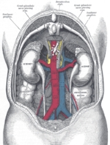
The renal arteries normally arise off the side of the abdominal aorta
, immediately below the superior mesenteric artery
, and supply the kidney
s with blood
. Each is directed across the crus of the diaphragm, so as to form nearly a right angle with the aorta.
The renal arteries carry a large portion of total blood flow to the kidneys. Up to a third of total cardiac output can pass through the renal arteries to be filtered by the kidneys.
The arterial supply of the kidneys is variable and there may be one or more renal arteries supplying each kidney. It is located above the renal vein. Supernumerary renal arteries(two or more arteries to a single kidney) are the most common renovascular anomaly, occurrence ranging from 25% to 40% of kidneys.
It has a radius of approximately 0.25 cm, 0.26 cm at the root. The measured mean diameter can differ depending on the imaging method used. For example, the diameter was found to be 5.04 ± 0.74 mm using ultrasound, but 5.68 ± 1.19 mm using angiography.
and the kidneys in the body, the right renal artery is normally longer than the left renal artery.
, each artery divides into four or five branches; the greater number of these (anterior branches) lie between the renal vein and ureter
, the vein being in front, the ureter behind, but one or more branches (posterior branches) are usually situated behind the ureter.
Each vessel gives off some small inferior suprarenal branches to the suprarenal gland, the ureter
, and the surrounding cellular tissue and muscles.
One or two accessory renal arteries are frequently found, especially on the left side since they usually arise from the aorta, and may come off above (more common) or below the main artery. Instead of entering the kidney at the hilus, they usually pierce the upper or lower part of the organ.
, or narrowing of one or both renal arteries will lead to hypertension as the affected kidneys release renin
to increase blood pressure to preserve perfusion to the kidneys. RAS is typically diagnosed with duplex ultrasonography of the renal arteries. It is treated with the use of balloon angioplasty and stents, if necessary.
Atherosclerosis
can also affect the renal arteries and can lead to poor perfusion of the kidneys leading to reduced kidney function and, possibly, renal failure
.
Abdominal aorta
The abdominal aorta is the largest artery in the abdominal cavity. As part of the aorta, it is a direct continuation of the descending aorta .-Path:...
, immediately below the superior mesenteric artery
Superior mesenteric artery
In human anatomy, the superior mesenteric artery arises from the anterior surface of the abdominal aorta, just inferior to the origin of the celiac trunk, and supplies the intestine from the lower part of the duodenum through two-thirds of the transverse colon, as well as the pancreas.-Location...
, and supply the kidney
Kidney
The kidneys, organs with several functions, serve essential regulatory roles in most animals, including vertebrates and some invertebrates. They are essential in the urinary system and also serve homeostatic functions such as the regulation of electrolytes, maintenance of acid–base balance, and...
s with blood
Blood
Blood is a specialized bodily fluid in animals that delivers necessary substances such as nutrients and oxygen to the cells and transports metabolic waste products away from those same cells....
. Each is directed across the crus of the diaphragm, so as to form nearly a right angle with the aorta.
The renal arteries carry a large portion of total blood flow to the kidneys. Up to a third of total cardiac output can pass through the renal arteries to be filtered by the kidneys.
The arterial supply of the kidneys is variable and there may be one or more renal arteries supplying each kidney. It is located above the renal vein. Supernumerary renal arteries(two or more arteries to a single kidney) are the most common renovascular anomaly, occurrence ranging from 25% to 40% of kidneys.
It has a radius of approximately 0.25 cm, 0.26 cm at the root. The measured mean diameter can differ depending on the imaging method used. For example, the diameter was found to be 5.04 ± 0.74 mm using ultrasound, but 5.68 ± 1.19 mm using angiography.
Asymmetries before reaching kidney
Due to the position of the aorta, the inferior vena cavaInferior vena cava
The inferior vena cava , also known as the posterior vena cava, is the large vein that carries de-oxygenated blood from the lower half of the body into the right atrium of the heart....
and the kidneys in the body, the right renal artery is normally longer than the left renal artery.
- The right passes behind the inferior vena cavaInferior vena cavaThe inferior vena cava , also known as the posterior vena cava, is the large vein that carries de-oxygenated blood from the lower half of the body into the right atrium of the heart....
, the right renal veinRenal veinThe renal veins are veins that drain the kidney. They connect the kidney to the inferior vena cava.It is usually singular to each kidney, except in the condition "multiple renal veins".It also divides into 2 divisions upon entering the kidney:...
, the head of the pancreas, and the descending part of the duodenumDuodenumThe duodenum is the first section of the small intestine in most higher vertebrates, including mammals, reptiles, and birds. In fish, the divisions of the small intestine are not as clear and the terms anterior intestine or proximal intestine may be used instead of duodenum...
. - The left is somewhat higher than the right; it lies behind the left renal vein, the body of the pancreas and the splenic veinSplenic veinIn anatomy, the splenic vein is the blood vessel that drains blood from the spleen.It joins with the superior mesenteric vein, to form the hepatic portal vein and follows a course superior to the pancreas, alongside of the similarly named artery, the splenic artery.It collects branches from the...
, and is crossed by the inferior mesenteric veinInferior mesenteric veinIn human anatomy, the inferior mesenteric vein is a blood vessel that drains blood from the large intestine. It usually terminates when reaching the splenic vein, which goes on to form the portal vein with the superior mesenteric vein...
.
At kidney
Before reaching the hilus of the kidneyHilum of kidney
The renal hilum or renal pedicle of the kidney is the recessed central fissure. The medial border of the kidney is concave in the center and convex toward either extremity; it is directed forward and a little downward. Its central part presents a deep longitudinal fissure, bounded by prominent...
, each artery divides into four or five branches; the greater number of these (anterior branches) lie between the renal vein and ureter
Ureter
In human anatomy, the ureters are muscular tubes that propel urine from the kidneys to the urinary bladder. In the adult, the ureters are usually long and ~3-4 mm in diameter....
, the vein being in front, the ureter behind, but one or more branches (posterior branches) are usually situated behind the ureter.
Each vessel gives off some small inferior suprarenal branches to the suprarenal gland, the ureter
Ureter
In human anatomy, the ureters are muscular tubes that propel urine from the kidneys to the urinary bladder. In the adult, the ureters are usually long and ~3-4 mm in diameter....
, and the surrounding cellular tissue and muscles.
One or two accessory renal arteries are frequently found, especially on the left side since they usually arise from the aorta, and may come off above (more common) or below the main artery. Instead of entering the kidney at the hilus, they usually pierce the upper or lower part of the organ.
Diseases of the renal arteries
Renal artery stenosisRenal artery stenosis
Renal artery stenosis is the narrowing of the renal artery, most often caused by atherosclerosis or fibromuscular dysplasia. This narrowing of the renal artery can impede blood flow to the target kidney...
, or narrowing of one or both renal arteries will lead to hypertension as the affected kidneys release renin
Renin
Renin , also known as an angiotensinogenase, is an enzyme that participates in the body's renin-angiotensin system -- also known as the Renin-Angiotensin-Aldosterone Axis -- that mediates extracellular volume , and arterial vasoconstriction...
to increase blood pressure to preserve perfusion to the kidneys. RAS is typically diagnosed with duplex ultrasonography of the renal arteries. It is treated with the use of balloon angioplasty and stents, if necessary.
Atherosclerosis
Atherosclerosis
Atherosclerosis is a condition in which an artery wall thickens as a result of the accumulation of fatty materials such as cholesterol...
can also affect the renal arteries and can lead to poor perfusion of the kidneys leading to reduced kidney function and, possibly, renal failure
Renal failure
Renal failure or kidney failure describes a medical condition in which the kidneys fail to adequately filter toxins and waste products from the blood...
.