

Projectional radiography
Encyclopedia
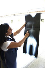
Projectional radiography or plain film radiography is the practice of producing two-dimensional images using x-ray radiation. Radiographic exams are typically performed by Radiologic Technologists, highly trained medical professionals who specialize in the usage of radiographic equipment, patient care, and radiation safety. Projectional radiography is the cornerstone of modern medical imaging, and can be used to image almost every part of the human body. Mammography
and Dental radiography
are also considered to be specialized variants of projectional radiography.
 Projectional radiography relies on the characteristics of x-ray radiation and knowledge of how it interacts with human tissue to create diagnostic images. X-rays are a form of ionizing radiation
, meaning it has sufficient energy to potentially remove electrons from an atom, thus giving it a charge and making it an ‘ion’. Ionizing radiation has sufficient energy to penetrate human tissue.

)
Abdomen - Usually a single AP supine (KUB—kidney, bladder, and ureter) projection. Special projections include a PA prone, Lateral Decubitus, upright AP, and Lateral Cross-Table (with the patient supine) A minimal acute obstructive series (for the purpose of ruling out small bowel obstruction) would include two views: typically, a supine view and an upright view (which would be sufficient to detect air-fluid levels), although a lateral decubitus could be substituted for the upright.
Cervical Spine - Five or six projections are common; a Lateral, two 45 degree obliques, an AP axial (Cephalad), an AP "Open Mouth" for C1-C2, and Cervicothoracic Lateral (Swimmer's) to better visualize C7-T1 if necessary. Special projections include a Lateral with Flexion and Extension of the cervical spine, an Axial for C1-C2 (Fuchs or Judd method), and an AP Axial (Caudad) for articular pillars.
Thoracic Spine - An AP and Lateral are basic projections. Obliques 20 degrees from lateral may be ordered to better visualize the zygapophysial joint
Lumbar Spine - Basic projections include an AP, two Obliques, a Lateral, and a Lateral L5-S1 spot to better visualize the L5-S1 interspace. Special projections are AP Right and Left bending, and Laterals with Flexion and Extension.
Sacrum and Coccyx - If both bones are to be examined separate cephalad and caudad AP axial projections are obtained for the sacrum and coccyx respectively as well as a single Lateral of both bones.
Sternum - The two basic projections are a 15 to 20 degree Right Anterior Oblique and a Lateral.
Sternoclavicular Joints - Are usually ordered as a single PA and a Right and Left 15 degree Right Anterior Obliques.
Ribs - Common rib projections are based on the location of the area of interest. These are obtained with shorter wavelengths/higher frequencies/higher levels of radiation than a standard CXR.
Radiology & Medical Imaging Web forums in Arabic
Mammography
Mammography is the process of using low-energy-X-rays to examine the human breast and is used as a diagnostic and a screening tool....
and Dental radiography
Dental radiography
Dental radiographs, commonly referred to as X-ray films, or informally, X-rays, are pictures of the teeth, bones, and surrounding soft tissues to screen for and help identify problems with the teeth, mouth, and jaw. X-ray pictures can show cavities, cancerous or benign masses, hidden dental...
are also considered to be specialized variants of projectional radiography.
Imaging principles
Ionizing radiation
Ionizing radiation is radiation composed of particles that individually have sufficient energy to remove an electron from an atom or molecule. This ionization produces free radicals, which are atoms or molecules containing unpaired electrons...
, meaning it has sufficient energy to potentially remove electrons from an atom, thus giving it a charge and making it an ‘ion’. Ionizing radiation has sufficient energy to penetrate human tissue.
X-ray attenuation
When an exposure is made, x-ray radiation exits the tube as what is known as the primary beam. When the primary beam passes through the body, some of the radiation is absorbed in a process known as attenuation. Anatomy that is denser has a higher rate of attenuation than anatomy that is less dense, so bone will absorb more x-rays than soft tissue. What remains of the primary beam after attenuation is known as the remnant beam. The remnant beam is responsible for exposing the image receptor. Areas on the image receptor that receive the most radiation (portions of the remnant beam experiencing the least attenuation) will be more heavily exposed, and therefore will be processed as being darker. Conversely, areas on the image receptor that receive the least radiation (portions of the remnant beam experience the most attenuation) will be less exposed and will be processed as being lighter. This is why bone, which is very dense, process as being ‘white’ on radio graphs, and the lungs, which contain mostly air and is the least dense, shows up as ‘black’.Density
Radiographic density is the measure of overall darkening of the image. Density is a logarithmic unit that describes the ratio between light hitting the film and light being transmitted through the film. A higher radiographic density represents more opaque areas of the film, and lower density more transparent areas of the film.Contrast
Contrast is defined as the difference in radiographic density between adjacent portions of the image. The range between black and white on the final radiograph. High contrast, or narrow latitude, means there is little gray on the radiograph, and there are fewer gray shades between black and white. Low contrast, or wide latitude, means there is much gray on the radiograph, and there are many gray shades between black and white.Divisions of the skeleton
The human skeleton is divided into two categories:- Axial skeletonAxial skeletonThe axial skeleton consists of the 80 bones along the central axis of the human body. It is composed of six parts; the human skull, the ossicles of the middle ear, the hyoid bone of the throat, the rib cage, sternum and the vertebral column...
- Appendicular skeletonAppendicular skeletonThe Appendicular skeleton is composed of 126 bones in the human body. The word appendicular is the adjective of the noun appendage, which itself means a part that is joined to something larger...
Axial skeleton
- SkullHuman skullThe human skull is a bony structure, skeleton, that is in the human head and which supports the structures of the face and forms a cavity for the brain.In humans, the adult skull is normally made up of 22 bones...
and Facial skeleton - ChestChestThe chest is a part of the anatomy of humans and various other animals. It is sometimes referred to as the thorax or the bosom.-Chest anatomy - Humans and other hominids:...
- Cervical Spine
- Thoracic spine
- Lumbar spine
- SacrumSacrumIn vertebrate anatomy the sacrum is a large, triangular bone at the base of the spine and at the upper and back part of the pelvic cavity, where it is inserted like a wedge between the two hip bones. Its upper part connects with the last lumbar vertebra, and bottom part with the coccyx...
and CoccyxCoccyxThe coccyx , commonly referred to as the tailbone, is the final segment of the vertebral column. Comprising three to five separate or fused vertebrae below the sacrum, it is attached to the sacrum by a fibrocartilaginous joint, the sacrococcygeal symphysis, which permits limited movement between... - AbdomenAbdomenIn vertebrates such as mammals the abdomen constitutes the part of the body between the thorax and pelvis. The region enclosed by the abdomen is termed the abdominal cavity...
Appendicular skeleton
- Pectoral girdle
- HumerusHumerusThe humerus is a long bone in the arm or forelimb that runs from the shoulder to the elbow....
- ElbowElbowThe human elbow is the region surrounding the elbow-joint—the ginglymus or hinge joint in the middle of the arm. Three bones form the elbow joint: the humerus of the upper arm, and the paired radius and ulna of the forearm....
- RadiusRadiusIn classical geometry, a radius of a circle or sphere is any line segment from its center to its perimeter. By extension, the radius of a circle or sphere is the length of any such segment, which is half the diameter. If the object does not have an obvious center, the term may refer to its...
and UlnaUlnaThe ulna is one of the two long bones in the forearm, the other being the radius. It is prismatic in form and runs parallel to the radius, which is shorter and smaller. In anatomical position The ulna is one of the two long bones in the forearm, the other being the radius. It is prismatic in form... - WristWristIn human anatomy, the wrist is variously defined as 1) the carpus or carpal bones, the complex of eight bones forming the proximal skeletal segment of the hand;...
- HandHandA hand is a prehensile, multi-fingered extremity located at the end of an arm or forelimb of primates such as humans, chimpanzees, monkeys, and lemurs...
- FingerFingerA finger is a limb of the human body and a type of digit, an organ of manipulation and sensation found in the hands of humans and other primates....
s / ThumbThumbThe thumb is the first digit of the hand. When a person is standing in the medical anatomical position , the thumb is the lateral-most digit... - Pelvic girdle
- FemurFemurThe femur , or thigh bone, is the most proximal bone of the leg in tetrapod vertebrates capable of walking or jumping, such as most land mammals, birds, many reptiles such as lizards, and amphibians such as frogs. In vertebrates with four legs such as dogs and horses, the femur is found only in...
- KneeKneeThe knee joint joins the thigh with the leg and consists of two articulations: one between the fibula and tibia, and one between the femur and patella. It is the largest joint in the human body and is very complicated. The knee is a mobile trocho-ginglymus , which permits flexion and extension as...
- TibiaTibiaThe tibia , shinbone, or shankbone is the larger and stronger of the two bones in the leg below the knee in vertebrates , and connects the knee with the ankle bones....
and Fibula - AnkleAnkleThe ankle joint is formed where the foot and the leg meet. The ankle, or talocrural joint, is a synovial hinge joint that connects the distal ends of the tibia and fibula in the lower limb with the proximal end of the talus bone in the foot...
- Calcaneum
- FootFootThe foot is an anatomical structure found in many vertebrates. It is the terminal portion of a limb which bears weight and allows locomotion. In many animals with feet, the foot is a separate organ at the terminal part of the leg made up of one or more segments or bones, generally including claws...
/ Toes
Projectional radiography terminology
NOTE: The word 'view' is often used erroneously to describe a radiographic projection.- AP - Antero-Posterior
- PA - Postero-Anterior
- Lateral - Projection taken with the central ray perpendicular to the midsaggital plane
- Oblique - Projection taken with the central ray at an angle to any of the body planes. Described by the angle of obliquity and the portion of the body the X-ray beam exits; right or left and posterior or anterior. For example a 45 degree Right Anterior Oblique of the Cevical Spine.
- Flexion - Joint is radiographed while in flexion
- Extension - Joint is radiographed while in extension
- Stress Views - Typically taken of joints held in a 'stressed' position. Test of stability.
- HBL, HRL, HCR or CTL - Horizontal Beam Lateral, Horizontal Ray Lateral, Horizontal Central Ray, or Cross Table Lateral. Used to obtain a lateral projection usually when patients are unable to move.
- Prone - Patient lies on their front, also known as "planking"
- Supine - Patient lies on the back
- Decubitus - Patient laying down. Further described by the downside body surface: dorsal (backside down), ventral (frontside down), or lateral (left or right side down).
- OM - occipito-mental, an imaginary positioning line extending from the menti (chin) to the occiput (particularly the external occiputal protuberance)
- Cranial or Cephalad - Tube angulation towards the head
- Caudal - Tube angulation towards the feet
Equipment Used in Projectional Radiography
- Ceiling or Floor Mounted X-ray tubeX-ray tubeAn X-ray tube is a vacuum tube that produces X-rays. They are used in X-ray machines. X-rays are part of the electromagnetic spectrum, an ionizing radiation with wavelengths shorter than ultraviolet light...
- Height adjustable table
- Bucky or Digital Detector
- User Interface
- Image Receptor - Film / Screen Cassette or CRCR-Other political places:* Castle Rock * Cedar Rapids, Iowa* Crawford County, Kansas* Province of Cremona in Northern Italy-Other places:* College of the Redwoods* public toilet, washroom, or comfort room...
Plate / DR Detectors - Processor or Image Reader
- Chest Stand
Routine projections used in the UK
- Chest - PA Only. Lateral on request by a Radiologist
- Abdomen - AP Only. Decubitus on special request
- Kidney, Ureter, Bladder (KUB) - AP Only.
- Cervical Spine - AP and Lateral. Peg projection with trauma only. Obliques and Flexion and Extension on special request
- Thoracic Spine - AP and Lateral
- Lumbar Spine - AP and Lateral +/- L5/S1 view. Obliques and Flexion and Extension requests are rare
- Pelvis - AP only. SIJ projections (prone) on special request
- Hip - AP and Lateral
- Skull - None for trauma, patient goes to CT. Only on request for skeletal suvery in cases for example like multiple myeloma
- Sinus - OM with open mouth
- Facial Bones - OM and OM 30°
- Shoulder - AP and Lateral Scapula or Axillary Projection. Other Special projections available on request
- Clavicle - AP and AP Cranial
- Humerus - AP and Lateral
- Elbow - AP and Lateral. Radial head projections available on request
- Radius and Ulna - AP and Lateral
- Wrist - AP and Lateral
- Scaphoid - AP with Ulna deviation, Lateral, Oblique and AP with 30° angulation
- Hand - AP and Oblique
- Fingers / Thumb - AP and Lateral
- Femur - AP and Lateral
- Knee - AP and Lateral. Intra Condular projections on request
- Patella - Skyline Projection
- Tibia and Fibula - AP and Lateral
- Ankle - AP/Mortice and Lateral
- Calcaneum - Axial and Lateral
- Foot / Toes - AP and Oblique
Routine projections used in the US
Chest - (CXR) Includes a PA and Lateral with the patient standing or sitting up. Special projections include an AP in cases where the image needs to be obtained stat and with a portable device, particularly when a patient cannot be safely positioned upright. Lateral Decubitus may be used for visualization of air-fluid levels if an upright image cannot be obtained. AP Axial Lordotic projects the clavicles above the lung fields, allowing better visualization of the apices (which is extremely useful when looking for evidence of primary tuberculosisTuberculosis
Tuberculosis, MTB, or TB is a common, and in many cases lethal, infectious disease caused by various strains of mycobacteria, usually Mycobacterium tuberculosis. Tuberculosis usually attacks the lungs but can also affect other parts of the body...
)
Abdomen - Usually a single AP supine (KUB—kidney, bladder, and ureter) projection. Special projections include a PA prone, Lateral Decubitus, upright AP, and Lateral Cross-Table (with the patient supine) A minimal acute obstructive series (for the purpose of ruling out small bowel obstruction) would include two views: typically, a supine view and an upright view (which would be sufficient to detect air-fluid levels), although a lateral decubitus could be substituted for the upright.
Cervical Spine - Five or six projections are common; a Lateral, two 45 degree obliques, an AP axial (Cephalad), an AP "Open Mouth" for C1-C2, and Cervicothoracic Lateral (Swimmer's) to better visualize C7-T1 if necessary. Special projections include a Lateral with Flexion and Extension of the cervical spine, an Axial for C1-C2 (Fuchs or Judd method), and an AP Axial (Caudad) for articular pillars.
Thoracic Spine - An AP and Lateral are basic projections. Obliques 20 degrees from lateral may be ordered to better visualize the zygapophysial joint
Zygapophysial joint
A zygapophysial joint is a synovial joint between the superior articular process of one vertebra and the inferior articular process of the vertebra directly above it...
Lumbar Spine - Basic projections include an AP, two Obliques, a Lateral, and a Lateral L5-S1 spot to better visualize the L5-S1 interspace. Special projections are AP Right and Left bending, and Laterals with Flexion and Extension.
Sacrum and Coccyx - If both bones are to be examined separate cephalad and caudad AP axial projections are obtained for the sacrum and coccyx respectively as well as a single Lateral of both bones.
Sternum - The two basic projections are a 15 to 20 degree Right Anterior Oblique and a Lateral.
Sternoclavicular Joints - Are usually ordered as a single PA and a Right and Left 15 degree Right Anterior Obliques.
Ribs - Common rib projections are based on the location of the area of interest. These are obtained with shorter wavelengths/higher frequencies/higher levels of radiation than a standard CXR.
- Anterior area of interest - a PA chest X-ray, a PA projection of the ribs, and a 45 degree Anterior Oblique with the non-interest side closest to the image receptor.
- Posterior area of interest - a PA chest X-ray, an AP projection of the ribs, and a 45 degree Posterior Oblique with the side of interest closest to the image receptor.
See also
- RadiographyRadiographyRadiography is the use of X-rays to view a non-uniformly composed material such as the human body. By using the physical properties of the ray an image can be developed which displays areas of different density and composition....
- Medical imagingMedical imagingMedical imaging is the technique and process used to create images of the human body for clinical purposes or medical science...
- X-rayX-rayX-radiation is a form of electromagnetic radiation. X-rays have a wavelength in the range of 0.01 to 10 nanometers, corresponding to frequencies in the range 30 petahertz to 30 exahertz and energies in the range 120 eV to 120 keV. They are shorter in wavelength than UV rays and longer than gamma...
- Radiologic technologistRadiologic technologistA radiologic technologist, also known as medical radiation technologist and as radiographer, performs imaging of the human body for diagnosis or treating medical problems...
- Computed radiographyComputed radiographyComputed Radiography uses very similar equipment to conventional radiography except that in place of a film to create the image, an imaging plate made of photostimulable phosphor is used. The imaging plate housed in a special cassette and placed under the body part or object to be examined and...
- Digital radiography
- TomographyTomographyTomography refers to imaging by sections or sectioning, through the use of any kind of penetrating wave. A device used in tomography is called a tomograph, while the image produced is a tomogram. The method is used in radiology, archaeology, biology, geophysics, oceanography, materials science,...
- Anatomical terms of locationAnatomical terms of locationStandard anatomical terms of location are designations employed in science that deal with the anatomy of animals to avoid ambiguities that might otherwise arise. They are not language-specific, and thus require no translation...
External links
- Online Radiography Positioning Manual
- Radiographers Forum Website - Discuss Imaging Projections
- Nice Guidelines
- Philips Medical
- The Human Skeleton
- RADIOGRAPHY WIKI A fledgling radiography specific wiki
Radiology & Medical Imaging Web forums in Arabic