

Polysomnography
Encyclopedia
Polysomnography also known as a sleep study, is a multi-parametric
test used in the study of sleep
and as a diagnostic tool in sleep medicine
. The test result is called a polysomnogram, also abbreviated PSG. The name is derived from Greek
and Latin roots: the Greek πολύς (polus for "many, much", indicating many channels), the Latin somnus ("sleep"), and the Greek γράφειν (graphein, "to write").
Polysomnography is a comprehensive recording of the biophysiological changes that occur during sleep. It is usually performed at night, when most people sleep, though some labs can accommodate shift workers and people with circadian rhythm sleep disorder
s and do the test at other times of day. The PSG monitors many body functions including brain
(EEG
), eye
movements (EOG), muscle activity or skeletal muscle
activation (EMG
) and heart
rhythm (ECG) during sleep. After the identification of the sleep disorder
sleep apnea
in the 1970s, the breathing functions respiratory
airflow and respiratory effort indicators were added along with peripheral pulse oximetry
.
s including narcolepsy, periodic limb movement disorder (PLMD), REM behavior disorder, parasomnias, and sleep apnea. It is often ordered for patients with complaints of daytime fatigue or sleepiness that may be caused by interrupted sleep. Although it is not directly useful in diagnosing circadian rhythm sleep disorders, it may be used to rule out other sleep disorders.
. This movement is equated to effort and produces a low-frequency sinusoidal waveform as the patient inhales and exhales. Because movement is equated to effort, this system of measurement can produce false positives. It is possible, especially during obstructive apneas, for effort to be made without measurable movement.
Wires for each channel of recorded data lead from the patient and converge into a central box, which in turn is connected to a computer system for recording, storing and displaying the data. During sleep the computer monitor can display multiple channels continuously. In addition, most labs have a small video camera in the room so the technician can observe the patient visually from an adjacent room.
The electroencephalogram (EEG) will generally use six "exploring" electrodes and two "reference" electrodes, unless a seizure disorder is suspected, in which case more electrodes will be applied to document the appearance of seizure activity. The exploring electrodes are usually attached to the scalp near the frontal, central (top) and occipital (back) portions of the brain via a paste that will conduct electrical signals originating from the neurons of the cortex. These electrodes will provide a readout of the brain activity that can be "scored" into different stages of sleep (N1, N2, N3 which combined are referred to as NREM sleep, and Stage R which is rapid eye movement sleep or REM, and Wakefulness). The EEG electrodes are placed according to the International 10-20 system.
The electrooculogram (EOG) uses two electrodes; one that is placed 1 cm above the outer canthus
of the right eye and one that is placed 1 cm below the outer canthus of the left eye. These electrodes pick up the activity of the eyes in virtue of the electropotential difference between the cornea and the retina (the cornea is positively charged relative to the retina). This helps to determine when REM sleep occurs, of which rapid eye movements are characteristic, and also essentially aids in determining when sleep occurs.
The electromyogram (EMG) typically uses four electrodes to measure muscle tension in the body as well as to monitor for an excessive amount of leg movements during sleep (which may be indicative of periodic limb movement disorder
, PLMD). Two leads are placed on the chin with one above the jaw line and one below. This, like the EOG, helps determine when sleep occurs as well as REM sleep. Sleep generally includes relaxation and so a marked decrease in muscle tension occurs. A further decrease in skeletal muscle tension occurs in REM sleep. A person becomes partially paralyzed to make acting out of dreams impossible, although people that do not have this paralysis can suffer from REM behavior disorder. Finally, two more leads are placed on the anterior tibialis
of each leg to measure leg movements.
Though a typical electrocardiogram
(ECG or EKG) would use ten electrodes, only two or three are used for a polysomnogram. They can either be placed under the collar bone on each side of the chest, or one under the collar bone and the other six inches above the waist on either side of the body. These electrodes measure the electrical activity of the heart as it contracts and expands, recording such features as the "P" wave, "QRS" complex, and "T" wave. These can be analyzed for any abnormalities that might be indicative of an underlying heart pathology.
Nasal and oral airflow can be measured using pressure transducers, and/or a thermocouple, fitted in or near the nostrils; the pressure transducer is considered the more sensitive. This allows the clinician/researcher to measure the rate of respiration and identify interruptions in breathing. Respiratory effort is also measured in concert with nasal/oral airflow by the use of belts. These belts expand and contract upon breathing effort. However, this method of respiration may also produce false positives. Some patients will open and close their mouth while obstructive apneas occur. This forces air in and out of the mouth while no air enters the airway and lungs. Thus, the pressure transducer and thermocouple will detect this diminished airflow and the respiratory event may be falsely identified as a hypopnea, or a period of reduced airflow, instead of an obstructive apnea.
Pulse oximetry determines changes in blood oxygen levels that often occur with sleep apnea and other respiratory problems. The pulse oximeter fits over a finger tip or an ear lobe.
Snoring
may be recorded with a sound probe over the neck, though more commonly the sleep technician will just note snoring as "mild", "moderate" or "loud" or give a numerical estimate on a scale of 1 to 10. Also, snoring indicates airflow and can be used during hypopneas to determine whether the hypopnea may be an obstructive apnea.
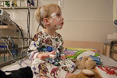 For the standard test the patient comes to a sleep lab in the early evening, and over the next 1–2 hours is introduced to the setting and "wired up" so that multiple channels of data can be recorded when he/she falls asleep. The sleep lab may be in a hospital, a free-standing medical office, or in a hotel. A sleep technician should always be in attendance and is responsible for attaching the electrodes to the patient and monitoring the patient during the study.
During the study, the technician observes sleep activity by looking at the video monitor and the computer screen that displays all the data second by second. In most labs the test is completed and the patient is discharged home by 7 a.m. unless a Multiple Sleep Latency Test
(MSLT) is to be done during the day to test for excessive daytime sleepiness
.
Most recently, physicians may prescribe home studies to enhance patient comfort and reduce expense. The patient is given instructions after a screening tool is used, uses the equipment at home and returns it the next day. Most screening tools consist of an airflow measuring device (thermistor) and a blood oxygen monitoring device (pulse oximeter). The patient would sleep with the screening device for one to several days, then return the device to the physician. The physician would retrieve data from the device and could make assumptions based on the information given ex. series of drastic blood oxygen desaturations during night periods may indicate some form of respiratory event (apnea). The equipment monitors, at a minimum, oxygen saturation.
 After the test is completed a "scorer" analyzes the data by reviewing the study in 30 second "epochs".
The score consists of the following information:
Once scored, the test recording and the scoring data are sent to the sleep medicine physician for interpretation. Ideally, interpretation is done in conjunction with the medical history, a complete list of drugs the patient is taking, and any other relevant information that might impact the study such as napping done before the test.
Once interpreted, the sleep physician writes a report which is sent to the referring physician, usually with specific recommendations based on the test results.
. CPAP is continuous positive airway pressure and is delivered via a mask to the patient's nose or the patient's nose and mouth. (Some masks cover one, some both). CPAP is typically prescribed after the diagnosis of OSA is made from a sleep study (i.e., after a PSG test). To determine the correct amount of pressure and the right mask type and size, and also to make sure the patient can tolerate this therapy, a "CPAP titration study" is recommended. This is the same as a "PSG", but with the addition of the mask applied, so the technician can increase the airway pressure inside the mask as needed, until all, or most, of the patient's airway obstructions are eliminated.
The above report recommends Mr. J---- return for a CPAP titration study, which means a return to the lab for a second all-night PSG (this one with the mask applied). Often, however, when a patient manifests OSA in the first 2 or 3 hours of the initial PSG, the technician will interrupt the study and apply the mask right then and there; the patient is awakened and fitted for a mask. The rest of the sleep study is then a "CPAP titration." When both the diagnostic PSG and a CPAP titration are done the same night, the entire study is called "Split Night".
The split-night study has these advantages:
The split-night study has these disadvantages:
Because of costs, more and more studies for "sleep apnea" are attempted as split-night studies when there is early evidence for OSA. (Note that both types of study, with and without a CPAP mask, are still polysomnograms.) When the CPAP mask is worn, however, the flow-measurement lead in the patient's nose is removed. Instead, the CPAP machine relays all flow-measurement data to the computer.
Parameter
Parameter from Ancient Greek παρά also “para” meaning “beside, subsidiary” and μέτρον also “metron” meaning “measure”, can be interpreted in mathematics, logic, linguistics, environmental science and other disciplines....
test used in the study of sleep
Sleep
Sleep is a naturally recurring state characterized by reduced or absent consciousness, relatively suspended sensory activity, and inactivity of nearly all voluntary muscles. It is distinguished from quiet wakefulness by a decreased ability to react to stimuli, and is more easily reversible than...
and as a diagnostic tool in sleep medicine
Sleep medicine
Sleep medicine is a medical specialty or subspecialty devoted to the diagnosis and therapy of sleep disturbances and disorders. From the middle of the 20th century, research has provided increasing knowledge and answered many questions about sleep-wake functioning. The rapidly evolving field has...
. The test result is called a polysomnogram, also abbreviated PSG. The name is derived from Greek
Greek language
Greek is an independent branch of the Indo-European family of languages. Native to the southern Balkans, it has the longest documented history of any Indo-European language, spanning 34 centuries of written records. Its writing system has been the Greek alphabet for the majority of its history;...
and Latin roots: the Greek πολύς (polus for "many, much", indicating many channels), the Latin somnus ("sleep"), and the Greek γράφειν (graphein, "to write").
Polysomnography is a comprehensive recording of the biophysiological changes that occur during sleep. It is usually performed at night, when most people sleep, though some labs can accommodate shift workers and people with circadian rhythm sleep disorder
Circadian rhythm sleep disorder
Circadian rhythm sleep disorders are a family of sleep disorders affecting, among other things, the timing of sleep. People with circadian rhythm sleep disorders are unable to sleep and wake at the times required for normal work, school, and social needs. They are generally able to get enough sleep...
s and do the test at other times of day. The PSG monitors many body functions including brain
Human brain
The human brain has the same general structure as the brains of other mammals, but is over three times larger than the brain of a typical mammal with an equivalent body size. Estimates for the number of neurons in the human brain range from 80 to 120 billion...
(EEG
EEG
EEG commonly refers to electroencephalography, a measurement of the electrical activity of the brain.EEG may also refer to:* Emperor Entertainment Group, a Hong Kong-based entertainment company...
), eye
Human eye
The human eye is an organ which reacts to light for several purposes. As a conscious sense organ, the eye allows vision. Rod and cone cells in the retina allow conscious light perception and vision including color differentiation and the perception of depth...
movements (EOG), muscle activity or skeletal muscle
Skeletal muscle
Skeletal muscle is a form of striated muscle tissue existing under control of the somatic nervous system- i.e. it is voluntarily controlled. It is one of three major muscle types, the others being cardiac and smooth muscle...
activation (EMG
Electromyography
Electromyography is a technique for evaluating and recording the electrical activity produced by skeletal muscles. EMG is performed using an instrument called an electromyograph, to produce a record called an electromyogram. An electromyograph detects the electrical potential generated by muscle...
) and heart
Human heart
The human heart is a muscular organ that provides a continuous blood circulation through the cardiac cycle and is one of the most vital organs in the human body...
rhythm (ECG) during sleep. After the identification of the sleep disorder
Sleep disorder
A sleep disorder, or somnipathy, is a medical disorder of the sleep patterns of a person or animal. Some sleep disorders are serious enough to interfere with normal physical, mental and emotional functioning...
sleep apnea
Sleep apnea
Sleep apnea is a sleep disorder characterized by abnormal pauses in breathing or instances of abnormally low breathing, during sleep. Each pause in breathing, called an apnea, can last from a few seconds to minutes, and may occur 5 to 30 times or more an hour. Similarly, each abnormally low...
in the 1970s, the breathing functions respiratory
Respiration (physiology)
'In physiology, respiration is defined as the transport of oxygen from the outside air to the cells within tissues, and the transport of carbon dioxide in the opposite direction...
airflow and respiratory effort indicators were added along with peripheral pulse oximetry
Pulse oximetry
Pulse oximetry is a non-invasive method allowing the monitoring of the oxygenation of a patient's hemoglobin.A sensor is placed on a thin part of the patient's body, usually a fingertip or earlobe, or in the case of an infant, across a foot....
.
Indications
Polysomnography is used to diagnose, or rule out, many types of sleep disorderSleep disorder
A sleep disorder, or somnipathy, is a medical disorder of the sleep patterns of a person or animal. Some sleep disorders are serious enough to interfere with normal physical, mental and emotional functioning...
s including narcolepsy, periodic limb movement disorder (PLMD), REM behavior disorder, parasomnias, and sleep apnea. It is often ordered for patients with complaints of daytime fatigue or sleepiness that may be caused by interrupted sleep. Although it is not directly useful in diagnosing circadian rhythm sleep disorders, it may be used to rule out other sleep disorders.
Mechanism
A polysomnogram will typically record a minimum of twelve channels requiring a minimum of 22 wire attachments to the patient. These channels vary in every lab and may be adapted to meet the doctor's requests. There is a minimum of three channels for the EEG, one or two measure airflow, one or two are for chin muscle tone, one or more for leg movements, two for eye movements (EOG), one or two for heart rate and rhythm, one for oxygen saturation and one each for the belts which measure chest wall movement and upper abdominal wall movement. The movement of the belts is typically measured with piezoelectric sensors or respiratory inductance plethysmographyRespiratory inductance plethysmography
Respiratory Inductance Plethysmography is a method of evaluating pulmonary ventilation by measuring the movement of the chest and abdominal wall....
. This movement is equated to effort and produces a low-frequency sinusoidal waveform as the patient inhales and exhales. Because movement is equated to effort, this system of measurement can produce false positives. It is possible, especially during obstructive apneas, for effort to be made without measurable movement.
Wires for each channel of recorded data lead from the patient and converge into a central box, which in turn is connected to a computer system for recording, storing and displaying the data. During sleep the computer monitor can display multiple channels continuously. In addition, most labs have a small video camera in the room so the technician can observe the patient visually from an adjacent room.
The electroencephalogram (EEG) will generally use six "exploring" electrodes and two "reference" electrodes, unless a seizure disorder is suspected, in which case more electrodes will be applied to document the appearance of seizure activity. The exploring electrodes are usually attached to the scalp near the frontal, central (top) and occipital (back) portions of the brain via a paste that will conduct electrical signals originating from the neurons of the cortex. These electrodes will provide a readout of the brain activity that can be "scored" into different stages of sleep (N1, N2, N3 which combined are referred to as NREM sleep, and Stage R which is rapid eye movement sleep or REM, and Wakefulness). The EEG electrodes are placed according to the International 10-20 system.
The electrooculogram (EOG) uses two electrodes; one that is placed 1 cm above the outer canthus
Canthus (anatomy)
Canthus is either corner of the eye where the upper and lower eyelids meet. More specifically, the medial and lateral canthi would be described as the medial and lateral ends/angles of the palpebral fissure....
of the right eye and one that is placed 1 cm below the outer canthus of the left eye. These electrodes pick up the activity of the eyes in virtue of the electropotential difference between the cornea and the retina (the cornea is positively charged relative to the retina). This helps to determine when REM sleep occurs, of which rapid eye movements are characteristic, and also essentially aids in determining when sleep occurs.
The electromyogram (EMG) typically uses four electrodes to measure muscle tension in the body as well as to monitor for an excessive amount of leg movements during sleep (which may be indicative of periodic limb movement disorder
Nocturnal myoclonus
Periodic limb movement disorder , previously known as nocturnal myoclonus, is a sleep disorder where the patient moves limbs involuntarily during sleep, and has symptoms or problems related to the movement....
, PLMD). Two leads are placed on the chin with one above the jaw line and one below. This, like the EOG, helps determine when sleep occurs as well as REM sleep. Sleep generally includes relaxation and so a marked decrease in muscle tension occurs. A further decrease in skeletal muscle tension occurs in REM sleep. A person becomes partially paralyzed to make acting out of dreams impossible, although people that do not have this paralysis can suffer from REM behavior disorder. Finally, two more leads are placed on the anterior tibialis
Tibialis anterior muscle
In human anatomy, the tibialis anterior is a muscle that originates in the upper two-thirds of the lateral surface of the tibia and inserts into the medial cuneiform and first metatarsal bones of the foot. Its acts to dorsiflex and invert the foot. This muscle is mostly located near the shin.It is...
of each leg to measure leg movements.
Though a typical electrocardiogram
Electrocardiogram
Electrocardiography is a transthoracic interpretation of the electrical activity of the heart over a period of time, as detected by electrodes attached to the outer surface of the skin and recorded by a device external to the body...
(ECG or EKG) would use ten electrodes, only two or three are used for a polysomnogram. They can either be placed under the collar bone on each side of the chest, or one under the collar bone and the other six inches above the waist on either side of the body. These electrodes measure the electrical activity of the heart as it contracts and expands, recording such features as the "P" wave, "QRS" complex, and "T" wave. These can be analyzed for any abnormalities that might be indicative of an underlying heart pathology.
Nasal and oral airflow can be measured using pressure transducers, and/or a thermocouple, fitted in or near the nostrils; the pressure transducer is considered the more sensitive. This allows the clinician/researcher to measure the rate of respiration and identify interruptions in breathing. Respiratory effort is also measured in concert with nasal/oral airflow by the use of belts. These belts expand and contract upon breathing effort. However, this method of respiration may also produce false positives. Some patients will open and close their mouth while obstructive apneas occur. This forces air in and out of the mouth while no air enters the airway and lungs. Thus, the pressure transducer and thermocouple will detect this diminished airflow and the respiratory event may be falsely identified as a hypopnea, or a period of reduced airflow, instead of an obstructive apnea.
Pulse oximetry determines changes in blood oxygen levels that often occur with sleep apnea and other respiratory problems. The pulse oximeter fits over a finger tip or an ear lobe.
Snoring
Snoring
Snoring is the vibration of respiratory structures and the resulting sound, due to obstructed air movement during breathing while sleeping. In some cases the sound may be soft, but in other cases, it can be loud and unpleasant...
may be recorded with a sound probe over the neck, though more commonly the sleep technician will just note snoring as "mild", "moderate" or "loud" or give a numerical estimate on a scale of 1 to 10. Also, snoring indicates airflow and can be used during hypopneas to determine whether the hypopnea may be an obstructive apnea.
Procedure
During the study, the technician observes sleep activity by looking at the video monitor and the computer screen that displays all the data second by second. In most labs the test is completed and the patient is discharged home by 7 a.m. unless a Multiple Sleep Latency Test
Multiple Sleep Latency Test
The Multiple Sleep Latency Test is a sleep disorder diagnostic tool. It is used to measure the time elapsed from the start of a daytime nap period to the first signs of sleep, called sleep latency...
(MSLT) is to be done during the day to test for excessive daytime sleepiness
Excessive daytime sleepiness
Excessive daytime sleepiness is characterized by persistent sleepiness, and often a general lack of energy, even after apparently adequate night time sleep...
.
Most recently, physicians may prescribe home studies to enhance patient comfort and reduce expense. The patient is given instructions after a screening tool is used, uses the equipment at home and returns it the next day. Most screening tools consist of an airflow measuring device (thermistor) and a blood oxygen monitoring device (pulse oximeter). The patient would sleep with the screening device for one to several days, then return the device to the physician. The physician would retrieve data from the device and could make assumptions based on the information given ex. series of drastic blood oxygen desaturations during night periods may indicate some form of respiratory event (apnea). The equipment monitors, at a minimum, oxygen saturation.
Interpretation
The score consists of the following information:
- Onset of sleep from time the lights were turned off; this is called "sleep onset latencySleep onset latencyIn sleep science, sleep onset latency is the length of time that it takes to accomplish the transition from full wakefulness to sleep, normally to the lightest of the non-REM sleep stages.-Sleep latency studies:...
" and normally is less than 20 minutes. (Note that determining "sleep" and "awake" is based solely on the EEG. Patients sometimes feel they were awake when the EEG shows they were sleeping. This may be because of sleep state misperception, drug effects on brain waves, or individual differences in brain waves.) - Sleep efficiency: the number of minutes of sleep divided by the number of minutes in bed. Normal is approximately 85 to 90% or higher.
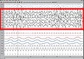
- Sleep stages; these are based on 3 sources of data coming from 7 channels: EEG (4 channels usually), EOG (2) and chin EMG (1). From this information each 30-second epoch is scored as "awake" or one of 4 sleep stages: 1, 2, 3, and REM or Rapid Eye Movement sleep. Stages 1–3 are together called non-REM sleep. Non-REM sleep is distinguished from REM sleep, which is altogether different. Within non-REM sleep, stage 3 is called "slow wave" sleep because of the relatively wide brain waves compared to other stages; another name for stage 3 is "deep sleep". By contrast, stage 1 and 2 are "light sleep". The figures show stage 3 sleep and REM sleep; each figure is a 30-second epoch from an overnight PSG.
(The percentage of each sleep stage varies by age, with decreasing amounts of REM and deep sleep in older people. The majority of sleep at all ages (except infancy) is Stage 2. REM normally occupies about 20-25% of sleep time. Many factors besides age can affect both the amount and percentage of each sleep stage, including drugs (particularly anti-depressants and pain meds), alcohol taken before bed time, and sleep deprivation.)
- Any breathing irregularities; mainly apneas and hypopneas. Apnea is a complete or near complete cessation of airflow for at least 10 seconds followed by an arousal and/or 3% oxygen desaturation; hypopnea is a 50% decrease in airflow for at least 10 seconds followed by an arousal and/or 3% oxygen desaturation. (Medicare requires a 4% desaturation in order to include the event in the report.)
- "Arousals" are sudden shifts in brain wave activity. They may be caused by numerous factors, including breathing abnormalities, leg movements, environmental noises, etc. An abnormal number of arousals indicates "interrupted sleep" and may explain a person's daytime symptoms of fatigue and/or sleepiness.
- Cardiac rhythm abnormalities.
- Leg movements.
- Body position during sleep.
- Oxygen saturation during sleep.
Once scored, the test recording and the scoring data are sent to the sleep medicine physician for interpretation. Ideally, interpretation is done in conjunction with the medical history, a complete list of drugs the patient is taking, and any other relevant information that might impact the study such as napping done before the test.
Once interpreted, the sleep physician writes a report which is sent to the referring physician, usually with specific recommendations based on the test results.
Example of summary report
"Split night" study
The above report mentions CPAP as treatment for obstructive sleep apneaSleep apnea
Sleep apnea is a sleep disorder characterized by abnormal pauses in breathing or instances of abnormally low breathing, during sleep. Each pause in breathing, called an apnea, can last from a few seconds to minutes, and may occur 5 to 30 times or more an hour. Similarly, each abnormally low...
. CPAP is continuous positive airway pressure and is delivered via a mask to the patient's nose or the patient's nose and mouth. (Some masks cover one, some both). CPAP is typically prescribed after the diagnosis of OSA is made from a sleep study (i.e., after a PSG test). To determine the correct amount of pressure and the right mask type and size, and also to make sure the patient can tolerate this therapy, a "CPAP titration study" is recommended. This is the same as a "PSG", but with the addition of the mask applied, so the technician can increase the airway pressure inside the mask as needed, until all, or most, of the patient's airway obstructions are eliminated.
The above report recommends Mr. J---- return for a CPAP titration study, which means a return to the lab for a second all-night PSG (this one with the mask applied). Often, however, when a patient manifests OSA in the first 2 or 3 hours of the initial PSG, the technician will interrupt the study and apply the mask right then and there; the patient is awakened and fitted for a mask. The rest of the sleep study is then a "CPAP titration." When both the diagnostic PSG and a CPAP titration are done the same night, the entire study is called "Split Night".
The split-night study has these advantages:
- The patient only has to come to the lab once, so it is less disruptive than is coming two different nights;
- It is "half as expensive" to whoever is paying for the study.
The split-night study has these disadvantages:
- There is less time to make a diagnosis of OSA (Medicare requires a minimum of 2 hours of diagnosis time before the mask can be applied); and
- There is less time to assure an adequate CPAP titration. If the titration begins with only a few hours of sleep left, the remaining time may not assure a proper CPAP titration, and the patient may still have to return to the lab.
Because of costs, more and more studies for "sleep apnea" are attempted as split-night studies when there is early evidence for OSA. (Note that both types of study, with and without a CPAP mask, are still polysomnograms.) When the CPAP mask is worn, however, the flow-measurement lead in the patient's nose is removed. Instead, the CPAP machine relays all flow-measurement data to the computer.
Example of summary report from a "split night" study
See also
- Sleep medicineSleep medicineSleep medicine is a medical specialty or subspecialty devoted to the diagnosis and therapy of sleep disturbances and disorders. From the middle of the 20th century, research has provided increasing knowledge and answered many questions about sleep-wake functioning. The rapidly evolving field has...
- Sleep disorderSleep disorderA sleep disorder, or somnipathy, is a medical disorder of the sleep patterns of a person or animal. Some sleep disorders are serious enough to interfere with normal physical, mental and emotional functioning...
- Polysomnographic technician
- Respiratory monitoringRespiratory monitoringMonitoring a patient's respiratory status usually takes place in a hospital setting and may be the primary purpose for a patient being observed or admitted to a medical setting....
Further reading
- Iber C, Ancoli-Israel S, Chesson A, and Quan SF for the American Academy of Sleep Medicine. The AASM Manual for the Scoring of Sleep and Associated Events: Rules, Terminology and Technical Specifications, 1st ed.: Westchester, Illinois: American Academy of Sleep Medicine, 2007.