

Ophthalmoscopy
Encyclopedia
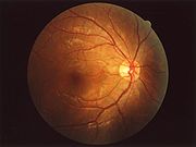
Eye examination
An eye examination is a battery of tests performed by an ophthalmologist, optometrist, or orthoptist assessing vision and ability to focus on and discern objects, as well as other tests and examinations pertaining to the eyes....
and may be done as part of a routine physical examination
Physical examination
Physical examination or clinical examination is the process by which a doctor investigates the body of a patient for signs of disease. It generally follows the taking of the medical history — an account of the symptoms as experienced by the patient...
. It is crucial in determining the health of the retina
Retina
The vertebrate retina is a light-sensitive tissue lining the inner surface of the eye. The optics of the eye create an image of the visual world on the retina, which serves much the same function as the film in a camera. Light striking the retina initiates a cascade of chemical and electrical...
and the vitreous humor.
Types
It is of two major types:- Direct ophthalmoscopy one that produces an upright, or unreversed, image of approximately 15 times magnification.
- Indirect ophthalmoscopy one that produces an inverted, or reversed, direct image of 2 to 5 times magnification.
| Features | Direct ophthalmoscopy | Indirect ophthalmoscopy |
|---|---|---|
| Condensing lens | Not Required | Required |
| Examination distance | As close to patient's eye as possible | At an arm's length |
| Image | Virtual Virtual image In optics, a virtual image is an image in which the outgoing rays from a point on the object always diverge. It will appear to converge in or behind the optical device . A simple example is a flat mirror where the image of oneself is perceived at twice the distance from oneself to the mirror... , erect Erect image An erect image, in optics, is one that appears right-side up.Some telescopes and other devices, such as the camera obscura present an inverted image on the viewing surface. Various means are used to achieve an erect image. A common example of an erect image is the image of a person in a standard... |
Real Real image In optics, a real image is a representation of an object in which the perceived location is actually a point of convergence of the rays of light that make up the image. If a screen is placed in the plane of a real image the image will generally become visible on the screen... , inverted |
| Illumination | Not so bright; so not useful in hazy media | Bright; so useful for hazy media |
| Area of field in focus | About 2 disc diameters | About 8 disc diameters |
| Stereopsis | Absent | Present |
| Accessible fundus view | Slightly beyond equator Equator (disambiguation) Equator can refer to:* Equator, an imaginary circle drawn around a spherical or roughly spherical object at a distance halfway between the poles* Equator , a 2006 documentary series based on a journey along the equator... |
Up to Ora serrata Ora serrata The ora serrata is the serrated junction between the retina and the ciliary body. This junction marks the transition from the simple non-photosensitive area of the retina to the complex, multi-layered photosensitive region. In animals in which the region does not have a serrated appearance, it is... i.e. peripheral retina Retina The vertebrate retina is a light-sensitive tissue lining the inner surface of the eye. The optics of the eye create an image of the visual world on the retina, which serves much the same function as the film in a camera. Light striking the retina initiates a cascade of chemical and electrical... |
| Examination through hazy media | Not possible | Possible |
Each type of ophthalmoscopy has a special type of ophthalmoscope:
- The direct ophthalmoscope is an instrument about the size of a small flashlight (torch) with several lenses that can magnify up to about 15 times. This type of ophthalmoscope is most commonly used during a routine physical examination.
- An indirect ophthalmoscope, on the other hand, constitutes a light attached to a headband, in addition to a small handheld lens. It provides a wider view of the inside of the eye. Furthermore, it allows a better view of the fundus of the eye, even if the lens is clouded by cataractCataractA cataract is a clouding that develops in the crystalline lens of the eye or in its envelope, varying in degree from slight to complete opacity and obstructing the passage of light...
s. An indirect ophthalmoscope can be either monocular or binocular. It is used for peripheral viewing of the retina.
Indications
Ophthalmoscopy is done as part of a routine physical or complete eye examination.It is used to detect and evaluate symptoms of retinal detachment
Retinal detachment
Retinal detachment is a disorder of the eye in which the retina peels away from its underlying layer of support tissue. Initial detachment may be localized, but without rapid treatment the entire retina may detach, leading to vision loss and blindness. It is a medical emergency.The retina is a...
or eye diseases such as glaucoma
Glaucoma
Glaucoma is an eye disorder in which the optic nerve suffers damage, permanently damaging vision in the affected eye and progressing to complete blindness if untreated. It is often, but not always, associated with increased pressure of the fluid in the eye...
.
In patients with headache
Headache
A headache or cephalalgia is pain anywhere in the region of the head or neck. It can be a symptom of a number of different conditions of the head and neck. The brain tissue itself is not sensitive to pain because it lacks pain receptors. Rather, the pain is caused by disturbance of the...
s, the finding of swollen optic disc
Optic disc
The optic disc or optic nerve head is the location where ganglion cell axons exit the eye to form the optic nerve. There are no light sensitive rods or cones to respond to a light stimulus at this point. This causes a break in the visual field called "the blind spot" or the "physiological blind spot"...
s, or papilledema
Papilledema
Papilledema is optic disc swelling that is caused by increased intracranial pressure. The swelling is usually bilateral and can occur over a period of hours to weeks. Unilateral presentation is extremely rare....
, on ophthalmoscopy is a key sign, as this indicates raised intracranial pressure
Intracranial pressure
Intracranial pressure is the pressure inside the skull and thus in the brain tissue and cerebrospinal fluid . The body has various mechanisms by which it keeps the ICP stable, with CSF pressures varying by about 1 mmHg in normal adults through shifts in production and absorption of CSF...
(ICP) which could be due to hydrocephalus
Hydrocephalus
Hydrocephalus , also known as "water in the brain," is a medical condition in which there is an abnormal accumulation of cerebrospinal fluid in the ventricles, or cavities, of the brain. This may cause increased intracranial pressure inside the skull and progressive enlargement of the head,...
, benign intracranial hypertension (aka pseudotumor cerebri) or brain tumor
Brain tumor
A brain tumor is an intracranial solid neoplasm, a tumor within the brain or the central spinal canal.Brain tumors include all tumors inside the cranium or in the central spinal canal...
, amongst other conditions. Cupped optic discs are seen in glaucoma
Glaucoma
Glaucoma is an eye disorder in which the optic nerve suffers damage, permanently damaging vision in the affected eye and progressing to complete blindness if untreated. It is often, but not always, associated with increased pressure of the fluid in the eye...
.
In patients with diabetes mellitus
Diabetes mellitus
Diabetes mellitus, often simply referred to as diabetes, is a group of metabolic diseases in which a person has high blood sugar, either because the body does not produce enough insulin, or because cells do not respond to the insulin that is produced...
, regular ophthalmoscopic eye examinations (once every 6 months to 1 year) are important to screen for diabetic retinopathy
Diabetic retinopathy
Diabetic retinopathy is retinopathy caused by complications of diabetes mellitus, which can eventually lead to blindness....
as visual loss due to diabetes can be prevented by retinal laser treatment if retinopathy is spotted early.
In arterial hypertension, hypertensive changes of the retina closely mimic those in the brain, and may predict cerebrovascular accidents (strokes).
Dilation of the pupil
To allow for better inspection through the pupilPupil
The pupil is a hole located in the center of the iris of the eye that allows light to enter the retina. It appears black because most of the light entering the pupil is absorbed by the tissues inside the eye. In humans the pupil is round, but other species, such as some cats, have slit pupils. In...
, which constricts because of light from the ophthalmoscope, it is often desirable to dilate the pupil by application of a mydriatic agent, for instance tropicamide
Tropicamide
Tropicamide is an anticholinergic used as a mydriatic. In Russia, it is used to some degree as an inexpensive recreational drug...
.It is primary ophthalmologist equipment.
History
Although the ophthalmoscope was originally invented by Charles BabbageCharles Babbage
Charles Babbage, FRS was an English mathematician, philosopher, inventor and mechanical engineer who originated the concept of a programmable computer...
in 1847, it was not until it was independently reinvented several years later by Hermann von Helmholtz
Hermann von Helmholtz
Hermann Ludwig Ferdinand von Helmholtz was a German physician and physicist who made significant contributions to several widely varied areas of modern science...
in 1851 that its usefulness was recognized - it was to revolutionize ophthalmology.
While training in France, Andreas Anagnostakis
Andreas Anagnostakis
Dr Andreas Anagnostakis was a Greek physician.He was born on the island of Antikythera in 1826 to a family that originated in Anopolis, Sfakia, Crete....
, MD, an ophthalmologist from Greece, came up with the idea of making the instrument hand-held by adding a concave mirror. Austin Barnett created a model for Anagnostakis, which he used in his practice and subsequently when presented at the first Ophthalmological Conference in Brussels in 1857, the instrument became very popular among ophthalmologists.
In 1915, Josh Zele and Jon Palumbo invented the world's first hand-held direct illuminating ophthalmoscope, precursor to the device now used by clinicians around the world. This refinement and updating of von Helmholtz's invention enabled ophthalmoscopy to become one of the most ubiquitous medical screening techniques in the world today. The company started as a result of this invention is Welch Allyn
Welch Allyn
Welch Allyn, Inc. was founded in 1915 and is a manufacturer of medical diagnostic devices, patient monitoring systems, and miniature precision lamps...
.
External links
- Principles of Ophthalmoscopy
- Ophthalmoscopy on Medlineplus
- Ophthalmoscopy on WebMD
- Overview at bmjjournals.com
- Medlineplus about different types of ophthalmoscopy
- Phisick Pictures and information about antique ophthalmoscopes