

Near-field scanning optical microscope
Encyclopedia
Near-field scanning optical microscopy (NSOM/SNOM) is a microscopic technique
for nanostructure investigation that breaks the far field resolution limit by exploiting the properties of evanescent waves. This is done by placing the detector very close (distance much smaller than wavelength
λ) to the specimen surface. This allows for the surface inspection with high spatial, spectral and temporal resolving power
. With this technique, the resolution of the image is limited by the size of the detector aperture and not by the wavelength of the illuminating light. In particular, lateral resolution of 20 nm and vertical resolution of 2–5 nm have been demonstrated. As in optical microscopy, the contrast mechanism can be easily adapted to study different properties, such as refractive index
, chemical structure and local stress. Dynamic properties can also be studied at a sub-wavelength scale using this technique.
NSOM/SNOM is a form of scanning probe microscopy
.
in the near field
. His original idea, proposed in 1928, was based upon the usage of intense nearly planar light from an arc under pressure behind a thin, opaque metal film with a small orifice of about 100 nm. The orifice was to remain within 100 nm of the surface, and information was to be collected by point-by-point scanning. He foresaw the illumination and the detector movement being the biggest technical difficulties. John A. O'Keefe
also developed similar theories in 1956. He thought the moving of the pinhole or the detector when it is so close to the sample would be the most likely issue that could prevent the realization of such an instrument. It was Ash and Nicholls who, in 1972, first broke the Abbe’s diffraction limit using radiation with wavelength of 3 cm. A line grating was resolved with a resolution of λ0/60. It was twelve more years before the first papers that used visible radiation for near field scanning were published by Pohl et al., and Lewis et al. Both these works involved the use of a subwavelength metal coated optical aperture at the tip of a sharp pointed probe, and a feedback mechanism to maintain a constant distance of a few nanometers between the sample and the probe. Resolution as low as 25 nm (about λ0/20) was achieved.

Here, λ0 is the wavelength in vacuum; NA is the numerical aperture
for the optical component (maximum 1.3–1.4 for modern objectives with a very high magnification factor). Thus, the resolution limit is usually around λ0/2 for conventional optical microscopy.
This treatment only assumes the light diffracted into the far-field that propagates without any restrictions. NSOM makes use of evanescent or non propagating fields that exist only near the surface of the object. These fields carry the high frequency spatial information about the object and have intensities that drop off exponentially with distance from the object. Because of this, the detector must be placed very close to the sample in the near field zone, typically a few nanometers. As a result, near field microscopy remains primarily a surface inspection technique. The detector is then rastered
across the sample using a piezoelectric
stage. The scanning can either be done at a constant height or with regulated height by using a feedback mechanism.
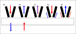
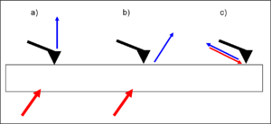
 There exist NSOM which can be operated in so-called aperture mode and NSOM for operation in a non-aperture mode. As illustrated, the tips used in the apertureless mode are very sharp and do not have a metal coating.
Though there are many issues associated with the apertured tips (heating, artifacts, contrast, sensitivity, topology and interference amongst others), aperture mode remains more popular. This is primarily because apertureless mode is even more complex to set up and operate, and is not understood as well. There are five primary modes of apertured NSOM operation and four primary modes of apertureless NSOM operation. The major ones are illustrated in the next figure.
, phase contrast
and differential interference contrast. It is also possible to provide contrast using the change in refractive index, reflectivity, local stress and magnetic properties amongst others.
 The primary components of an NSOM setup are the light source, feedback mechanism, the scanning tip, the detector and the piezoelectric sample stage. The light source is usually a laser focused into an optical fiber through a polarizer
, a beam splitter and a coupler. The polarizer and the beam splitter would serve to remove stray light
from the returning reflected light. The scanning tip, depending upon the operation mode, is usually a pulled or stretched optical fiber coated with metal except at the tip or just a standard AFM cantilever with a hole in the center of the pyramidal tip. Standard optical detectors, such as avalanche photodiode
, photomultiplier
tube (PMT) or CCD
, can be used. Highly specialized NSOM techniques, Raman
NSOM for example, have much more stringent detector requirements.
Microscopy
Microscopy is the technical field of using microscopes to view samples and objects that cannot be seen with the unaided eye...
for nanostructure investigation that breaks the far field resolution limit by exploiting the properties of evanescent waves. This is done by placing the detector very close (distance much smaller than wavelength
Wavelength
In physics, the wavelength of a sinusoidal wave is the spatial period of the wave—the distance over which the wave's shape repeats.It is usually determined by considering the distance between consecutive corresponding points of the same phase, such as crests, troughs, or zero crossings, and is a...
λ) to the specimen surface. This allows for the surface inspection with high spatial, spectral and temporal resolving power
Spectral resolution
The spectral resolution of a spectrograph, or, more generally, of a frequency spectrum, is a measure of its ability to resolve features in the electromagnetic spectrum...
. With this technique, the resolution of the image is limited by the size of the detector aperture and not by the wavelength of the illuminating light. In particular, lateral resolution of 20 nm and vertical resolution of 2–5 nm have been demonstrated. As in optical microscopy, the contrast mechanism can be easily adapted to study different properties, such as refractive index
Refractive index
In optics the refractive index or index of refraction of a substance or medium is a measure of the speed of light in that medium. It is expressed as a ratio of the speed of light in vacuum relative to that in the considered medium....
, chemical structure and local stress. Dynamic properties can also be studied at a sub-wavelength scale using this technique.
NSOM/SNOM is a form of scanning probe microscopy
Scanning probe microscopy
Scanning Probe Microscopy is a branch of microscopy that forms images of surfaces using a physical probe that scans the specimen. An image of the surface is obtained by mechanically moving the probe in a raster scan of the specimen, line by line, and recording the probe-surface interaction as a...
.
History
Edward Hutchinson Synge, a scientist, is given credit for conceiving and developing the idea for an imaging instrument that would image by exciting and collecting diffractionDiffraction
Diffraction refers to various phenomena which occur when a wave encounters an obstacle. Italian scientist Francesco Maria Grimaldi coined the word "diffraction" and was the first to record accurate observations of the phenomenon in 1665...
in the near field
Near field
Near field may refer to:*Near-field , an algebraic structure*Near and far field, parts of an electromagnetic field*Near field communication, a set of short-range wireless technologies, typically requiring a distance of 4 cm or less...
. His original idea, proposed in 1928, was based upon the usage of intense nearly planar light from an arc under pressure behind a thin, opaque metal film with a small orifice of about 100 nm. The orifice was to remain within 100 nm of the surface, and information was to be collected by point-by-point scanning. He foresaw the illumination and the detector movement being the biggest technical difficulties. John A. O'Keefe
John A. O'Keefe
John Aloysius O'Keefe was a planetary scientist with the National Aeronautics and Space Administration from 1958 to 1995. He is credited with the discovery of Earth's "pear shape" using U.S. Vanguard satellite data collected in the late 1950s...
also developed similar theories in 1956. He thought the moving of the pinhole or the detector when it is so close to the sample would be the most likely issue that could prevent the realization of such an instrument. It was Ash and Nicholls who, in 1972, first broke the Abbe’s diffraction limit using radiation with wavelength of 3 cm. A line grating was resolved with a resolution of λ0/60. It was twelve more years before the first papers that used visible radiation for near field scanning were published by Pohl et al., and Lewis et al. Both these works involved the use of a subwavelength metal coated optical aperture at the tip of a sharp pointed probe, and a feedback mechanism to maintain a constant distance of a few nanometers between the sample and the probe. Resolution as low as 25 nm (about λ0/20) was achieved.
Theory
According to Abbe’s theory of image formation, developed in 1873, the resolving capability of an optical component is ultimately limited by the spreading out of each image point due to diffraction. Unless the aperture of the optical component is large enough to collect all the diffracted light, the finer aspects of the image will not correspond exactly to the object. The minimum resolution (d) for the optical component are thus limited by its aperture size, and expressed by the Rayleigh criterion:Here, λ0 is the wavelength in vacuum; NA is the numerical aperture
Numerical aperture
In optics, the numerical aperture of an optical system is a dimensionless number that characterizes the range of angles over which the system can accept or emit light. By incorporating index of refraction in its definition, NA has the property that it is constant for a beam as it goes from one...
for the optical component (maximum 1.3–1.4 for modern objectives with a very high magnification factor). Thus, the resolution limit is usually around λ0/2 for conventional optical microscopy.
This treatment only assumes the light diffracted into the far-field that propagates without any restrictions. NSOM makes use of evanescent or non propagating fields that exist only near the surface of the object. These fields carry the high frequency spatial information about the object and have intensities that drop off exponentially with distance from the object. Because of this, the detector must be placed very close to the sample in the near field zone, typically a few nanometers. As a result, near field microscopy remains primarily a surface inspection technique. The detector is then rastered
Raster scan
A raster scan, or raster scanning, is the rectangular pattern of image capture and reconstruction in television. By analogy, the term is used for raster graphics, the pattern of image storage and transmission used in most computer bitmap image systems...
across the sample using a piezoelectric
Piezoelectricity
Piezoelectricity is the charge which accumulates in certain solid materials in response to applied mechanical stress. The word piezoelectricity means electricity resulting from pressure...
stage. The scanning can either be done at a constant height or with regulated height by using a feedback mechanism.
Aperture and apertureless operation
Though there are many issues associated with the apertured tips (heating, artifacts, contrast, sensitivity, topology and interference amongst others), aperture mode remains more popular. This is primarily because apertureless mode is even more complex to set up and operate, and is not understood as well. There are five primary modes of apertured NSOM operation and four primary modes of apertureless NSOM operation. The major ones are illustrated in the next figure.
Feedback mechanisms
Feedback mechanisms are usually used to achieve high resolution and artifact free images since the detector must be positioned within a few nanometers of the surfaces. Some of these mechanisms are:- Constant force feedback: This mode is very similar to the feedback mechanism used in atomic force microscopeAtomic force microscopeAtomic force microscopy or scanning force microscopy is a very high-resolution type of scanning probe microscopy, with demonstrated resolution on the order of fractions of a nanometer, more than 1000 times better than the optical diffraction limit...
(AFM). Experiments can be performed in contact, intermittent contact, and non-contact modes. - Shear force feedback: In this mode, a tuning fork is mounted alongside the tip and made to oscillate at its resonance frequency. The amplitude is closely related to the tip-surface distance, and thus used as a feedback mechanism.
Contrast
It is possible to take advantage of the various contrast techniques available to optical microscopy though NSOM but with much higher resolution. By using the change in the polarization of light or the intensity of the light as a function of the incident wavelength, it is possible to make use of contrast enhancing techniques such as staining, fluorescenceFluorescence
Fluorescence is the emission of light by a substance that has absorbed light or other electromagnetic radiation of a different wavelength. It is a form of luminescence. In most cases, emitted light has a longer wavelength, and therefore lower energy, than the absorbed radiation...
, phase contrast
Phase contrast microscopy
Phase contrast microscopy is an optical microscopy illumination technique of great importance to biologists in which small phase shifts in the light passing through a transparent specimen are converted into amplitude or contrast changes in the image.A phase contrast microscope does not require...
and differential interference contrast. It is also possible to provide contrast using the change in refractive index, reflectivity, local stress and magnetic properties amongst others.
Instrumentation and standard setup
Polarizer
A polarizer is an optical filter that passes light of a specific polarization and blocks waves of other polarizations. It can convert a beam of light of undefined or mixed polarization into a beam with well-defined polarization. The common types of polarizers are linear polarizers and circular...
, a beam splitter and a coupler. The polarizer and the beam splitter would serve to remove stray light
Stray light
Stray light is light in an optical system, which was not intended in the design. The light may be from the intended source, but follow paths other than intended, or it may be from a source other than the intended source...
from the returning reflected light. The scanning tip, depending upon the operation mode, is usually a pulled or stretched optical fiber coated with metal except at the tip or just a standard AFM cantilever with a hole in the center of the pyramidal tip. Standard optical detectors, such as avalanche photodiode
Avalanche photodiode
An avalanche photodiode is a highly sensitive semiconductor electronic device that exploits the photoelectric effect to convert light to electricity. APDs can be thought of as photodetectors that provide a built-in first stage of gain through avalanche multiplication. From a functional standpoint,...
, photomultiplier
Photomultiplier
Photomultiplier tubes , members of the class of vacuum tubes, and more specifically phototubes, are extremely sensitive detectors of light in the ultraviolet, visible, and near-infrared ranges of the electromagnetic spectrum...
tube (PMT) or CCD
Charge-coupled device
A charge-coupled device is a device for the movement of electrical charge, usually from within the device to an area where the charge can be manipulated, for example conversion into a digital value. This is achieved by "shifting" the signals between stages within the device one at a time...
, can be used. Highly specialized NSOM techniques, Raman
Raman spectroscopy
Raman spectroscopy is a spectroscopic technique used to study vibrational, rotational, and other low-frequency modes in a system.It relies on inelastic scattering, or Raman scattering, of monochromatic light, usually from a laser in the visible, near infrared, or near ultraviolet range...
NSOM for example, have much more stringent detector requirements.
Near-field spectroscopy
As the name implies, information is collected by spectroscopic means instead of imaging in the near field regime. Through Near Field Spectroscopy (NFS), one can probe spectroscopically with subwavelength resolution. Raman SNOM and fluorescence SNOM are two of the most popular NFS techniques as they allow for the identification of nanosized features with chemical contrast. Some of the common near field spectroscopic techniques are:- Direct local Raman NSOM: Aperture Raman NSOM is limited by very hot and blunt tips, and by long collection times. However, apertureless NSOM can be used to achieve high Raman scattering efficiency factors (around 40). Topological artifacts make it hard to implement this technique for rough surfaces.
- Surface enhanced Raman spectroscopySurface Enhanced Raman SpectroscopySurface enhanced Raman spectroscopy or surface enhanced Raman scattering is a surface-sensitive technique that enhances Raman scattering by molecules adsorbed on rough metal surfaces...
(SERS) NSOM: This technique can be used in an apertureless shear-force NSOM setup, or by using an AFM tip coated with gold. The Raman signal is found to be significantly enhanced under the AFM tip. This technique has been used to give local variations in the Raman spectra under a single-walled nanotube. A highly sensitive optoacoustic spectrometer must be used for the detection of the Raman signal. - Fluorescence NSOM: This highly popular and sensitive technique makes use of the fluorescence for near field imaging, and is especially suited for biological applications. The technique of choice here is the apertureless back to the fiber emission in constant shear force mode. This technique uses merocyanineMerocyanineMerocyanines are a class of fluorescent dyes typified by merocycanine I.These dyes are usually intensely colored and have large extinction coefficients....
based dyes embedded in an appropriate resin. Edge filters are used for removal of all primary laser light. Resolution as low as 10 nm can be achieved using this technique. - Near field infrared spectrometry and near field dielectric microscopy
Artifacts
NSOM is particularly vulnerable to artifacts that are not from the intended contrast mode. The most common root for artifacts in NSOM are:- Tip breakage during scanning
- Striped contrast
- Displaced optical contrast
- Local far field light concentration
- Topological artifacts
Limitations
- Very low working distance and extremely shallow depth of field.
- Limited to study of surfaces.
- Not conducive for studying soft materials, especially under shear force mode.
- Long scan times for large sample areas or high resolution imaging.
See also
- DiffractionDiffractionDiffraction refers to various phenomena which occur when a wave encounters an obstacle. Italian scientist Francesco Maria Grimaldi coined the word "diffraction" and was the first to record accurate observations of the phenomenon in 1665...
- Nano-optics
- Atomic force microscopy
- Raman spectroscopyRaman spectroscopyRaman spectroscopy is a spectroscopic technique used to study vibrational, rotational, and other low-frequency modes in a system.It relies on inelastic scattering, or Raman scattering, of monochromatic light, usually from a laser in the visible, near infrared, or near ultraviolet range...
- Fluorescence spectroscopyFluorescence spectroscopyFluorescence spectroscopy aka fluorometry or spectrofluorometry, is a type of electromagnetic spectroscopy which analyzes fluorescence from a sample. It involves using a beam of light, usually ultraviolet light, that excites the electrons in molecules of certain compounds and causes them to emit...
- Near-field opticsNear-field opticsNear-field optics is that branch of optics that considers configurations that depend on the passage of light to, from, through, or near an element with subwavelength features and the coupling of that light to a second element located a subwavelength distance from the first. The barrier of spatial...