

Myofibril
Encyclopedia
Actin
Actin is a globular, roughly 42-kDa moonlighting protein found in all eukaryotic cells where it may be present at concentrations of over 100 μM. It is also one of the most highly-conserved proteins, differing by no more than 20% in species as diverse as algae and humans...
, myosin
Myosin
Myosins comprise a family of ATP-dependent motor proteins and are best known for their role in muscle contraction and their involvement in a wide range of other eukaryotic motility processes. They are responsible for actin-based motility. The term was originally used to describe a group of similar...
, and titin
Titin
Titin , also known as connectin, is a protein that in humans is encoded by the TTN gene. Titin is a giant protein that functions as a molecular spring which is responsible for the passive elasticity of muscle. It is composed of 244 individually folded protein domains connected by unstructured...
, and other proteins that hold them together. These proteins are organized into thin filaments and thick filaments, which repeat along the length of the myofibril in sections called sarcomeres. Muscles contract by sliding the thin (actin) and thick (myosin) filaments along each other.
Actomyosin motors are important in muscle contraction
Muscle contraction
Muscle fiber generates tension through the action of actin and myosin cross-bridge cycling. While under tension, the muscle may lengthen, shorten, or remain the same...
(relying in this case on "classical myosins") as well as other processes like retraction of membrane blebs, filiopod retraction, and uropodium advancement (relying in this case on "nonclassical myosins").
Structure
The filaments of myofibrils, myofilamentMyofilament
Myofilaments, the filaments of myofibrils constructed from proteins,. The principal types of muscle are striated muscle, obliquely striated muscle and smooth muscle. Various arrangements of myofilaments create different muscles. Striated muscle has transverse bands of filaments...
s, consist of two types, thick and thin.
- Thin filaments consist primarily of the protein actinActinActin is a globular, roughly 42-kDa moonlighting protein found in all eukaryotic cells where it may be present at concentrations of over 100 μM. It is also one of the most highly-conserved proteins, differing by no more than 20% in species as diverse as algae and humans...
, coiled with nebulinNebulinNebulin is an actin-binding protein which is localized to the I-band of the sarcomeres in skeletal muscle. It is a very large protein and binds as many as 200 actin monomers. Because its length is proportional to thin filament length, it is believed that nebulin acts as a thin filament "ruler"...
filaments. - Thick filaments consist primarily of the protein myosinMyosinMyosins comprise a family of ATP-dependent motor proteins and are best known for their role in muscle contraction and their involvement in a wide range of other eukaryotic motility processes. They are responsible for actin-based motility. The term was originally used to describe a group of similar...
, held in place by titinTitinTitin , also known as connectin, is a protein that in humans is encoded by the TTN gene. Titin is a giant protein that functions as a molecular spring which is responsible for the passive elasticity of muscle. It is composed of 244 individually folded protein domains connected by unstructured...
filaments.
The protein complex composed of actin and myosin is sometimes referred to as "actomyosin."
In striated muscle, such as skeletal
Skeletal muscle
Skeletal muscle is a form of striated muscle tissue existing under control of the somatic nervous system- i.e. it is voluntarily controlled. It is one of three major muscle types, the others being cardiac and smooth muscle...
and cardiac muscle
Cardiac muscle
Cardiac muscle is a type of involuntary striated muscle found in the walls and histologic foundation of the heart, specifically the myocardium. Cardiac muscle is one of three major types of muscle, the others being skeletal and smooth muscle...
, the actin and myosin filaments each have a specific and constant length on the order of a few micrometers, far less than the length of the elongated muscle cell (a few millimeters in the case of human skeletal muscle cells). The filaments are organized into repeated subunits along the length of the myofibril. These subunits are called sarcomere
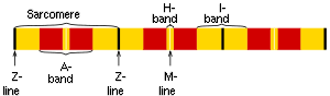
Sarcomere
A sarcomere is the basic unit of a muscle. Muscles are composed of tubular muscle cells . Muscle cells are composed of tubular myofibrils. Myofibrils are composed of repeating sections of sarcomeres, which appear under the microscope as dark and light bands...
s. The muscle cell is nearly filled with myofibrils running parallel to each other on the long axis of the cell. The sarcomeric subunits of one myofibril are in nearly perfect alignment with those of the myofibrils next to it. This alignment gives rise to certain optical properties which cause the cell to appear striped or striated. In smooth muscle cells, this alignment is absent, hence there are no apparent striations and the cells are called smooth.
Appearance
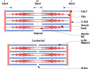
The names of the various sub-regions of the sarcomere are based on their relatively lighter or darker appearance when viewed through the light microscope. Each sarcomere is delimited by two very dark colored bands called Z-discs or Z-lines (from the German zwischen meaning between). These Z-discs are dense protein discs that do not easily allow the passage of light. The T-tubule is present in this area. The area between the Z-discs is further divided into two lighter colored bands at either end called the I-bands, and a darker, grayish band in the middle called the A band.The I bands appear lighter because these regions of the sarcomere mainly contain the thin actin filaments, whose smaller diameter allows the passage of light between them. The A band, on the other hand, contains mostly myosin
Myosin
Myosins comprise a family of ATP-dependent motor proteins and are best known for their role in muscle contraction and their involvement in a wide range of other eukaryotic motility processes. They are responsible for actin-based motility. The term was originally used to describe a group of similar...
filaments whose larger diameter restricts the passage of light. A stands for anisotropic and I for isotropic, referring to the optical properties of living muscle as demonstrated with polarized light microscopy.
The parts of the A band that abut the I bands are occupied by the both actin and myosin filaments (where they interdigitate as described above). Also within the A band is a relatively brighter central region called the H-zone (from the German helle, meaning bright) in which there is no actin/myosin overlap when the muscle is in a relaxed state. Finally, the A band is bisected by a dark central line called the M-line (from the German mittel meaning middle).
Action
When a muscle contracts, the actin is pulled along myosinMyosin
Myosins comprise a family of ATP-dependent motor proteins and are best known for their role in muscle contraction and their involvement in a wide range of other eukaryotic motility processes. They are responsible for actin-based motility. The term was originally used to describe a group of similar...
toward the center of the sarcomere
Sarcomere
A sarcomere is the basic unit of a muscle. Muscles are composed of tubular muscle cells . Muscle cells are composed of tubular myofibrils. Myofibrils are composed of repeating sections of sarcomeres, which appear under the microscope as dark and light bands...
until the actin
Actin
Actin is a globular, roughly 42-kDa moonlighting protein found in all eukaryotic cells where it may be present at concentrations of over 100 μM. It is also one of the most highly-conserved proteins, differing by no more than 20% in species as diverse as algae and humans...
and myosin filaments are completely overlapped. The H zone becomes smaller and smaller due to the increasing overlap of actin and myosin filaments, and the muscle shortens. Thus when the muscle is fully contracted, the H zone is no longer visible (as in the bottom diagram, left). Note that the actin and myosin filaments themselves do not change length, but instead slide past each other. This is known as the sliding filament theory of muscle contraction.
External links
- http://www.nismat.org/physcor/muscle.html
- http://msjensen.cehd.umn.edu/1135/Links/Animations/Flash/0008-swf_sarcomere_shor.swf
(animation of sarcomeres contraction)