

Mammography
Encyclopedia
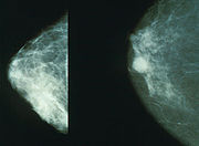
Mammography is the process of using low-energy-X-rays (usually around 30 kVp) to examine the human breast
and is used as a diagnostic and a screening tool.
The goal of mammography is the early detection of breast cancer
, typically through detection of characteristic masses and/or microcalcification
s. Most doctors believe that mammography reduces deaths from breast cancer
, although a minority do not.
In many countries routine mammography of older women is encouraged as a screening method to diagnose early breast cancer. In 2009, the U.S. Preventive Services Task Force (USPSTF) recommended that women with no risk factors have screening mammographies every 2 years between age 50 and 74. They found that the information was insufficient to recommend for or against screening between age 40 and 49 or above age 74. Altogether clinical trials have found a relative reduction in breast cancer mortality of 20%. Some doctors believe that mammographies do not reduce deaths from breast cancer, or at least that the evidence does not demonstrate it.
Like all x-rays, mammograms use doses of ionizing radiation
to create images. Radiologists then analyze the image for any abnormal findings. It is normal to use lower energy X-rays (typically Mo-K) than those used for radiography
of bone
s.
At this time, mammography along with physical breast examination is the modality of choice for screening for early breast cancer
. Ultrasound
, ductography, positron emission mammography
(PEM), and magnetic resonance imaging
are adjuncts to mammography. Ultrasound
is typically used for further evaluation of masses found on mammography or palpable masses not seen on mammograms. Ductograms are still used in some institutions for evaluation of bloody nipple discharge when the mammogram is non-diagnostic. MRI
can be useful for further evaluation of questionable findings as well as for screening pre-surgical evaluation in patients with known breast cancer to detect any additional lesions that might change the surgical approach, for instance from breast-conserving lumpectomy
to mastectomy
. New procedures, not yet approved for use in the general public, including breast tomosynthesis may offer benefits in years to come.
Breast self-examination
(BSE) was once promoted as a means of finding cancer at a more curable stage, however, it has been shown to be ineffective, and is no longer routinely recommended by health authorities for general use. Awareness of breast health and familiarity with one's own body is typically promoted instead of self-exams.
Mammography has a false-negative (missed cancer) rate of at least 10 percent. This is partly due to dense tissues obscuring the cancer and the fact that the appearance of cancer on mammograms has a large overlap with the appearance of normal tissues.
 During the procedure, the breast is compressed using a dedicated mammography unit. Parallel-plate compression evens out the thickness of breast tissue
to increase image quality by reducing the thickness of tissue that x-rays must penetrate, decreasing the amount of scattered radiation (scatter degrades image quality), reducing the required radiation dose, and holding the breast still (preventing motion blur
). In screening mammography, both head-to-foot (craniocaudal, CC) view and angled side-view (mediolateral oblique, MLO) images of the breast are taken. Diagnostic mammography may include these and other views, including geometrically magnified and spot-compressed views of the particular area of concern. Deodorant
, talcum powder or lotion
may show up on the X-ray as calcium
spots, and women are discouraged from applying these on the day of their exam.
Until some years ago, mammography was typically performed with screen-film cassettes. Now, mammography is undergoing transition to digital detectors, known as digital mammography
or Full Field Digital Mammography (FFDM). The first FFDM system was approved by the FDA in the U.S. in 2000. This progress is some years later than in general radiology. This is due to several factors:
As of March 1, 2010, 62% of facilities in the United States and its territories have at least one FFDM unit. (The FDA includes computed radiography units in this figure.)
In order to encourage the use of mammograms as a screening measure for breast cancer, a number of hospitals, cancer centers and other healthcare groups have started mobile mammography vans to bring affordable, accessible and convenient mammograms to their communities. Many mobile mammography vans prioritize serving uninsured, low-income and/or non-English-speaking women who otherwise could not otherwise afford a mammogram or who are unaccustomed to seeing a doctor. Many offer free or low-cost mammograms to women who are uninsured and/or cannot afford a mammogram.
when necessary, often performed via stereotactic core biopsy
or ultrasound
-guided core biopsy. After a screening mammogram, some women may have areas of concern which can't be resolved with only the information available from the screening mammogram. They would then be called back for a "diagnostic mammogram". This phrase essentially means a problem-solving mammogram. During this session, the radiologist will be monitoring each of the additional films as they are taken by a technologist. Depending on the nature of the finding, ultrasound
may often be used at this point, as well.
Generally the cause of the unusual appearance is found to be benign
. If the cause cannot be determined to be benign with sufficient certainty, a biopsy will be recommended. The biopsy procedure will be used to obtain actual tissue from the site for the pathologist to examine microscopically to determine the precise cause of the abnormality. In the past, biopsies were most frequently done in surgery, under local or general anesthesia
. The majority are now done with needles using either ultrasound or mammographic guidance to be sure that the area of concern is the area that is biopsied. These core biopsies require only local anesthesia, similar to what would be given during a small dental procedure.
 Often women are quite distressed to be called back for a diagnostic mammogram. Most of these recalls will be false positive
results. It helps to know these approximate statistics: of every 1,000 U.S. women who are screened, about 7% (70) will be called back for a diagnostic session (although some studies estimate the number closer to 10%–15%). About 10 of these individuals will be referred for a biopsy; the remaining 60 are found to be of benign cause. Of the 10 referred for biopsy, about 3.5 will have a cancer and 6.5 will not. Of the 3.5 who have cancer, about 2 have a low stage cancer that will be essentially cured after treatment. Mammogram results are often expressed in terms of the BI-RADS
Assessment Category, often called a "BI-RADS score." The categories range from 0 (Incomplete) to 6 (Known biopsy – proven malignancy). In the UK mammograms are scored on a scale from 1-5 (1 = normal, 2 = benign, 3 = indeterminate, 4 = suspicious of malignancy, 5 = malignant).
Mammography may also produce false negatives. Estimates of the numbers of cancers missed by mammography are usually around 10%–30%. This means that of the 350 per 100,000 women who have breast cancer, about 35–105 will not be detected by mammography. Reasons for not seeing the cancer include observer error, but more frequently it is because the cancer is hidden by other dense tissue in the breast and even after retrospective review of the mammogram, cannot be seen. Furthermore, one form of breast cancer, lobular cancer, has a growth pattern that produces shadows on the mammogram which are indistinguishable from normal breast tissue.
Computer-aided diagnosis
(CAD) are being tested to decrease the number of cases of cancer that are missed in mammograms. In one test, a computer identified 71% of the cases of cancer that had been missed by physicians. However, the computer also flagged twice as many non-cancerous masses than the physicians did. In a second study of a larger set of mammograms, a computer recommended six biopsies that physicians did not. All six turned out to be cancers that would have been missed. Generally, CAD systems in screening mammography have poor specificity and compare poorly to double reading.
While data are accumulating suggesting that CAD can find a few additional cancers, this should be put in perspective. The additional find rate was 20%, thus in a group of 10,000 women who will have about 40 cancers, CAD may help find an additional 8. The types of additional cancers that may be found are likely to be early and small. As of 2006, there have been no data to show that finding these additional cancers will have any effect on survival rate. Some feel that these cancers are likely to be found at the next screening, still at a curable stage, and therefore it remains to be proven whether CAD will be eventually found to have any effect on patient outcome.
A study released October 1, 2008, by British researchers revealed that using CAD in conjunction with a single reading by a physician may be as beneficial as a second reading by a physician. The study of 31,000 women, the largest of its kind to date, determined that the find rate for a single physician in conjunction with CAD as compared to two physicians was nearly identical. Out of 227 cancers found, the CAD method found just one fewer than the 199 cancers found using two separate physicians.
Only between 3% and 13% of breast cancers detected by screening mammography fall into this last category. Clinical trial data suggests that 1 woman per 1,000 healthy women screened over 10 years fall into this category as well. Screening mammography produces no benefit to any of the remaining 87% to 97% of women.
Research shows that false-positive mammograms may affect women's well-being and behavior. Some women who receive false-positive results may be more likely to return for routine screening or perform breast self-examinations more frequently. However, some women who receive false-positive results become anxious, worried and distressed about the possibility of having breast cancer, feelings that can last for many years.
False positives also mean greater expense, both for the individual woman, and for the screening program. Since follow-up screening is typically much more expensive than initial screening, more false positives that must receive follow-up means fewer woman may be screened for a given amount of money. Thus as sensitivity increases, a screening program will cost more, or be able to screen a smaller number of women.
Dr. H. Gilbert Welch, a researcher at Dartmouth College, states that "in screen-detected breast and prostate cancer survivors are more likely to have been overdiagnosed than actually helped by the test."
cannot be performed on every woman who has had a mammogram to determine the false negative rate accurately. Estimates of the false negative rate depend on close follow-up of a large number of patients for many years. This is difficult in practice, because many women do not return for regular mammography making it impossible to know if they ever developed a cancer. Dr. Samuel S. Epstein, in his book, The Politics of Cancer, claims that in women ages 40 to 49, one in four instances of cancer is missed at each mammography. Researchers have found that breast tissue is denser among younger women, making it difficult to detect tumors. For this reason, false negatives are twice as likely to occur in premenopausal mammograms(Prate.) This is why the screening program in the UK does not start calling women for screening mammograms until the age of 50.
The importance of these missed cancers is not clear, particularly if the woman is getting yearly mammograms. Research on a closely related situation has shown that small cancers that are not acted upon immediately, but are observed over periods of even several years, will have good outcomes. A group of 3,184 women had mammograms which were formally classified as "probably benign." This classification is for patients who are not clearly normal but have some area of minor concern. This results, not in the patient being biopsied, but having early follow up mammography every six months for three years to guarantee no change. Of these 3,184 women, 17 (0.5%) did have cancers. Most importantly, when the diagnosis was finally made, they were all still stage 0 or 1, the earliest stages. Five years after treatment, none of these 17 women had evidence of recurrence. Thus, small early cancers, even though not acted on immediately, were still entirely curable (Sickles, Radiology, 179:463-468, 1991).
and United States Preventive Task Force take such risks into account when formulating screening guidelines.
The majority of health experts agree that the risk of breast cancer for asymptomatic women under 35 is not high enough to warrant the risk of radiation exposure. For this reason, and because the radiation sensitivity of the breast in women under 35 is possibly greater than in older women, most radiologists will not perform screening mammography in women under 40. However, if there is a significant risk of cancer in a particular patient (BRCA positive, very positive family history, palpable mass), mammography may still be important. Often, the radiologist will try to avoid mammography by using ultrasound or MRI imaging.
The statistics about mammography and women between the ages of 40 and 55 are the most contentious. A 1992 Canadian National Breast Cancer Study showed that mammography (conducted in the 1980s) had no positive effect on mortality for women between the ages of 50 and 60. This study, however, is the only study to find this result. The study's critics pointed out that there were very serious design flaws in the study that invalidated these results.
There is a body of evidence that clearly shows that there is overdiagnosis
of cancer when women are screened. These cancers would never have affected these women in their lifetimes. An estimate of this overdiagnosis is 10 breast cancers diagnosed and unnecessarily treated per life saved when 2000 women are screened for 10 years.
While screening between 40 and 50 is still controversial, the preponderance of the evidence indicates that there is some small benefit in terms of early detection. Currently, the American Cancer Society
, the National Cancer Institute
, and the American College of Radiology
encourage mammograms every two years for women ages 40 to 49. In contrast, the American College of Physicians
, a large internist group, has recently encouraged individualized screening plans as opposed to wholesale biannual screening of women aged 40 to 49. In 2009, the U.S. Preventive Services Task Force recommended that screening of those age 40 to 49 be based on individual's risk factors and values, and that screening should not be routine in this age group. Their report says that the benefits of screenings before the age of 50 don't outweigh the risks.
Finally, a significant recent article points out that a successful screening program should result in an increase in the number of early breast cancers, followed by a decrease in the number of late-stage cancers. However this is not happening with current mammography screening.
At this time MQSA applies only to traditional mammography and not related scans such as breast ultrasound, stereotactic breast biospy, or breast MRI.
Breast
The breast is the upper ventral region of the torso of a primate, in left and right sides, which in a female contains the mammary gland that secretes milk used to feed infants.Both men and women develop breasts from the same embryological tissues...
and is used as a diagnostic and a screening tool.
The goal of mammography is the early detection of breast cancer
Breast cancer
Breast cancer is cancer originating from breast tissue, most commonly from the inner lining of milk ducts or the lobules that supply the ducts with milk. Cancers originating from ducts are known as ductal carcinomas; those originating from lobules are known as lobular carcinomas...
, typically through detection of characteristic masses and/or microcalcification
Microcalcification
Microcalcifications are tiny specks of mineral deposits , that can be scattered throughout the mammary gland, or occur in clusters. When found on a mammogram, a radiologist will then decide whether the specks are of concern - usually, this is not the case...
s. Most doctors believe that mammography reduces deaths from breast cancer
Breast cancer
Breast cancer is cancer originating from breast tissue, most commonly from the inner lining of milk ducts or the lobules that supply the ducts with milk. Cancers originating from ducts are known as ductal carcinomas; those originating from lobules are known as lobular carcinomas...
, although a minority do not.
In many countries routine mammography of older women is encouraged as a screening method to diagnose early breast cancer. In 2009, the U.S. Preventive Services Task Force (USPSTF) recommended that women with no risk factors have screening mammographies every 2 years between age 50 and 74. They found that the information was insufficient to recommend for or against screening between age 40 and 49 or above age 74. Altogether clinical trials have found a relative reduction in breast cancer mortality of 20%. Some doctors believe that mammographies do not reduce deaths from breast cancer, or at least that the evidence does not demonstrate it.
Like all x-rays, mammograms use doses of ionizing radiation
Ionizing radiation
Ionizing radiation is radiation composed of particles that individually have sufficient energy to remove an electron from an atom or molecule. This ionization produces free radicals, which are atoms or molecules containing unpaired electrons...
to create images. Radiologists then analyze the image for any abnormal findings. It is normal to use lower energy X-rays (typically Mo-K) than those used for radiography
Radiography
Radiography is the use of X-rays to view a non-uniformly composed material such as the human body. By using the physical properties of the ray an image can be developed which displays areas of different density and composition....
of bone
Bone
Bones are rigid organs that constitute part of the endoskeleton of vertebrates. They support, and protect the various organs of the body, produce red and white blood cells and store minerals. Bone tissue is a type of dense connective tissue...
s.
At this time, mammography along with physical breast examination is the modality of choice for screening for early breast cancer
Breast cancer
Breast cancer is cancer originating from breast tissue, most commonly from the inner lining of milk ducts or the lobules that supply the ducts with milk. Cancers originating from ducts are known as ductal carcinomas; those originating from lobules are known as lobular carcinomas...
. Ultrasound
Ultrasound
Ultrasound is cyclic sound pressure with a frequency greater than the upper limit of human hearing. Ultrasound is thus not separated from "normal" sound based on differences in physical properties, only the fact that humans cannot hear it. Although this limit varies from person to person, it is...
, ductography, positron emission mammography
Positron emission mammography
Positron emission mammography is a modality used to detect breast cancer.It is approved for patients with a history of breast cancer....
(PEM), and magnetic resonance imaging
Magnetic resonance imaging
Magnetic resonance imaging , nuclear magnetic resonance imaging , or magnetic resonance tomography is a medical imaging technique used in radiology to visualize detailed internal structures...
are adjuncts to mammography. Ultrasound
Ultrasound
Ultrasound is cyclic sound pressure with a frequency greater than the upper limit of human hearing. Ultrasound is thus not separated from "normal" sound based on differences in physical properties, only the fact that humans cannot hear it. Although this limit varies from person to person, it is...
is typically used for further evaluation of masses found on mammography or palpable masses not seen on mammograms. Ductograms are still used in some institutions for evaluation of bloody nipple discharge when the mammogram is non-diagnostic. MRI
Magnetic resonance imaging
Magnetic resonance imaging , nuclear magnetic resonance imaging , or magnetic resonance tomography is a medical imaging technique used in radiology to visualize detailed internal structures...
can be useful for further evaluation of questionable findings as well as for screening pre-surgical evaluation in patients with known breast cancer to detect any additional lesions that might change the surgical approach, for instance from breast-conserving lumpectomy
Lumpectomy
Lumpectomy is a common surgical procedure designed to remove a discrete lump, usually a benign tumor or breast cancer, from an affected man or woman's breast...
to mastectomy
Mastectomy
Mastectomy is the medical term for the surgical removal of one or both breasts, partially or completely. Mastectomy is usually done to treat breast cancer; in some cases, women and some men believed to be at high risk of breast cancer have the operation prophylactically, that is, to prevent cancer...
. New procedures, not yet approved for use in the general public, including breast tomosynthesis may offer benefits in years to come.
Breast self-examination
Breast self-examination
Breast self-examination is a screening method used in an attempt to detect early breast cancer. The method involves the woman herself looking at and feeling each breast for possible lumps, distortions or swelling....
(BSE) was once promoted as a means of finding cancer at a more curable stage, however, it has been shown to be ineffective, and is no longer routinely recommended by health authorities for general use. Awareness of breast health and familiarity with one's own body is typically promoted instead of self-exams.
Mammography has a false-negative (missed cancer) rate of at least 10 percent. This is partly due to dense tissues obscuring the cancer and the fact that the appearance of cancer on mammograms has a large overlap with the appearance of normal tissues.
Procedure
Biological tissue
Tissue is a cellular organizational level intermediate between cells and a complete organism. A tissue is an ensemble of cells, not necessarily identical, but from the same origin, that together carry out a specific function. These are called tissues because of their identical functioning...
to increase image quality by reducing the thickness of tissue that x-rays must penetrate, decreasing the amount of scattered radiation (scatter degrades image quality), reducing the required radiation dose, and holding the breast still (preventing motion blur
Motion blur
Motion blur is the apparent streaking of rapidly moving objects in a still image or a sequence of images such as a movie or animation. It results when the image being recorded changes during the recording of a single frame, either due to rapid movement or long exposure.- Photography :When a camera...
). In screening mammography, both head-to-foot (craniocaudal, CC) view and angled side-view (mediolateral oblique, MLO) images of the breast are taken. Diagnostic mammography may include these and other views, including geometrically magnified and spot-compressed views of the particular area of concern. Deodorant
Deodorant
Deodorants are substances applied to the body to affect body odor caused by bacterial growth and the smell associated with bacterial breakdown of perspiration in armpits, feet and other areas of the body. A subgroup of deodorants, antiperspirants, affect odor as well as prevent sweating by...
, talcum powder or lotion
Lotion
A lotion is a low- to medium-viscosity, topical preparation intended for application to unbroken skin. By contrast, creams and gels have higher viscosity.Lotions are usually applied to external skin with bare hands, a clean cloth, cotton wool or gauze...
may show up on the X-ray as calcium
Calcium
Calcium is the chemical element with the symbol Ca and atomic number 20. It has an atomic mass of 40.078 amu. Calcium is a soft gray alkaline earth metal, and is the fifth-most-abundant element by mass in the Earth's crust...
spots, and women are discouraged from applying these on the day of their exam.
Until some years ago, mammography was typically performed with screen-film cassettes. Now, mammography is undergoing transition to digital detectors, known as digital mammography
Digital mammography
Digital mammography is a specialized form of mammography that uses digital receptors and computers instead of x-ray film to help examine breast tissue for breast cancer. The electrical signals can be read on computer screens, permitting more manipulation of images to theoretically allow...
or Full Field Digital Mammography (FFDM). The first FFDM system was approved by the FDA in the U.S. in 2000. This progress is some years later than in general radiology. This is due to several factors:
- the higher spatial resolution demands of mammography,
- significantly increased expense of the equipment,
- concern by the FDA that digital mammography equipment demonstrate that it is at least as good as screen-film mammography at detecting breast cancers without increasing breast dose or the number of women recalled for further evaluation.
As of March 1, 2010, 62% of facilities in the United States and its territories have at least one FFDM unit. (The FDA includes computed radiography units in this figure.)
In order to encourage the use of mammograms as a screening measure for breast cancer, a number of hospitals, cancer centers and other healthcare groups have started mobile mammography vans to bring affordable, accessible and convenient mammograms to their communities. Many mobile mammography vans prioritize serving uninsured, low-income and/or non-English-speaking women who otherwise could not otherwise afford a mammogram or who are unaccustomed to seeing a doctor. Many offer free or low-cost mammograms to women who are uninsured and/or cannot afford a mammogram.
"Work-up" process
In the past several years, the "work-up" process has become quite formalized. It generally consists of screening mammography, diagnostic mammography, and biopsyBiopsy
A biopsy is a medical test involving sampling of cells or tissues for examination. It is the medical removal of tissue from a living subject to determine the presence or extent of a disease. The tissue is generally examined under a microscope by a pathologist, and can also be analyzed chemically...
when necessary, often performed via stereotactic core biopsy
Stereotactic biopsy
Stereotactic biopsy, also known as stereotactic core biopsy, is a biopsy procedure that uses a computer and imaging performed in at least two planes to localize a target lesion in three-dimensional space and guide the removal of tissue for examination by a pathologist under a microscope...
or ultrasound
Ultrasound
Ultrasound is cyclic sound pressure with a frequency greater than the upper limit of human hearing. Ultrasound is thus not separated from "normal" sound based on differences in physical properties, only the fact that humans cannot hear it. Although this limit varies from person to person, it is...
-guided core biopsy. After a screening mammogram, some women may have areas of concern which can't be resolved with only the information available from the screening mammogram. They would then be called back for a "diagnostic mammogram". This phrase essentially means a problem-solving mammogram. During this session, the radiologist will be monitoring each of the additional films as they are taken by a technologist. Depending on the nature of the finding, ultrasound
Ultrasound
Ultrasound is cyclic sound pressure with a frequency greater than the upper limit of human hearing. Ultrasound is thus not separated from "normal" sound based on differences in physical properties, only the fact that humans cannot hear it. Although this limit varies from person to person, it is...
may often be used at this point, as well.
Generally the cause of the unusual appearance is found to be benign
Benign
A benign tumor is a tumor that lacks the ability to metastasize. Common examples of benign tumors include moles and uterine fibroids.The term "benign" implies a mild and nonprogressive disease. Indeed, many kinds of benign tumors are harmless to human health...
. If the cause cannot be determined to be benign with sufficient certainty, a biopsy will be recommended. The biopsy procedure will be used to obtain actual tissue from the site for the pathologist to examine microscopically to determine the precise cause of the abnormality. In the past, biopsies were most frequently done in surgery, under local or general anesthesia
Anesthesia
Anesthesia, or anaesthesia , traditionally meant the condition of having sensation blocked or temporarily taken away...
. The majority are now done with needles using either ultrasound or mammographic guidance to be sure that the area of concern is the area that is biopsied. These core biopsies require only local anesthesia, similar to what would be given during a small dental procedure.
Results
Type I and type II errors
In statistical test theory the notion of statistical error is an integral part of hypothesis testing. The test requires an unambiguous statement of a null hypothesis, which usually corresponds to a default "state of nature", for example "this person is healthy", "this accused is not guilty" or...
results. It helps to know these approximate statistics: of every 1,000 U.S. women who are screened, about 7% (70) will be called back for a diagnostic session (although some studies estimate the number closer to 10%–15%). About 10 of these individuals will be referred for a biopsy; the remaining 60 are found to be of benign cause. Of the 10 referred for biopsy, about 3.5 will have a cancer and 6.5 will not. Of the 3.5 who have cancer, about 2 have a low stage cancer that will be essentially cured after treatment. Mammogram results are often expressed in terms of the BI-RADS
BI-RADS
BI-RADS is an acronym for Breast Imaging-Reporting and Data System, a quality assurance tool originally designed for use with mammography. The system is a collaborative effort of many health groups but is published and trademarked by the American College of Radiology .The system is designed to...
Assessment Category, often called a "BI-RADS score." The categories range from 0 (Incomplete) to 6 (Known biopsy – proven malignancy). In the UK mammograms are scored on a scale from 1-5 (1 = normal, 2 = benign, 3 = indeterminate, 4 = suspicious of malignancy, 5 = malignant).
Mammography may also produce false negatives. Estimates of the numbers of cancers missed by mammography are usually around 10%–30%. This means that of the 350 per 100,000 women who have breast cancer, about 35–105 will not be detected by mammography. Reasons for not seeing the cancer include observer error, but more frequently it is because the cancer is hidden by other dense tissue in the breast and even after retrospective review of the mammogram, cannot be seen. Furthermore, one form of breast cancer, lobular cancer, has a growth pattern that produces shadows on the mammogram which are indistinguishable from normal breast tissue.
Computer-aided diagnosis
Computer-aided diagnosis
Computer-aided detection and computer-aided diagnosis are procedures in medicine that assist doctors in the interpretation of medical images. Imaging techniques in X-ray, MRI, and Ultrasound diagnostics yield a great deal of information, which the radiologist has to analyze and evaluate...
(CAD) are being tested to decrease the number of cases of cancer that are missed in mammograms. In one test, a computer identified 71% of the cases of cancer that had been missed by physicians. However, the computer also flagged twice as many non-cancerous masses than the physicians did. In a second study of a larger set of mammograms, a computer recommended six biopsies that physicians did not. All six turned out to be cancers that would have been missed. Generally, CAD systems in screening mammography have poor specificity and compare poorly to double reading.
While data are accumulating suggesting that CAD can find a few additional cancers, this should be put in perspective. The additional find rate was 20%, thus in a group of 10,000 women who will have about 40 cancers, CAD may help find an additional 8. The types of additional cancers that may be found are likely to be early and small. As of 2006, there have been no data to show that finding these additional cancers will have any effect on survival rate. Some feel that these cancers are likely to be found at the next screening, still at a curable stage, and therefore it remains to be proven whether CAD will be eventually found to have any effect on patient outcome.
A study released October 1, 2008, by British researchers revealed that using CAD in conjunction with a single reading by a physician may be as beneficial as a second reading by a physician. The study of 31,000 women, the largest of its kind to date, determined that the find rate for a single physician in conjunction with CAD as compared to two physicians was nearly identical. Out of 227 cancers found, the CAD method found just one fewer than the 199 cancers found using two separate physicians.
Likelihood of saving a life
Women whose breast cancer was detected by screening mammography before the appearance of a lump or other symptoms commonly assume that the mammogram "saved their lives". In practice, the vast majority of these women received no practical benefit from the mammogram. There are four categories of cancers found by mammography:- cancers that are so easily treated that a later detection would have produced the same total cure (woman would have lived even without mammography);
- cancers so aggressive that even "early" detection is too late (woman dies despite detection by mammography);
- cancers that would have receded on their own or are so slow-growing that the woman would die of other causes before the cancer produces symptoms (mammography results in overdiagnosisOverdiagnosisOverdiagnosis is the diagnosis of "disease" that will never cause symptoms or death during a patient's lifetime. Overdiagnosis is the least familiar side effect of testing for early forms of disease – and, arguably, the most important...
and overtreatment of this class); and - the small number of breast cancers that are detected by screening mammography and whose treatment outcome improves as a result of earlier detection.
Only between 3% and 13% of breast cancers detected by screening mammography fall into this last category. Clinical trial data suggests that 1 woman per 1,000 healthy women screened over 10 years fall into this category as well. Screening mammography produces no benefit to any of the remaining 87% to 97% of women.
False positives
The goal of any screening procedure is to examine a large population of patients and find the small number most likely to have a serious condition. These patients are then referred for further, usually more invasive, testing. Thus a screening exam is not intended to be definitive: It is intended to have sufficient sensitivity to detect a useful proportion of cancers. The cost of higher sensitivity is a larger number of results that would be regarded as suspicious in patients without disease. This is true of mammography. The patients without disease who are called back for further testing from a screening session (about 7%) are sometimes referred to as "false positives". There is a trade-off between the number of patients with disease found, and the much larger number of patients without disease that must be re-screened.Research shows that false-positive mammograms may affect women's well-being and behavior. Some women who receive false-positive results may be more likely to return for routine screening or perform breast self-examinations more frequently. However, some women who receive false-positive results become anxious, worried and distressed about the possibility of having breast cancer, feelings that can last for many years.
False positives also mean greater expense, both for the individual woman, and for the screening program. Since follow-up screening is typically much more expensive than initial screening, more false positives that must receive follow-up means fewer woman may be screened for a given amount of money. Thus as sensitivity increases, a screening program will cost more, or be able to screen a smaller number of women.
Dr. H. Gilbert Welch, a researcher at Dartmouth College, states that "in screen-detected breast and prostate cancer survivors are more likely to have been overdiagnosed than actually helped by the test."
False negatives
At the same time, mammograms also have a rate of missed tumors, or "false negatives." Accurate data regarding the number of false negatives are very difficult to obtain, simply because mastectomiesMastectomy
Mastectomy is the medical term for the surgical removal of one or both breasts, partially or completely. Mastectomy is usually done to treat breast cancer; in some cases, women and some men believed to be at high risk of breast cancer have the operation prophylactically, that is, to prevent cancer...
cannot be performed on every woman who has had a mammogram to determine the false negative rate accurately. Estimates of the false negative rate depend on close follow-up of a large number of patients for many years. This is difficult in practice, because many women do not return for regular mammography making it impossible to know if they ever developed a cancer. Dr. Samuel S. Epstein, in his book, The Politics of Cancer, claims that in women ages 40 to 49, one in four instances of cancer is missed at each mammography. Researchers have found that breast tissue is denser among younger women, making it difficult to detect tumors. For this reason, false negatives are twice as likely to occur in premenopausal mammograms(Prate.) This is why the screening program in the UK does not start calling women for screening mammograms until the age of 50.
The importance of these missed cancers is not clear, particularly if the woman is getting yearly mammograms. Research on a closely related situation has shown that small cancers that are not acted upon immediately, but are observed over periods of even several years, will have good outcomes. A group of 3,184 women had mammograms which were formally classified as "probably benign." This classification is for patients who are not clearly normal but have some area of minor concern. This results, not in the patient being biopsied, but having early follow up mammography every six months for three years to guarantee no change. Of these 3,184 women, 17 (0.5%) did have cancers. Most importantly, when the diagnosis was finally made, they were all still stage 0 or 1, the earliest stages. Five years after treatment, none of these 17 women had evidence of recurrence. Thus, small early cancers, even though not acted on immediately, were still entirely curable (Sickles, Radiology, 179:463-468, 1991).
Other risks
The radiation exposure associated with mammography is a potential risk of screening. The risk of exposure appears to be greater in younger women. The largest study of radiation risk from mammography concluded that for women 40 years of age or older, the risk of radiation-induced breast cancer was minuscule, particularly compared with the potential benefit of mammographic screening, with a benefit-to-risk ratio of 48.5 lives saved for each life lost due to radiation exposure. Organizations such as the National Cancer InstituteNational Cancer Institute
The National Cancer Institute is part of the National Institutes of Health , which is one of 11 agencies that are part of the U.S. Department of Health and Human Services. The NCI coordinates the U.S...
and United States Preventive Task Force take such risks into account when formulating screening guidelines.
The majority of health experts agree that the risk of breast cancer for asymptomatic women under 35 is not high enough to warrant the risk of radiation exposure. For this reason, and because the radiation sensitivity of the breast in women under 35 is possibly greater than in older women, most radiologists will not perform screening mammography in women under 40. However, if there is a significant risk of cancer in a particular patient (BRCA positive, very positive family history, palpable mass), mammography may still be important. Often, the radiologist will try to avoid mammography by using ultrasound or MRI imaging.
The statistics about mammography and women between the ages of 40 and 55 are the most contentious. A 1992 Canadian National Breast Cancer Study showed that mammography (conducted in the 1980s) had no positive effect on mortality for women between the ages of 50 and 60. This study, however, is the only study to find this result. The study's critics pointed out that there were very serious design flaws in the study that invalidated these results.
There is a body of evidence that clearly shows that there is overdiagnosis
Overdiagnosis
Overdiagnosis is the diagnosis of "disease" that will never cause symptoms or death during a patient's lifetime. Overdiagnosis is the least familiar side effect of testing for early forms of disease – and, arguably, the most important...
of cancer when women are screened. These cancers would never have affected these women in their lifetimes. An estimate of this overdiagnosis is 10 breast cancers diagnosed and unnecessarily treated per life saved when 2000 women are screened for 10 years.
While screening between 40 and 50 is still controversial, the preponderance of the evidence indicates that there is some small benefit in terms of early detection. Currently, the American Cancer Society
American Cancer Society
The American Cancer Society is the "nationwide community-based voluntary health organization" dedicated, in their own words, "to eliminating cancer as a major health problem by preventing cancer, saving lives, and diminishing suffering from cancer, through research, education, advocacy, and...
, the National Cancer Institute
National Cancer Institute
The National Cancer Institute is part of the National Institutes of Health , which is one of 11 agencies that are part of the U.S. Department of Health and Human Services. The NCI coordinates the U.S...
, and the American College of Radiology
American College of Radiology
The American College of Radiology , founded in 1923, is a non-profit professional medical association composed of diagnostic radiologists, radiation oncologists, interventional radiologists, nuclear medicine physicians, and medical physicists. It is based in Reston, Virginia, with offices in...
encourage mammograms every two years for women ages 40 to 49. In contrast, the American College of Physicians
American College of Physicians
The American College of Physicians is a national organization of doctors of internal medicine —physicians who specialize in the prevention, detection, and treatment of illnesses in adults. With 130,000 members, ACP is the largest medical-specialty organization and second-largest physician group in...
, a large internist group, has recently encouraged individualized screening plans as opposed to wholesale biannual screening of women aged 40 to 49. In 2009, the U.S. Preventive Services Task Force recommended that screening of those age 40 to 49 be based on individual's risk factors and values, and that screening should not be routine in this age group. Their report says that the benefits of screenings before the age of 50 don't outweigh the risks.
Critique of screening mammography
The use of mammography as a screening tool for the detection of early breast cancer continues to be debated. Critics point out that a large number of women need to be screened to find cancer. Kopans reminds us that since 1990, the death rate from breast cancer has decreased by almost 30% and points to studies in Sweden and the Netherlands that show two-thirds of the decrease in cancer deaths is due to mammography screening. Keen and Keen indicated that repeated mammography starting at age 50 saves about 1.8 lives over 15 years for every 1,000 women screened. This result has to be seen against the negatives of errors in diagnosis, overtreatment, and radiation exposure. Countercritics argue that the benefit is greater. The Cochrane analysis of screening indicates that it is "not clear whether screening does more good than harm". According to their analysis one in 2,000 women will have her life prolonged by 10 years of screening, however, another 10 healthy women will undergo unnecessary breast cancer treatment. Additionally, 200 women will suffer from significant psychological stress due to false posivitive results. Newman points out that screening mammography does not reduce death overall, but causes significant harm by inflicting cancer scare and unnecessary surgical interventions.Finally, a significant recent article points out that a successful screening program should result in an increase in the number of early breast cancers, followed by a decrease in the number of late-stage cancers. However this is not happening with current mammography screening.
Research alternatives to mammography
While the cost of mammography is relatively low, its sensitivity is not ideal, with reports listing the range from 45% to about 90% depending on factors such as the density of the breast. Neither is the X-ray based technology completely benign, as noted above. Therefore there is considerable ongoing research into the use of alternative technologies.- Breast MRIBreast MRIOne alternative to mammography, Breast MRI or contrast enhanced magnetic resonance imaging , has shown substantial progress in the detection of breast cancer.-Operation:...
- Breast thermography, infrared imaging
- Molecular breast imagingMolecular breast imagingMolecular breast imaging can refer to:* Scintimammography* Positron emission mammography...
(MBI), is a new technology used for breast imaging. MBI identifies tumors in dense breast tissue that are often not visible with X-ray based analog or digital mammography.
Regulation
Mammography facilities in the United States and its territories (including military bases) are subject to the Mammography Quality Standards Act (MQSA). The act requires annual inspections and accredition every 3 years through an FDA-approved body. Facilities found deficient during the inspection or accreditation process can be barred from performing mammograms until corrective action has been verified or, in extreme cases, can be required to notify past patients that their exams were sub-standard and should not be trusted.At this time MQSA applies only to traditional mammography and not related scans such as breast ultrasound, stereotactic breast biospy, or breast MRI.
See also
- Computed Tomography Laser MammographyComputed Tomography Laser MammographyComputed Tomography Laser Mammography is the trademark of Imaging Diagnostic Systems, Inc. for its optical tomographic technique for female breast imaging....
- Digital mammographyDigital mammographyDigital mammography is a specialized form of mammography that uses digital receptors and computers instead of x-ray film to help examine breast tissue for breast cancer. The electrical signals can be read on computer screens, permitting more manipulation of images to theoretically allow...
- Picture archiving and communication systemPicture archiving and communication systemA picture archiving and communication system is a medical imaging technology which provides economical storage of, and convenient access to, images from multiple modalities . Electronic images and reports are transmitted digitally via PACS; this eliminates the need to manually file, retrieve, or...
- XeromammographyXeromammographyXeromammography is a photoelectric method of recording an x-ray image on a coated metal plate, using low-energy photon beams, long exposure time, and dry chemical developers.It is a form of xeroradiography....
- Molecular breast imagingMolecular breast imagingMolecular breast imaging can refer to:* Scintimammography* Positron emission mammography...
External links
- Mammographic Image Analysis Homepage
- National Cancer Institute Statement on Mammography Screening
- Screening Mammograms: Questions and Answers, from the National Cancer InstituteNational Cancer InstituteThe National Cancer Institute is part of the National Institutes of Health , which is one of 11 agencies that are part of the U.S. Department of Health and Human Services. The NCI coordinates the U.S...
- Screening Mammograms: Questions and Answers, from the National Cancer Institute
- American Cancer Society: Mammograms and Other Breast Imaging Procedures
- U.S. Preventive Task Force recommendations on screening mammography