
Lipid bilayer fusion
Encyclopedia
Fusion is involved in many cellular processes, particularly in eukaryotes since the eukaryotic cell is extensively sub-divided by lipid bilayer membranes. Exocytosis
Exocytosis
Exocytosis , also known as 'The peni-cytosis', is the durable process by which a cell directs the contents of secretory vesicles out of the cell membrane...
, fertilization of an egg
Egg (biology)
An egg is an organic vessel in which an embryo first begins to develop. In most birds, reptiles, insects, molluscs, fish, and monotremes, an egg is the zygote, resulting from fertilization of the ovum, which is expelled from the body and permitted to develop outside the body until the developing...
by sperm
Sperm
The term sperm is derived from the Greek word sperma and refers to the male reproductive cells. In the types of sexual reproduction known as anisogamy and oogamy, there is a marked difference in the size of the gametes with the smaller one being termed the "male" or sperm cell...
and transport of waste products to the lysozome are a few of the many eukaryotic processes that rely on some form of fusion. Fusion is also an important mechanism for transport of lipids from their site of synthesis to the membrane where they are needed. Even the entry of pathogens can be governed by fusion, as many bilayer-coated viruses have dedicated fusion proteins to gain entry into the host cell.
Lipid mechanism
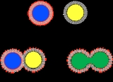
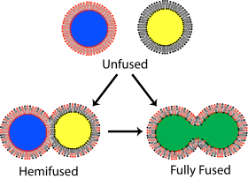
There are four fundamental steps in the fusion process, although each of these steps actually represents a complex sequence of events. First, the involved membranes must aggregate, approaching each other to within several nanometers. Second, the two bilayers must come into very close contact (within a few angstroms). To achieve this close contact, the two surfaces must become at least partially dehydrated, as the bound surface water normally present causes bilayers to strongly repel at this distance. Third, a destabilization must nucleate at one point between the two bilayers, inducing a highly localized rearrangement of the two bilayers. Finally, as this point defect grows, the components of the two bilayers mix and diffuse away from the site of contact. Depending on whether hemifusion or full fusion occurs, the internal contents of the membranes may mix at this point as well.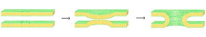
Phosphatidylserine
Phosphatidylserine is a phospholipid component, usually kept on the inner-leaflet of cell membranes by an enzyme called flippase...
, phosphatidylglycerol
Phosphatidylglycerol
Phosphatidylglycerol is a glycerophospholipid found in pulmonary surfactant.The general structure of phosphatidylglycerol consists of a L-glycerol 3-phosphate backbone ester-bonded to either saturated or unsaturated fatty acids on carbons 1 and 2. The head group substituent glycerol is bonded...
and cardiolipin
Cardiolipin
Cardiolipin is an important component of the inner mitochondrial membrane, where it constitutes about 20% of the total lipid composition. The only other place that cardiolipin can be found is in the membranes of most bacteria. The name ‘cardiolipin’ is derived from the fact that it was first...
. One role on these ions in the fusion process is to shield the negative charge on the surface of the bilayer, diminishing electrostatic repulsion and allowing the membranes to approach each other. This is clearly not the only role, however, since there is an extensively documented difference in the ability of Mg2+ versus Ca2+ to induce fusion. Although Mg2+ will induce extensive aggregation it will not induce fusion, while Ca2+ induces both. It has been proposed that this discrepancy is due to a difference in extent of dehydration. Under this theory, calcium ions bind more strongly to charged lipids, but less strongly to water. The resulting displacement of calcium for water destabilizes the lipid-water interface and promotes intimate interbilayer contact. A recently proposed alternative hypothesis is that the binding of calcium induces a destabilizing lateral tension. Whatever the mechanism of calcium-induced fusion, the initial interaction is clearly electrostatic, since zwitterionic lipids are not susceptible to this effect.
The role of lipid headgroup in the fusion process extends beyond charge density and can affect dehydration and defect nucleation independent of the effect of ions. The presence of the uncharged headgroup phosphatidylethanolamine (PE) increases fusion when incorporated into a phosphatidylcholine bilayer. This phenomenon has been explained by some as a dehydration effect similar to the influence of calcium. The PE headgroup binds water less tightly than PC and therefore may allow close apposition more easily. An alternate explanation is that the physical rather than chemical nature of PE may help induce fusion. According to the stalk hypothesis of fusion, a highly curved bridge must form between the two bilayers for fusion to occur. Since PE has a small headgroup and readily forms inverted micelle
Micelle
A micelle is an aggregate of surfactant molecules dispersed in a liquid colloid. A typical micelle in aqueous solution forms an aggregate with the hydrophilic "head" regions in contact with surrounding solvent, sequestering the hydrophobic single tail regions in the micelle centre. This phase is...
phases it should, according to the stalk model, promote the formation of these stalks. Further evidence cited in favor of this theory is the fact that certain lipid mixtures have been shown to only support fusion when raised above the transition temperature of these inverted phases. This topic also remains controversial, and even if there is a curved structure present in the fusion process, there is debate in the literature over whether it is a cubic, hexagonal or more exotic extended phase.
Fusion proteins
Membrane protein
A membrane protein is a protein molecule that is attached to, or associated with the membrane of a cell or an organelle. More than half of all proteins interact with membranes.-Function:...
. The first of these proteins to be studied were the viral fusion proteins, which allow an enveloped virus
Virus
A virus is a small infectious agent that can replicate only inside the living cells of organisms. Viruses infect all types of organisms, from animals and plants to bacteria and archaea...
to insert its genetic material into the host cell (enveloped viruses are those surrounded by a lipid bilayer; some others have only a protein coat). Broadly, there are two classes of viral fusion proteins: acidic and pH-independent. pH
PH
In chemistry, pH is a measure of the acidity or basicity of an aqueous solution. Pure water is said to be neutral, with a pH close to 7.0 at . Solutions with a pH less than 7 are said to be acidic and solutions with a pH greater than 7 are basic or alkaline...
independent fusion proteins can function under neutral conditions and fuse with the plasma membrane, allowing viral entry into the cell. Viruses utilizing this scheme included HIV
HIV
Human immunodeficiency virus is a lentivirus that causes acquired immunodeficiency syndrome , a condition in humans in which progressive failure of the immune system allows life-threatening opportunistic infections and cancers to thrive...
, measles
Measles
Measles, also known as rubeola or morbilli, is an infection of the respiratory system caused by a virus, specifically a paramyxovirus of the genus Morbillivirus. Morbilliviruses, like other paramyxoviruses, are enveloped, single-stranded, negative-sense RNA viruses...
and herpes. Acidic fusion proteins such as those found on influenza
Influenza
Influenza, commonly referred to as the flu, is an infectious disease caused by RNA viruses of the family Orthomyxoviridae , that affects birds and mammals...
are only activated when in the low pH of acidic endosomes and must first be endocytosed
Endocytosis
Endocytosis is a process by which cells absorb molecules by engulfing them. It is used by all cells of the body because most substances important to them are large polar molecules that cannot pass through the hydrophobic plasma or cell membrane...
to gain entry into the cell.
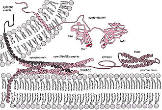
Eukaryotic cells use entirely different classes of fusion proteins, the best studied of which are the SNAREs
SNARE (protein)
SNARE proteins are a large protein superfamily consisting of more than 60 members in yeast and mammalian cells....
. SNARE proteins are used to direct all vesicular
Vesicle (biology)
A vesicle is a bubble of liquid within another liquid, a supramolecular assembly made up of many different molecules. More technically, a vesicle is a small membrane-enclosed sack that can store or transport substances. Vesicles can form naturally because of the properties of lipid membranes , or...
intracellular trafficking. Despite years of study, much is still unknown about the function of this protein class. In fact, there is still an active debate regarding whether SNAREs are linked to early docking or participate later in the fusion process by facilitating hemifusion. Even once the role of SNAREs or other specific proteins is illuminated, a unified understanding of fusion proteins is unlikely as there is an enormous diversity of structure and function within these classes, and very few themes are conserved.
Fusion in laboratory practice
In studies of molecular and cellular biology it is often desirable to artificially induce fusion. Although this can be accomplished with the addition of calcium as discussed earlier, this procedure is often not feasible because calcium regulates many other biochemical processes and its addition would be a strong confound. Also, as mentioned, calcium induces massive aggregation as well as fusion. The addition of polyethylene glycolPolyethylene glycol
Polyethylene glycol is a polyether compound with many applications from industrial manufacturing to medicine. It has also been known as polyethylene oxide or polyoxyethylene , depending on its molecular weight, and under the tradename Carbowax.-Available forms:PEG, PEO, or POE refers to an...
(PEG) causes fusion without significant aggregation or biochemical disruption. This procedure is now used extensively, for example by fusing B-cells with melanoma
Melanoma
Melanoma is a malignant tumor of melanocytes. Melanocytes are cells that produce the dark pigment, melanin, which is responsible for the color of skin. They predominantly occur in skin, but are also found in other parts of the body, including the bowel and the eye...
cells. The resulting “hybridoma
Hybridoma
Hybridoma technology is a technology of forming hybrid cell lines by fusing a specific antibody-producing B cell with a myeloma cell that is selected for its ability to grow in tissue culture and for an absence of antibody chain synthesis...
” from this combination expresses a desired antibody
Antibody
An antibody, also known as an immunoglobulin, is a large Y-shaped protein used by the immune system to identify and neutralize foreign objects such as bacteria and viruses. The antibody recognizes a unique part of the foreign target, termed an antigen...
as determined by the B-cell involved, but is immortalized due to the melanoma component. The mechanism of PEG fusion has not been definitively identified, but some researchers believe that the PEG, by binding a large number of water molecules, effectively decreases the chemical activity of the water and thus dehydrates the lipid headgroups. Fusion can also be artificially induced through electroporation
Electroporation
Electroporation, or electropermeabilization, is a significant increase in the electrical conductivity and permeability of the cell plasma membrane caused by an externally applied electrical field...
in a process known as electrofusion. It is believed that this phenomenon results from the energetically active edges
Lipid bilayer mechanics
Lipid bilayer mechanics is the study of the physical material properties of lipid bilayers, classifying bilayer behavior with stress and strain rather than biochemical interactions. These properties are typically characterized in terms of three mechanical elastic modulus: the area compression...
formed during electroporation, which can act as the local defect point to nucleate stalk growth between two bilayers.
Assays to measure membrane fusion
There are two levels of fusion: mixing of membrane lipids and mixing of contents. Assays of membrane fusion report either the mixing of membrane lipids or the mixing of the aqueous contents of the fused entities.Assays for measuring lipid mixing
Assays evaluating lipid mixing make use of concentration dependent effects such as nonradioactive energy transfer, fluorescence quenching and pyrene eximer formation.1. NBD-Rhodamine Energy Transfer :
In this method, membrane labeled with both NBD (donor) and Rhodamine (acceptor) combine with unlabeled membrane. When NBD and Rhodamine are within a certain distance, the Fluorescence resonance energy transfer (FRET) happens. After fusion, resonance energy transfer (FRET) decreases when the average distance between probes increases, while NBD fluorescence increases.
2. Pyrene Excimer Formation:
Pyrene monomer and excimer emission wavelengths are different. The emission wavelength of monomer is around 400 nm and that of excimer is around 470 nm. In this method, membrane labeled with Pyrene combines with unlabeled membrane. Pyrene self associates in membrane and then excited pyrene excites other pyrene. Before fusion, the majority of the emission is excimer emission. After fusion, the distance between probes increases and the ratio of excimer emission decreases.
3. Octadecyl Rhodamine B Self-Quenching:This assay is based on self-quenching of octadecyl rhodamine B. Octadecyl rhodamine B self-quenching occurs when the probe is incorporated into membrane lipids at concentrations of 1-10 mole percent because Rhodamine dimmers quench fluorescence. In this method, membrane labeled Rhodamine combines with unlabeled membrane. Fusion with unlabeled membranes resulting in dilution of the probe, which is accompanies by increasing fluorescence. The major problem of this assay is spontaneous transfer.
Assays for measuring content mixing
Mixing of aqueous contents from vesicles as a result of lysis, fusion or physiological permeability can be detected fluorometrically using low molecular weight soluble tracers.1. Fluorescence quenching assays with ANTS/DPX:
ANTS is a polyanionic fluorophore, while DPX is a cationic quencher. The assay is based on the collisional quenching of them. Separate vesicle populations are loaded with ANTS or DPX, respectively. When content mixing happens, ANTS and DPX collide and fluorescence of ANTS monitored at 530 nm, with excitation at 360 nm is quenched. This method is performed at acidic pH and high concentration.
2. Fluorescence enhancement assays with Tb3+/DPA:
This method is based on the fact that chelate of Tb3+/DPA is 10,000 times more fluorescent than Tb3+ alone. In the Tb3+/DPA assay, separate vesicle populations are loaded with TbCl3 or DPA. The formation of Tb3+/DPA chelate can be used to indicate vesicle fusion. This method is good for protein free membranes.
3. Single molecule DNA assay. A DNA hairpin composed of 5 base pair stem and poly-thymidine loop that is labeled with a donor (Cy3) and an acceptor (Cy5) at the ends of the stem was encapsulated in the v-SNARE vesicle. We separately encapsulated multiple unlabeled poly-adenosine DNA strands in the t-SNARE vesicle. If the two vesicles, both ~100 nm in diameter, dock and a large enough fusion pore forms between them, the two DNA molecules should hybridize, opening up the stem region of the hairpin and switching the Förster resonance energy transfer (FRET) efficiency (E) between Cy3 and Cy5 from a high to a low value.