

Industrial radiography
Encyclopedia
Industrial Radiography is the use of ionizing radiation
to view objects in a way that cannot be seen otherwise. It is not to be confused with the use of ionizing radiation to change or modify objects; radiography's purpose is strictly viewing. Industrial radiography has grown out of engineering, and is a major element of nondestructive testing
. It is a method of inspecting materials for hidden flaws by using the ability of short X-rays and Gamma rays to penetrate various materials.
s (later also called Röntgen
rays after the man who first described their properties in detail), a type of electromagnetic radiation
. Soon after the discovery of X-rays, radioactivity was discovered. By using radioactive sources such as radium
, far higher photon
energies could be obtained than those from normal X-ray machines. Soon these found various applications, with one of the earliest users being Loughborough University
, from helping to fit shoes, more lasting medical uses and the examination of non-living objects. X-rays and gamma-rays were put to use very early, before the dangers of ionising radiation were discovered. After World War II
new isotopes such as caesium-137
, iridium-192 and cobalt-60
became available for industrial radiography, and the use of radium and radon decreased.
 Gamma radiation sources, most commonly Iridium-192 and Cobalt-60, are used to inspect a variety of materials. The vast majority of radiography concerns the testing and grading of welds on pressurized piping, pressure vessels, high-capacity storage containers, pipelines, and some structural welds. Other tested materials include concrete (locating rebar or conduit), welder's test coupons, machined parts, plate metal, or pipewall (locating anomalies due to corrosion or mechanical damage). Non-metal components such as ceramics used in the aerospace industries are also regularly tested. Theoretically, industrial radiographers could radiograph any solid, flat material (walls, ceilings, floors, square or rectangular containers) or any hollow cylindrical or spherical object.
For purposes of inspection, including weld inspection, there exist several exposure arrangements.
First, there is the panoramic, one of the four single wall exposure/single wall view (SWE/SWV) arrangements. This exposure is created when the radiographer places the source of radiation at the center of a sphere, cone, or cylinder (including tanks, vessels, and piping). Depending upon client requirements, the radiographer would then place film cassettes on the outside of the surface to be examined. This exposure arrangement is ideal - when properly arranged and exposed, all portions of all exposed film will be of the same approximate density. It also has the advantage of taking less time than other arrangements since the source must only penetrate the total wall thickness (WT) once and must only travel the radius of the inspection item, not its full diameter. The major disadvantage of the panoramic is that it may be impractical to reach the center of the item (enclosed pipe) or the source may be too weak to perform in this arrangement (large vessels or tanks).
The second SWE/SWV arrangement is an interior placement of the source in an enclosed inspection item without having the source centered up. The source does not come in direct contact with the item, but is placed a distance away, depending on client requirements. The third is an exterior placement with similar characteristics. The fourth is reserved for flat objects, such as plate metal, and is also radiographed without the source coming in direct contact with the item. In each case, the radiographic film is located on the opposite side of the inspection item from the source. In all four cases, only one wall is exposed, and only one wall is viewed on the radiograph.
Of the other exposure arrangements, only the contact shot has the source located on the inspection item. This type of radiograph exposes both walls, but only resolves the image on the wall nearest the film. This exposure arrangement takes more time than a panoramic, as the source must penetrate the WT twice and travel the entire outside diameter of the pipe or vessel to reach the film on the opposite side. This is a double wall exposure/single wall view DWE/SWV arrangement. Another is the superimposure (wherein the source is placed on one side of the item, not in direct contact with it, with the film on the opposite side). This arrangement is usually reserved for very small diameter piping or parts. The last DWE/SWV exposure arrangement is the elliptical, in which the source is offset from the plane of the inspection item (usually a weld in pipe) and the elliptical image of the weld furthest from the source is cast onto the film.
The beam of radiation must be directed to the middle of the section under examination and must be normal to the material surface at that point, except in special techniques where known defects are best revealed by a different alignment of the beam. The length of weld
under examination for each exposure shall be such that the thickness of the material at the diagnostic extremities, measured in the direction of the incident beam, does not exceed the actual thickness at that point by more than 6%. The specimen to be inspected is placed between the source of radiation and the detecting device, usually the film in a light tight holder or cassette, and the radiation is allowed to penetrate the part for the required length of time to be adequately recorded. Lead is often placed behind the film to reduce the 'back scattered' radiation which can lead to the film becoming over exposed.
The result is a two-dimensional projection of the part onto the film, producing a latent image of varying densities according to the amount of radiation
reaching each area. It is known as a radiograph, as distinct from a photograph produced by light. Because film is cumulative in its response (the exposure increasing as it absorbs more radiation), relatively weak radiation can be detected by prolonging the exposure until the film can record an image that will be visible after development. The radiograph is examined as a negative
, without printing as a positive as in photography. This is because, in printing, some of the detail is always lost and no useful purpose is served.
Before commencing a radiographic examination, it is always advisable to examine the component with one's own eyes, to eliminate any possible external defects. If the surface of a weld is too irregular, it may be desirable to grind
it to obtain a smooth finish, but this is likely to be limited to those cases in which the surface irregularities (which will be visible on the radiograph) may make detecting internal defects difficult.
After this visual examination, the operator will have a clear idea of the possibilities of access to the two faces of the weld, which is important both for the setting up of the equipment and for the choice of the most appropriate technique.
s using X-ray radiography. See airport security
for more details.
 Gamma Radiography and High-Energy X-ray radiography are currently used to scan intermodal freight
cargo containers in US and other countries. Also research is being done on adapting other types of radiography like Dual-Energy X-ray Radiography or Muon Radiography for scanning intermodal
cargo containers.
. One of the leading makers of radiographic equipment is the Source Production & Equipment Co., Inc. http://www.spec150.com
It might be possible to use Cs-137 as a photon source for radiography but this isotope is always diluted with inactive caesium isotopes. This makes it difficult to get a physically small source, and a large volume of the source makes it impossible to capture fine details in a radiographic examination.
Both cobalt-60 and caesium-137 have only a few gamma energies, which makes them close to monochromatic. The photon energy of cobalt-60 is higher than that of caesium-137, which allows cobalt sources to be used to examine thicker sections of metals than those that could be examined with Cs-137. Iridium-192 has a lower photon energy than cobalt-60 and its gamma spectrum is complex (many lines of very different energies), but this can be an advantage as this can give better contrast for the final photographs.
It has been known for many years that an inactive iridium
or cobalt
metal object can be machined to size. In the case of cobalt it is common to alloy
it with nickel to improve the mechanical properties. In the case of iridium a thin wire or rod could be used. These precursor materials can then be placed in stainless steel
containers that have been leak tested before being converted into radioactive sources. These objects can be processed by neutron activation
to form gamma emitting radioisotopes. The stainless steel has only a small ability to be activated and the small activity due to 55Fe and 63Ni are unlikely to pose a problem in the final application because these isotopes are beta
emitters, which have very weak gamma emission. The 59Fe which might form has a short half life, so by allowing a cobalt source to stand for a year much of this isotope will decay away.
The source is often a very small object, which must be transported to the work site in a shielded container. It is normal to place the film in industrial radiography, clear the area where the work is to be done, add shielding (collimators) to reduce the size of the controlled area
before exposing the radioactive source. A series of different designs have been developed for radiographic "cameras". Rather than the "camera" being a device that accepts photons to record a picture, the "camera" in industrial radiography is the radioactive photon source.
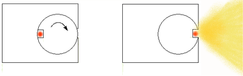
 Another design for a torch is where the source is placed in a metal wheel, which can turn inside the camera to move between the expose and storage positions.
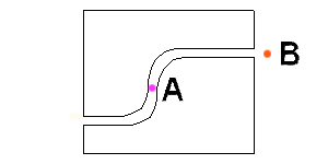
or depleted uranium
shielding that has a S shaped tube-like hole through the block. In the safe position the source is in the centre of the block and is attached to a metal wire that extends in both directions, to use the source a guide tube is attached to one side of the device while a drive cable is attached to the other end of the short cable. Using a hand operated winch the source is then pushed out of the shield and along the source guide tube to the tip of the tube to expose the film, then cranked back into its fully shielded position.
s and planar
cracks are difficult to detect using radiography, which is why penetrants are often used to enhance the contrast in the detection of such defects. Penetrants used include silver nitrate
, zinc iodide
, chloroform
and diiodomethane
. Choice of the penetrant is determined by the ease with which it can penetrate the cracks and also with which it can be removed. Diiodomethane has the advantages of high opacity
, ease of penetration
, and ease of removal because it evaporates relatively quickly. However, it can cause skin burns.
s. This type of radiography is called Neutron Radiography
(NR, Nray, N-Ray) or neutron imaging
. Neutron radiography can see very different things than X-rays, because neutrons can pass with ease through lead and steel but are stopped by plastics, water and oils. Neutron sources include radioactive (241Am/Be and Cf) sources, electrically driven D-T reactions in vacuum tubes and conventional critical nuclear reactors. It might be possible to use a neutron amplifier to increase the neutron flux.http://www.tfd.chalmers.se/~valeri/Mars/Mo-o-f10.pdf
Since the amount of radiation emerging from the opposite side of the material can be detected and measured, variations in this amount (or intensity) of radiation are used to determine thickness or composition of material. Penetrating radiations are those restricted to that part of the electromagnetic spectrum of wavelength less than about 10 nanometre
s.
The safety equipment usually includes four basic items: a radiation survey meter (such as a Geiger/Mueller counter), an alarming dosimeter or rate meter, a gas-charged dosimeter, and a film badge or thermoluminescent dosimeter (TLD). The easiest way to remember what each of these items does is to compare them to gauges on an automobile.
The survey meter could be compared to the speedometer, as it measures the speed, or rate, at which radiation is being picked up. When properly calibrated, used, and maintained, it allows the radiographer to see the current exposure to radiation at the meter. It can usually be set for different intensities, and is used to prevent the radiographer from being overexposed to the radioactive source, as well as for verifying the boundary that radiographers are required to maintain around the exposed source during radiographic operations.
The alarming dosimeter could be most closely compared with the tachometer, as it alarms when the radiographer "redlines" or is exposed to too much radiation. When properly calibrated, activated, and worn on the radiographer's person, it will emit an alarm when the meter measures a radiation level in excess of a preset threshold. This device is intended to prevent the radiographer from inadvertently walking up on an exposed source.
The gas-charged dosimeter is like a trip meter in that it measures the total radiation received, but can be reset. It is designed to help the radiographer measure his/her total periodic dose of radiation. When properly calibrated, recharged, and worn on the radiographer's person, it can tell the radiographer at a glance how much radiation to which the device has been exposed since it was last recharged. Radiographers in many states are required to log their radiation exposures and generate an exposure report. In many countries personal dosimeters are not required to be used by radiographers as the dose rates they show are not always correctly recorded.
The film badge or TLD is more like a car's odometer. It is actually a specialized piece of radiographic film in a rugged container. It is meant to measure the radiographer's total exposure over time (usually a month) and is used by regulating authorities to monitor the total exposure of certified radiographers in a certain jurisdiction. At the end of the month, the film badge is turned in and is processed. A report of the radiographer's total dose is generated and is kept on file.
When these safety devices are properly calibrated, maintained, and used, it is virtually impossible for a radiographer to be injured by a radioactive overexposure. Sadly, the elimination of just one of these devices can jeopardize the safety of the radiographer and all those who are nearby. Without the survey meter, the radiation received may be just below the threshold of the rate alarm, and it may be several hours before the radiographer checks the dosimeter, and up to a month or more before the film badge is developed to detect a low intensity overexposure. Without the rate alarm, one radiographer may inadvertently walk up on the source exposed by the other radiographer. Without the dosimeter, the radiographer may be unaware of an overexposure, or even a radiation burn, which may take weeks to result in noticeable injury. And without the film badge, the radiographer is deprived of an important tool designed to protect him or her from the effects of a long-term overexposure to occupationally obtained radiation, and thus may suffer long-term health problems as a result.
There are three ways a radiographer will ensure they are not exposed to higher than required levels of radiation, time, distance, shielding. The less time that a person is exposed to radiation the lower their dose will be. The further a person is from a radioactive source the lower the level of radiation they receive, this is largely due to the inverse square law. Lastly the more a radioactive source is shielded by either better or greater amounts of shielding the lower the levels of radiation that will escape from the testing area. The most commanly used shielding materials in use are sand, lead (sheets or shot), steel, spent (non-radioactive uranium) tungsten and in suitable situations water.
Industrial radiography appears to have one of the worst safety profiles of the radiation professions, possibly because there are many operators using strong gamma
sources (> 2 Ci) in remote sites with little supervision when compared with workers within the nuclear
industry or within hospitals.http://www-pub.iaea.org/MTCD/publications/PDF/P066_scr.pdf Due to the levels of radiation present whilst they are working many radiographers are also required to work late at night when there are few other people present as most industrial radiography is carried out 'in the open' rather than in purpose built exposure booths or rooms. Fatigue, carelessness and lack of proper training are the three most comman factors attributed to industrial radiography accidents. Many of the "lost source" accidents commented on by the International Atomic Energy Agency
involve radiography equipment. Lost source accidents have the potential to cause a considerable loss of human life. One scenario is that a passerby finds the radiography source and not knowing what it is, takes it home.http://www.irpa.net/irpa10/cdrom/00140.pdf The person shortly afterwards becomes ill and dies as a result of the radiation dose. The source remains in their home where it continues to irradiate other members of the household.http://ean.cepn.asso.fr/pdf/program5/session%201/5_biau.PDF Such an event occurred in March 1984 in Casablanca
(Mohammedia), which is part of Morocco
. This is related to the more famous Goiânia accident
, where a related chain of events caused members of the public to be exposed to radiation sources. Also see List of civilian radiation accidents.
Electromagnetic radiation
Electromagnetic radiation is a form of energy that exhibits wave-like behavior as it travels through space...
to view objects in a way that cannot be seen otherwise. It is not to be confused with the use of ionizing radiation to change or modify objects; radiography's purpose is strictly viewing. Industrial radiography has grown out of engineering, and is a major element of nondestructive testing
Nondestructive testing
Nondestructive testing or Non-destructive testing is a wide group of analysis techniques used in science and industry to evaluate the properties of a material, component or system without causing damage....
. It is a method of inspecting materials for hidden flaws by using the ability of short X-rays and Gamma rays to penetrate various materials.
History
Radiography started in 1895 with the discovery of X-rayX-ray
X-radiation is a form of electromagnetic radiation. X-rays have a wavelength in the range of 0.01 to 10 nanometers, corresponding to frequencies in the range 30 petahertz to 30 exahertz and energies in the range 120 eV to 120 keV. They are shorter in wavelength than UV rays and longer than gamma...
s (later also called Röntgen
Wilhelm Conrad Röntgen
Wilhelm Conrad Röntgen was a German physicist, who, on 8 November 1895, produced and detected electromagnetic radiation in a wavelength range today known as X-rays or Röntgen rays, an achievement that earned him the first Nobel Prize in Physics in 1901....
rays after the man who first described their properties in detail), a type of electromagnetic radiation
Electromagnetic radiation
Electromagnetic radiation is a form of energy that exhibits wave-like behavior as it travels through space...
. Soon after the discovery of X-rays, radioactivity was discovered. By using radioactive sources such as radium
Radium
Radium is a chemical element with atomic number 88, represented by the symbol Ra. Radium is an almost pure-white alkaline earth metal, but it readily oxidizes on exposure to air, becoming black in color. All isotopes of radium are highly radioactive, with the most stable isotope being radium-226,...
, far higher photon
Photon
In physics, a photon is an elementary particle, the quantum of the electromagnetic interaction and the basic unit of light and all other forms of electromagnetic radiation. It is also the force carrier for the electromagnetic force...
energies could be obtained than those from normal X-ray machines. Soon these found various applications, with one of the earliest users being Loughborough University
Loughborough College
Loughborough College is a college of Further Education in Leicestershire, England established in 1909. It is located opposite Loughborough University on Epinal Way, and adjacent to the Loughborough University School of Art and Design, situated next to the main entrance of the college...
, from helping to fit shoes, more lasting medical uses and the examination of non-living objects. X-rays and gamma-rays were put to use very early, before the dangers of ionising radiation were discovered. After World War II
World War II
World War II, or the Second World War , was a global conflict lasting from 1939 to 1945, involving most of the world's nations—including all of the great powers—eventually forming two opposing military alliances: the Allies and the Axis...
new isotopes such as caesium-137
Caesium-137
Caesium-137 is a radioactive isotope of caesium which is formed as a fission product by nuclear fission.It has a half-life of about 30.17 years, and decays by beta emission to a metastable nuclear isomer of barium-137: barium-137m . Caesium-137 is a radioactive isotope of caesium which is formed...
, iridium-192 and cobalt-60
Cobalt-60
Cobalt-60, , is a synthetic radioactive isotope of cobalt. Due to its half-life of 5.27 years, is not found in nature. It is produced artificially by neutron activation of . decays by beta decay to the stable isotope nickel-60...
became available for industrial radiography, and the use of radium and radon decreased.
Inspection of products
For purposes of inspection, including weld inspection, there exist several exposure arrangements.
First, there is the panoramic, one of the four single wall exposure/single wall view (SWE/SWV) arrangements. This exposure is created when the radiographer places the source of radiation at the center of a sphere, cone, or cylinder (including tanks, vessels, and piping). Depending upon client requirements, the radiographer would then place film cassettes on the outside of the surface to be examined. This exposure arrangement is ideal - when properly arranged and exposed, all portions of all exposed film will be of the same approximate density. It also has the advantage of taking less time than other arrangements since the source must only penetrate the total wall thickness (WT) once and must only travel the radius of the inspection item, not its full diameter. The major disadvantage of the panoramic is that it may be impractical to reach the center of the item (enclosed pipe) or the source may be too weak to perform in this arrangement (large vessels or tanks).
The second SWE/SWV arrangement is an interior placement of the source in an enclosed inspection item without having the source centered up. The source does not come in direct contact with the item, but is placed a distance away, depending on client requirements. The third is an exterior placement with similar characteristics. The fourth is reserved for flat objects, such as plate metal, and is also radiographed without the source coming in direct contact with the item. In each case, the radiographic film is located on the opposite side of the inspection item from the source. In all four cases, only one wall is exposed, and only one wall is viewed on the radiograph.
Of the other exposure arrangements, only the contact shot has the source located on the inspection item. This type of radiograph exposes both walls, but only resolves the image on the wall nearest the film. This exposure arrangement takes more time than a panoramic, as the source must penetrate the WT twice and travel the entire outside diameter of the pipe or vessel to reach the film on the opposite side. This is a double wall exposure/single wall view DWE/SWV arrangement. Another is the superimposure (wherein the source is placed on one side of the item, not in direct contact with it, with the film on the opposite side). This arrangement is usually reserved for very small diameter piping or parts. The last DWE/SWV exposure arrangement is the elliptical, in which the source is offset from the plane of the inspection item (usually a weld in pipe) and the elliptical image of the weld furthest from the source is cast onto the film.
The beam of radiation must be directed to the middle of the section under examination and must be normal to the material surface at that point, except in special techniques where known defects are best revealed by a different alignment of the beam. The length of weld
Weld
Weld most commonly refers to a joint formed by welding.Weld may also refer to:-People:* Weld family, an extended family of New England** Theodore Dwight Weld** Tuesday Weld* Weld-Blundell family* Cecil Weld-Forester, 1st Baron Forester...
under examination for each exposure shall be such that the thickness of the material at the diagnostic extremities, measured in the direction of the incident beam, does not exceed the actual thickness at that point by more than 6%. The specimen to be inspected is placed between the source of radiation and the detecting device, usually the film in a light tight holder or cassette, and the radiation is allowed to penetrate the part for the required length of time to be adequately recorded. Lead is often placed behind the film to reduce the 'back scattered' radiation which can lead to the film becoming over exposed.
The result is a two-dimensional projection of the part onto the film, producing a latent image of varying densities according to the amount of radiation
Radiation
In physics, radiation is a process in which energetic particles or energetic waves travel through a medium or space. There are two distinct types of radiation; ionizing and non-ionizing...
reaching each area. It is known as a radiograph, as distinct from a photograph produced by light. Because film is cumulative in its response (the exposure increasing as it absorbs more radiation), relatively weak radiation can be detected by prolonging the exposure until the film can record an image that will be visible after development. The radiograph is examined as a negative
Negative (photography)
In photography, a negative may refer to three different things, although they are all related.-A negative:Film for 35 mm cameras comes in long narrow strips of chemical-coated plastic or cellulose acetate. As each image is captured by the camera onto the film strip, the film strip advances so that...
, without printing as a positive as in photography. This is because, in printing, some of the detail is always lost and no useful purpose is served.
Before commencing a radiographic examination, it is always advisable to examine the component with one's own eyes, to eliminate any possible external defects. If the surface of a weld is too irregular, it may be desirable to grind
Grind
The grind of a blade refers to the shape of the cross-section of the blade. It is distinct from the type of blade , though different tools and blades may have lent their name to a particular grind.Grinding involves removing significant portions of metal from the blade and is thus distinct from...
it to obtain a smooth finish, but this is likely to be limited to those cases in which the surface irregularities (which will be visible on the radiograph) may make detecting internal defects difficult.
After this visual examination, the operator will have a clear idea of the possibilities of access to the two faces of the weld, which is important both for the setting up of the equipment and for the choice of the most appropriate technique.
Airport security
Both hold luggage and carry-on hand luggage are normally examined by X-ray machineX-ray machine
An X-ray generator is a device used to generate X-rays. These devices are commonly used by radiographers to acquire an x-ray image of the inside of an object but they are also used in sterilization or fluorescence....
s using X-ray radiography. See airport security
Airport security
Airport security refers to the techniques and methods used in protecting airports and aircraft from crime.Large numbers of people pass through airports. This presents potential targets for terrorism and other forms of crime due to the number of people located in a particular location...
for more details.
Non-intrusive Cargo Scanning (aka Non-Intrusive Inspection - NII)
Intermodal freight transport
Intermodal freight transport involves the transportation of freight in an intermodal container or vehicle, using multiple modes of transportation , without any handling of the freight itself when changing modes. The method reduces cargo handling, and so improves security, reduces damages and...
cargo containers in US and other countries. Also research is being done on adapting other types of radiography like Dual-Energy X-ray Radiography or Muon Radiography for scanning intermodal
Intermodal freight transport
Intermodal freight transport involves the transportation of freight in an intermodal container or vehicle, using multiple modes of transportation , without any handling of the freight itself when changing modes. The method reduces cargo handling, and so improves security, reduces damages and...
cargo containers.
X-ray sources
A high energy X-ray machine can be used. It is often important to use a high accelerating voltage to provide the electrons with a very high energy. This is because in a braking radiation source the maximum photon energy is determined by the energy of the charged particles.Radioisotope sources
These have the advantage that they do not need a supply of electrical power to function, but they do have the disadvantage that they can not be turned off. Also it is difficult using radioactivity to create a small and compact source that offers the photon flux possible with a normal sealed X-ray tubeX-ray tube
An X-ray tube is a vacuum tube that produces X-rays. They are used in X-ray machines. X-rays are part of the electromagnetic spectrum, an ionizing radiation with wavelengths shorter than ultraviolet light...
. One of the leading makers of radiographic equipment is the Source Production & Equipment Co., Inc. http://www.spec150.com
It might be possible to use Cs-137 as a photon source for radiography but this isotope is always diluted with inactive caesium isotopes. This makes it difficult to get a physically small source, and a large volume of the source makes it impossible to capture fine details in a radiographic examination.
Both cobalt-60 and caesium-137 have only a few gamma energies, which makes them close to monochromatic. The photon energy of cobalt-60 is higher than that of caesium-137, which allows cobalt sources to be used to examine thicker sections of metals than those that could be examined with Cs-137. Iridium-192 has a lower photon energy than cobalt-60 and its gamma spectrum is complex (many lines of very different energies), but this can be an advantage as this can give better contrast for the final photographs.
It has been known for many years that an inactive iridium
Iridium
Iridium is the chemical element with atomic number 77, and is represented by the symbol Ir. A very hard, brittle, silvery-white transition metal of the platinum family, iridium is the second-densest element and is the most corrosion-resistant metal, even at temperatures as high as 2000 °C...
or cobalt
Cobalt
Cobalt is a chemical element with symbol Co and atomic number 27. It is found naturally only in chemically combined form. The free element, produced by reductive smelting, is a hard, lustrous, silver-gray metal....
metal object can be machined to size. In the case of cobalt it is common to alloy
Alloy
An alloy is a mixture or metallic solid solution composed of two or more elements. Complete solid solution alloys give single solid phase microstructure, while partial solutions give two or more phases that may or may not be homogeneous in distribution, depending on thermal history...
it with nickel to improve the mechanical properties. In the case of iridium a thin wire or rod could be used. These precursor materials can then be placed in stainless steel
Stainless steel
In metallurgy, stainless steel, also known as inox steel or inox from French "inoxydable", is defined as a steel alloy with a minimum of 10.5 or 11% chromium content by mass....
containers that have been leak tested before being converted into radioactive sources. These objects can be processed by neutron activation
Neutron activation
Neutron activation is the process in which neutron radiation induces radioactivity in materials, and occurs when atomic nuclei capture free neutrons, becoming heavier and entering excited states. The excited nucleus often decays immediately by emitting particles such as neutrons, protons, or alpha...
to form gamma emitting radioisotopes. The stainless steel has only a small ability to be activated and the small activity due to 55Fe and 63Ni are unlikely to pose a problem in the final application because these isotopes are beta
Beta particle
Beta particles are high-energy, high-speed electrons or positrons emitted by certain types of radioactive nuclei such as potassium-40. The beta particles emitted are a form of ionizing radiation also known as beta rays. The production of beta particles is termed beta decay...
emitters, which have very weak gamma emission. The 59Fe which might form has a short half life, so by allowing a cobalt source to stand for a year much of this isotope will decay away.
The source is often a very small object, which must be transported to the work site in a shielded container. It is normal to place the film in industrial radiography, clear the area where the work is to be done, add shielding (collimators) to reduce the size of the controlled area
Controlled area
In telecommunication, the term controlled area is an area in which uncontrolled movement will not result in compromise of classified information, that is designed to provide administrative control and safety, or that serves as a buffer for controlling access to limited-access areas...
before exposing the radioactive source. A series of different designs have been developed for radiographic "cameras". Rather than the "camera" being a device that accepts photons to record a picture, the "camera" in industrial radiography is the radioactive photon source.
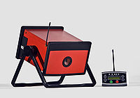
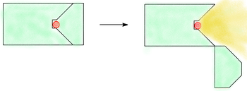
Torch design of radiographic cameras
One design is best thought of as being like a torch. The radioactive source is placed inside a shielded box, a hinge allows part of the shielding to be opened exposing the source, allowing photons to exit the radiography camera.Cable based design of radiographic cameras
One group of designs use a radioactive source, which connects to a drive cable contained shielded exposure device. In one design of equipment the source is stored in a block of leadLead
Lead is a main-group element in the carbon group with the symbol Pb and atomic number 82. Lead is a soft, malleable poor metal. It is also counted as one of the heavy metals. Metallic lead has a bluish-white color after being freshly cut, but it soon tarnishes to a dull grayish color when exposed...
or depleted uranium
Depleted uranium
Depleted uranium is uranium with a lower content of the fissile isotope U-235 than natural uranium . Uses of DU take advantage of its very high density of 19.1 g/cm3...
shielding that has a S shaped tube-like hole through the block. In the safe position the source is in the centre of the block and is attached to a metal wire that extends in both directions, to use the source a guide tube is attached to one side of the device while a drive cable is attached to the other end of the short cable. Using a hand operated winch the source is then pushed out of the shield and along the source guide tube to the tip of the tube to expose the film, then cranked back into its fully shielded position.
Contrast agents
Defects such as delaminationDelamination
Delamination is a mode of failure for composite materials. Modes of failure are also known as 'failure mechanisms'. In laminated materials, repeated cyclic stresses, impact, and so on can cause layers to separate, forming a mica-like structure of separate layers, with significant loss of mechanical...
s and planar
Planar
In computer graphics, planar is the method of representing pixel colours with several bitplanes of RAM. Each bit in a bitplane is related to one pixel on the screen...
cracks are difficult to detect using radiography, which is why penetrants are often used to enhance the contrast in the detection of such defects. Penetrants used include silver nitrate
Silver nitrate
Silver nitrate is an inorganic compound with chemical formula . This compound is a versatile precursor to many other silver compounds, such as those used in photography. It is far less sensitive to light than the halides...
, zinc iodide
Zinc iodide
Zinc iodide is a chemical compound of zinc and iodine, ZnI2. The anhydrous form is white and readily absorbs water from the atmosphere. It can be prepared by the direct reaction of zinc and iodine in refluxing ether...
, chloroform
Chloroform
Chloroform is an organic compound with formula CHCl3. It is one of the four chloromethanes. The colorless, sweet-smelling, dense liquid is a trihalomethane, and is considered somewhat hazardous...
and diiodomethane
Diiodomethane
Diiodomethane or methylene iodide, commonly abbreviated "MI", is a liquid organoiodine compound. It is insoluble in water, but soluble in ether and alcohol. It has a relatively high refractive index of 1.741, and a surface tension of 0.0508 N·m−1...
. Choice of the penetrant is determined by the ease with which it can penetrate the cracks and also with which it can be removed. Diiodomethane has the advantages of high opacity
Opacity (optics)
Opacity is the measure of impenetrability to electromagnetic or other kinds of radiation, especially visible light. In radiative transfer, it describes the absorption and scattering of radiation in a medium, such as a plasma, dielectric, shielding material, glass, etc...
, ease of penetration
Penetration
Penetration may refer to:* Penetration depth of light or any electromagnetic radiation.* Penetrating trauma* Sexual penetration, a term which may mean**sexual intercourse in general or,...
, and ease of removal because it evaporates relatively quickly. However, it can cause skin burns.
Neutrons
In some rare cases, radiography is done with neutronNeutron
The neutron is a subatomic hadron particle which has the symbol or , no net electric charge and a mass slightly larger than that of a proton. With the exception of hydrogen, nuclei of atoms consist of protons and neutrons, which are therefore collectively referred to as nucleons. The number of...
s. This type of radiography is called Neutron Radiography
Neutron Radiography
Neutron Radiography is the process by which film is exposed by first passing neutrons through an object to produce a visible image of the materials that make up the object. Primarily used in scientific investigations.- Brief History of Neutron Imaging :...
(NR, Nray, N-Ray) or neutron imaging
Neutron imaging
Neutron Imaging is the process of making a image with neutrons. The resulting image is based on the neutron attenuation properties of the imaged object...
. Neutron radiography can see very different things than X-rays, because neutrons can pass with ease through lead and steel but are stopped by plastics, water and oils. Neutron sources include radioactive (241Am/Be and Cf) sources, electrically driven D-T reactions in vacuum tubes and conventional critical nuclear reactors. It might be possible to use a neutron amplifier to increase the neutron flux.http://www.tfd.chalmers.se/~valeri/Mars/Mo-o-f10.pdf
Since the amount of radiation emerging from the opposite side of the material can be detected and measured, variations in this amount (or intensity) of radiation are used to determine thickness or composition of material. Penetrating radiations are those restricted to that part of the electromagnetic spectrum of wavelength less than about 10 nanometre
Nanometre
A nanometre is a unit of length in the metric system, equal to one billionth of a metre. The name combines the SI prefix nano- with the parent unit name metre .The nanometre is often used to express dimensions on the atomic scale: the diameter...
s.
Safety
Industrial radiographers are in many locations required by governing authorities to use certain types of safety equipment and to work in pairs. Depending on location industrial radiographers may have been required to obtain permits, licences and/or undertake special training. Prior to conducting any testing the nearby area should always first be cleared of all other persons and measures taken to ensure that people do not accidentally enter into an area that may expose them to a large dose of radiation.The safety equipment usually includes four basic items: a radiation survey meter (such as a Geiger/Mueller counter), an alarming dosimeter or rate meter, a gas-charged dosimeter, and a film badge or thermoluminescent dosimeter (TLD). The easiest way to remember what each of these items does is to compare them to gauges on an automobile.
The survey meter could be compared to the speedometer, as it measures the speed, or rate, at which radiation is being picked up. When properly calibrated, used, and maintained, it allows the radiographer to see the current exposure to radiation at the meter. It can usually be set for different intensities, and is used to prevent the radiographer from being overexposed to the radioactive source, as well as for verifying the boundary that radiographers are required to maintain around the exposed source during radiographic operations.
The alarming dosimeter could be most closely compared with the tachometer, as it alarms when the radiographer "redlines" or is exposed to too much radiation. When properly calibrated, activated, and worn on the radiographer's person, it will emit an alarm when the meter measures a radiation level in excess of a preset threshold. This device is intended to prevent the radiographer from inadvertently walking up on an exposed source.
The gas-charged dosimeter is like a trip meter in that it measures the total radiation received, but can be reset. It is designed to help the radiographer measure his/her total periodic dose of radiation. When properly calibrated, recharged, and worn on the radiographer's person, it can tell the radiographer at a glance how much radiation to which the device has been exposed since it was last recharged. Radiographers in many states are required to log their radiation exposures and generate an exposure report. In many countries personal dosimeters are not required to be used by radiographers as the dose rates they show are not always correctly recorded.
The film badge or TLD is more like a car's odometer. It is actually a specialized piece of radiographic film in a rugged container. It is meant to measure the radiographer's total exposure over time (usually a month) and is used by regulating authorities to monitor the total exposure of certified radiographers in a certain jurisdiction. At the end of the month, the film badge is turned in and is processed. A report of the radiographer's total dose is generated and is kept on file.
When these safety devices are properly calibrated, maintained, and used, it is virtually impossible for a radiographer to be injured by a radioactive overexposure. Sadly, the elimination of just one of these devices can jeopardize the safety of the radiographer and all those who are nearby. Without the survey meter, the radiation received may be just below the threshold of the rate alarm, and it may be several hours before the radiographer checks the dosimeter, and up to a month or more before the film badge is developed to detect a low intensity overexposure. Without the rate alarm, one radiographer may inadvertently walk up on the source exposed by the other radiographer. Without the dosimeter, the radiographer may be unaware of an overexposure, or even a radiation burn, which may take weeks to result in noticeable injury. And without the film badge, the radiographer is deprived of an important tool designed to protect him or her from the effects of a long-term overexposure to occupationally obtained radiation, and thus may suffer long-term health problems as a result.
There are three ways a radiographer will ensure they are not exposed to higher than required levels of radiation, time, distance, shielding. The less time that a person is exposed to radiation the lower their dose will be. The further a person is from a radioactive source the lower the level of radiation they receive, this is largely due to the inverse square law. Lastly the more a radioactive source is shielded by either better or greater amounts of shielding the lower the levels of radiation that will escape from the testing area. The most commanly used shielding materials in use are sand, lead (sheets or shot), steel, spent (non-radioactive uranium) tungsten and in suitable situations water.
Industrial radiography appears to have one of the worst safety profiles of the radiation professions, possibly because there are many operators using strong gamma
Gamma
Gamma is the third letter of the Greek alphabet. In the system of Greek numerals it has a value of 3. It was derived from the Phoenician letter Gimel . Letters that arose from Gamma include the Roman C and G and the Cyrillic letters Ge Г and Ghe Ґ.-Greek:In Ancient Greek, gamma represented a...
sources (> 2 Ci) in remote sites with little supervision when compared with workers within the nuclear
Nuclear power
Nuclear power is the use of sustained nuclear fission to generate heat and electricity. Nuclear power plants provide about 6% of the world's energy and 13–14% of the world's electricity, with the U.S., France, and Japan together accounting for about 50% of nuclear generated electricity...
industry or within hospitals.http://www-pub.iaea.org/MTCD/publications/PDF/P066_scr.pdf Due to the levels of radiation present whilst they are working many radiographers are also required to work late at night when there are few other people present as most industrial radiography is carried out 'in the open' rather than in purpose built exposure booths or rooms. Fatigue, carelessness and lack of proper training are the three most comman factors attributed to industrial radiography accidents. Many of the "lost source" accidents commented on by the International Atomic Energy Agency
International Atomic Energy Agency
The International Atomic Energy Agency is an international organization that seeks to promote the peaceful use of nuclear energy, and to inhibit its use for any military purpose, including nuclear weapons. The IAEA was established as an autonomous organization on 29 July 1957...
involve radiography equipment. Lost source accidents have the potential to cause a considerable loss of human life. One scenario is that a passerby finds the radiography source and not knowing what it is, takes it home.http://www.irpa.net/irpa10/cdrom/00140.pdf The person shortly afterwards becomes ill and dies as a result of the radiation dose. The source remains in their home where it continues to irradiate other members of the household.http://ean.cepn.asso.fr/pdf/program5/session%201/5_biau.PDF Such an event occurred in March 1984 in Casablanca
Casablanca
Casablanca is a city in western Morocco, located on the Atlantic Ocean. It is the capital of the Grand Casablanca region.Casablanca is Morocco's largest city as well as its chief port. It is also the biggest city in the Maghreb. The 2004 census recorded a population of 2,949,805 in the prefecture...
(Mohammedia), which is part of Morocco
Morocco
Morocco , officially the Kingdom of Morocco , is a country located in North Africa. It has a population of more than 32 million and an area of 710,850 km², and also primarily administers the disputed region of the Western Sahara...
. This is related to the more famous Goiânia accident
Goiânia accident
The Goiânia accident was a radioactive contamination accident that occurred on September 13, 1987, at Goiânia, in the Brazilian State of Goiás after an old radiotherapy source was taken from an abandoned hospital site in the city...
, where a related chain of events caused members of the public to be exposed to radiation sources. Also see List of civilian radiation accidents.