ABSOLUTEASTRONOMY
Create a free discussion account!
Signup
Login
x
Home
Search
Topics
Almanac
Science
Nature
People
History
Society
Signup
Login
ABSOLUTEASTRONOMY
HOME
TOPICS
ALMANAC
SCIENCE
NATURE
PEOPLE
HISTORY
SOCIETY
PHILOSOPHY
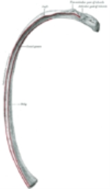
Human rib cage
Topic Home
Discussion
3
Overview
Terms for bones
Unanswered Questions
Does the rib cage open or seperate during any time of night?
What are the thorax bones protecting respiratory system and how they protect the respiratory system ?
WHen I was younger i used to have bad cramp. i went to see a doctor and they performed x rays. the doctor the told me i was born with 2 sets of ribs a...
More
x
OK