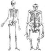
The
human legŁęg may refer to the following places in Poland:*A former name for the town of Ełk *Part of the Czyżyny district of Kraków*Łęg, Pleszew County in Greater Poland Voivodeship...
is the entire lower extremity or
limbA limb is a jointed, or prehensile , appendage of the human or other animal body....
of the
human bodyThe human body is the entire structure of a human organism, and consists of a head, neck, torso, two arms and two legs.By the time the human reaches adulthood, the body consists of close to 100 trillion cells, the basic unit of life...
, including the
footThe foot is an anatomical structure found in many vertebrates. It is the terminal portion of a limb which bears weight and allows locomotion. In many animals with feet, the foot is a separate organ at the terminal part of the leg made up of one or more segments or bones, generally including claws...
,
thighIn humans the thigh is the area between the pelvis and the knee. Anatomically, it is part of the lower limb.The single bone in the thigh is called the femur...
and even the hip or gluteal region; however, the precise definition in
human anatomyHuman anatomy is primarily the scientific study of the morphology of the human body. Anatomy is subdivided into gross anatomy and microscopic anatomy. Gross anatomy is the study of anatomical structures that can be seen by the naked eye...
refers only to the section of the lower limb extending from the
kneeThe knee joint joins the thigh with the leg and consists of two articulations: one between the fibula and tibia, and one between the femur and patella. It is the largest joint in the human body and is very complicated. The knee is a mobile trocho-ginglymus , which permits flexion and extension as...
to the
ankleThe ankle joint is formed where the foot and the leg meet. The ankle, or talocrural joint, is a synovial hinge joint that connects the distal ends of the tibia and fibula in the lower limb with the proximal end of the talus bone in the foot...
.
Legs are used for
standingStanding is a human position in which the body is held upright and supported only by the feet, referred to as an orthostatic state.Although quiet standing appears to be static, modern instrumentation shows it to be a process of rocking from the ankle in the sagittal plane...
,
walkingWalking is one of the main gaits of locomotion among legged animals, and is typically slower than running and other gaits. Walking is defined by an 'inverted pendulum' gait in which the body vaults over the stiff limb or limbs with each step...
,
jumpingJumping or leaping is a form of locomotion or movement in which an organism or non-living mechanical system propels itself through the air along a ballistic trajectory...
,
runningRunning is a means of terrestrial locomotion allowing humans and other animals to move rapidly on foot. It is simply defined in athletics terms as a gait in which at regular points during the running cycle both feet are off the ground...
, kicking, and similar activities, and constitute a significant portion of a person's mass.
Anatomy
In
human anatomical termsHuman anatomy is primarily the scientific study of the morphology of the human body. Anatomy is subdivided into gross anatomy and microscopic anatomy. Gross anatomy is the study of anatomical structures that can be seen by the naked eye...
, the leg is the part of the lower limb that lies between the
kneeThe knee joint joins the thigh with the leg and consists of two articulations: one between the fibula and tibia, and one between the femur and patella. It is the largest joint in the human body and is very complicated. The knee is a mobile trocho-ginglymus , which permits flexion and extension as...
and the
ankleThe ankle joint is formed where the foot and the leg meet. The ankle, or talocrural joint, is a synovial hinge joint that connects the distal ends of the tibia and fibula in the lower limb with the proximal end of the talus bone in the foot...
, the
thighIn humans the thigh is the area between the pelvis and the knee. Anatomically, it is part of the lower limb.The single bone in the thigh is called the femur...
is between the hip and
kneeThe knee joint joins the thigh with the leg and consists of two articulations: one between the fibula and tibia, and one between the femur and patella. It is the largest joint in the human body and is very complicated. The knee is a mobile trocho-ginglymus , which permits flexion and extension as...
and the term "lower limb" is used to describe the colloquial leg. This article generally follows the common usage.
The leg from the knee to the ankle is called the
cnemis 'niːmɪs or
crusCrus is the portion of the body starting from the ankle and ending at the knee. It is sometimes known as the gaiter...
. The
calfIn human anatomy the calf is the back portion of the lower leg . In terms of muscle systems, the calf corresponds to the posterior compartment of the leg. Within the posterior compartment, the two largest muscles are known together as the calf muscle and attach to the heel via the Achilles tendon...
is the back portion and the shin is the front.
The human body has two distinct features: the specialization of the upper limb for visually guided manipulation and the lower limb's development into a mechanism specifically adapted for efficient bipedal
gaitGait is the pattern of movement of the limbs of animals, including humans, during locomotion over a solid substrate. Most animals use a variety of gaits, selecting gait based on speed, terrain, the need to maneuver, and energetic efficiency...
. While the capacity to walk upright is not unique to humans, other
primateA primate is a mammal of the order Primates , which contains prosimians and simians. Primates arose from ancestors that lived in the trees of tropical forests; many primate characteristics represent adaptations to life in this challenging three-dimensional environment...
s can only achieve this for short periods and at a great expenditure of energy. The human adaption to bipedalism is not limited to the leg, however, but has also affected the location of the body's
center of gravityIn physics, the center of mass or barycenter of a system is the average location of all of its mass. In the case of a rigid body, the position of the center of mass is fixed in relation to the body...
, the reorganisation of
internal organsIn anatomy, a viscus is an internal organ, and viscera is the plural form. The viscera, when removed from a butchered animal, are known collectively as offal...
, and the form and
biomechanismBiomechanics is the application of mechanical principles to biological systems, such as humans, animals, plants, organs, and cells. Perhaps one of the best definitions was provided by Herbert Hatze in 1974: "Biomechanics is the study of the structure and function of biological systems by means of...
of the trunk. In humans, the double S-shaped
vertebral columnIn human anatomy, the vertebral column is a column usually consisting of 24 articulating vertebrae, and 9 fused vertebrae in the sacrum and the coccyx. It is situated in the dorsal aspect of the torso, separated by intervertebral discs...
acts as a shock-absorber which shifts the weight from the trunk over the load-bearing surface of the feet. The human legs are exceptionally long and powerful as a result of their exclusive specialization to support and locomotion — in
orangutanOrangutans are the only exclusively Asian genus of extant great ape. The largest living arboreal animals, they have proportionally longer arms than the other, more terrestrial, great apes. They are among the most intelligent primates and use a variety of sophisticated tools, also making sleeping...
s the leg length is 111% of the trunk; in
chimpanzeeChimpanzee, sometimes colloquially chimp, is the common name for the two extant species of ape in the genus Pan. The Congo River forms the boundary between the native habitat of the two species:...
s 128%, and in humans 171%. Many of the leg's muscles are also adapted to bipedalism, most substantially the
gluteal musclesThe gluteal muscles are the three muscles that make up the buttocks: the gluteus maximus muscle, gluteus medius muscle and gluteus minimus muscle.-The gluteal muscles:...
, the extensors of the knee joint, and the calf muscles.
- See also: Human skeletal changes due to bipedalism
The evolution of human bipedalism approximately four million years ago has led to morphological alterations to the human skeleton including changes to the arrangement and size of the bones of the foot, hip size and shape, knee size, leg length, and the shape and orientation of the vertebral column...
Skeleton
The major (long) bones of the human leg are the
femurThe femur , or thigh bone, is the most proximal bone of the leg in tetrapod vertebrates capable of walking or jumping, such as most land mammals, birds, many reptiles such as lizards, and amphibians such as frogs. In vertebrates with four legs such as dogs and horses, the femur is found only in...
(thighbone),
tibiaThe tibia , shinbone, or shankbone is the larger and stronger of the two bones in the leg below the knee in vertebrates , and connects the knee with the ankle bones....
(shinbone), and
fibula (the smaller, rear calf bone). The
patella (kneecap) is the bone in front of the knee. Most of the leg skeleton has bony prominences and margins that can be
palpatedPalpation is used as part of a physical examination in which an object is felt to determine its size, shape, firmness, or location...
, notable exceptions being the hip joint, and the
neckThe femur neck or neck of the femur is a flattened pyramidal process of bone, connecting the femoral head with the femoral shaft, and forming with the latter a wide angle opening medialward.- Angle of inclination :...
and
shaft of femurThe body of the femur , almost cylindrical in form, is a little broader above than in the center, broadest and somewhat flattened from before backward below...
. Many of these anatomical landmarks are used to define the extent of the leg: most notably the
anterior superior iliac spineThe anterior superior iliac spine is an important landmark of surface anatomy. It refers to the anterior extremity of the iliac crest of the pelvis, which provides attachment for the inguinal ligament, and the sartorius muscle...
, the
greater trochanterThe greater trochanter of the femur is a large, irregular, quadrilateral eminence and a part of the skeletal system.It is directed a little lateralward and backward, and, in the adult, is about 1 cm lower than the head...
, the superior margin of the
medial condyle of tibiaThe medial condyle is the medial portion of the upper extremity of tibia.It is the site of insertion for the Semimembranosus muscle....
, and the medial malleolus.
In the normal case, the large joints of the lower limb are aligned on a straight line which represents the mechanical longitudinal axis of the leg, the
MikuliczJan Mikulicz-Radecki was a Polish-Austrian surgeon. He was born May 16, 1850 in Czernowitz in the Austrian Empire and died June 4, 1905 in Breslau, German Empire .While his mother Freiin von Damnitz was Austrian, his parental ancestors of the Mikulicz...
line. This line stretches from the hip joint (or more precisely the
head of the femurThe femur head is the highest part of the thigh bone . It is supported by the neck of the femur.The head is globular and forms rather more than a hemisphere, is directed upward, medialward, and a little forward, the greater part of its convexity being above and in front.Its surface is smooth,...
), through the knee joint (the intercondylar eminence of the tibia), and down to the center of the
ankleThe ankle joint is formed where the foot and the leg meet. The ankle, or talocrural joint, is a synovial hinge joint that connects the distal ends of the tibia and fibula in the lower limb with the proximal end of the talus bone in the foot...
(the ankle mortise, the fork-like grip between the medial and lateral malleoli). In the
tibial shaft- Borders :The anterior crest or border, the most prominent of the three, commences above at the tuberosity, and ends below at the anterior margin of the medial malleolus...
, the mechanical and anatomical axes coincide, but in the
femoral shaftThe body of the femur , almost cylindrical in form, is a little broader above than in the center, broadest and somewhat flattened from before backward below...
they diverge 6°, resulting in the
femorotibial angle of 174° in a leg with normal axial alignment. A leg is considered straight when, with the feet brought together, both the medial malleoli of the ankle and the medial condyles of the knee are touching. Divergence from the normal femorotibial angle is called
genu varum if the center of the knee joint is lateral to the mechanical axis (intermalleolar distance exceeds 3 cm), and
genu valgumGenu valgum, commonly called "knock-knee", is a condition where the knees angle in and touch one another when the legs are straightened. Women have a wider pelvis than men and a relatively shorter length of the thigh bone, and as a result, have a greater static genu valgum than men...
if it is medial to the mechanical axis (intercondylar distance exceeds 5 cm). These conditions impose unbalanced loads on the joints and stretching of either the thigh's adductors and abductors.
The angle of inclination formed between the neck and shaft of the femur, the collodiaphysial angle, varies with age—about 150° in the newborn, it gradually decreases to 126-128° in adults, to reach 120° in old age. Pathological changes in this angle results in abnormal posture of the leg: A small angle produces
coxa varaCoxa vara is a deformity of the hip, whereby the angle between the ball and the shaft of the femur is reduced to less than 120 degrees. This results in the leg being shortened, and therefore a limp occurs. It is commonly caused by injury, such as a fracture...
and a large angle in
coxa valgaIt is a deformity of the hip where the angle formed between the head and neck of the femur and its shaft is increased, usually above 135 degrees. It is caused by a slipped epiphysis of the femoral head.-See also:*Coxa vara*Genu valgum*Genu varum...
; the latter is usually combined with genu varum and coxa vara leads genu valgum. Additionally, a line drawn through the femoral neck superimposed on a line drawn through the femoral condyles forms an angle, the
torsion angle, which makes it possible for flexion movements of the hip joint to be transposed into rotary movements of the femoral head. Abnormally increased torsion angles results in a limb turned inward and a decreased angle in a limb turned outward; both cases resulting in a reduced range of mobility.
Hip
Function of hip muscles
| Movement | Muscles (In order of importance) |
Lateral rotation |
• Sartorius
• Gluteus maximus
• Quadratus femoris
• Obturator internus
• Gluteus medius and minimus
• Iliopsoas (with psoas major♣)
• Obturator externus
• All functional adductors
except gracilis* and pectineus
• Piriformis |
Medial rotation |
• Gluteus medius and minimus (anterior fibers)
• Tensor fascia latae*
• Adductor magnus (long medial fibers)
• Pectineus (with leg abducted) |
| Extension |
• Gluteus maximus
• Gluteus medius and minimus (dorsal fibers)
• Adductor magnus
• Piriformis
• Semimembranosus*
• Semitendinousus*
• Biceps femoris* (long head) |
| Flexion |
• Iliopsoas (with psoas major♣)
• Tensor fascia latae*
• Pectineus
• Adductor longus
• Adductor brevis
• Gracilis*
• Rectus femoris*
• Sartorius* |
| Abduction |
• Gluteus medius
• Tensor fascia latae*
• Gluteus maximus (fibers to fascia lata)
• Gluteus minimus
• Piriformis
• Obturator internus |
| Adduction |
• Adductor magnus (with adductor minimus)
• Adductor longus
• Adductor brevis
• Gluteus maximus (fibers to gluteal tuberosity)
• Gracilis
• Pectineus
• Quadratus femoris
• Obturator externus
• Semitendinosus* |
| Notes |
♣ Also act on vertebral joints. * Also act on knee joint. |
There are several ways of classifying the muscles of the hip: (1) By location or innervation (ventral an dorsal divisions of the plexus layer); (2) by development on the basis of their points of insertion (a posterior group in two layers and an anterior group); and (3) by function (i.e. extensors, flexors, adductors, and abductors).
Some hip muscles also act on either the knee joint or on vertebral joints. Additionally, because the area of origin and insertion of many of these muscles are very extensive, these muscles are often involved in several very different movements. In the hip joint,
lateral and
medial rotation occur along the axis of the limb;
extension (also called dorsiflexion or retroversion) and
flexion (anteflexion or anteversion) occur along a transverse axis; and
abduction and
adduction occur about a sagittal axis.
The
anterior dorsal hip muscles are the
iliopsoas-External links:*...
, a group of two or three muscles with a shared insertion on the
lesser trochanterThe lesser trochanter of the femur is a conical eminence, which varies in size in different subjects-Anatomy:It projects from the lower and back part of the base of the femur neck.From its apex three well-marked borders extend:...
of the femur. The
psoas major originates from the last vertebra and along the lumbar spine to stretch down into the pelvis. The
iliacusThe iliacus is a flat, triangular muscle which fills the iliac fossa.- Course :The iliacus arises from the iliac fossa on the interior side of the hip bone, and also from the region of the anterior inferior iliac spine...
originates on the
iliac fossaThe iliac fossa is a large, smooth, concave surface located on the internal surface of the ilium...
on the interior side of the pelvis. The two muscles unite to form the iliopsoas muscle which is inserted on the
lesser trochanterThe lesser trochanter of the femur is a conical eminence, which varies in size in different subjects-Anatomy:It projects from the lower and back part of the base of the femur neck.From its apex three well-marked borders extend:...
of the femur. The
psoas minor, only present in about 50 per cent of subjects, originates above psoas major to stretch obliquely down to its insertion on the interior side of the major muscle.
The
posterior dorsal hip muscles are inserted on or directly below the
greater trochanterThe greater trochanter of the femur is a large, irregular, quadrilateral eminence and a part of the skeletal system.It is directed a little lateralward and backward, and, in the adult, is about 1 cm lower than the head...
of the femur. The
tensor fascia latae, stretching from the
anterior superior iliac spineThe anterior superior iliac spine is an important landmark of surface anatomy. It refers to the anterior extremity of the iliac crest of the pelvis, which provides attachment for the inguinal ligament, and the sartorius muscle...
down into the
iliotibial tractThe iliotibial tract or iliotibial band is a longitudinal fibrous reinforcement of the fascia lata. It is attached to the anterolateral iliac tubercle portion of the external lip of the iliac crest and to the lateral condyle of the tibia...
, presses the head of the femur into the
acetabulumThe acetabulum is a concave surface of the pelvis. The head of the femur meets with the pelvis at the acetabulum, forming the hip joint.-Structure:...
but also flexes, rotates medially, and abducts to hip joint. The
piriformis originates on the anterior pelvic surface of the
sacrumIn vertebrate anatomy the sacrum is a large, triangular bone at the base of the spine and at the upper and back part of the pelvic cavity, where it is inserted like a wedge between the two hip bones. Its upper part connects with the last lumbar vertebra, and bottom part with the coccyx...
, passes through the
greater sciatic foramen-Boundaries:It is bounded as follows:* anterolaterally by the greater sciatic notch of the illium* posteromedially by the sacrotuberous ligament* inferiorly by the sacrospinous ligament and the ischial spine* superiorly by the anterior sacroilliac ligament...
, and inserts on the posterior aspect of the tip of the greater trochanter. In a standing posture it is a lateral rotator, but it also assists extending the thigh. The
gluteus maximus has its origin between (and around) the
iliac crest-External links: - "Superficial muscles of the gluteal region and posterior thigh." - "Anterior Abdominal Wall: Osteology and Surface Anatomy " - "The Back, Posterior View" * *...
and the
coccyxThe coccyx , commonly referred to as the tailbone, is the final segment of the vertebral column. Comprising three to five separate or fused vertebrae below the sacrum, it is attached to the sacrum by a fibrocartilaginous joint, the sacrococcygeal symphysis, which permits limited movement between...
from where one part radiates into the iliotibial tract and the other stretches down to the
gluteal tuberosityThe lateral ridge of the linea aspera is very rough, and runs almost vertically upward to the base of the greater trochanter. It is termed the gluteal tuberosity, and gives attachment to part of the Glutæus maximus: its upper part is often elongated into a roughened crest, on which a more or less...
under the greater trochanter. The gluteus maximus is primarily an extensor and lateral rotator of the hip joint, and it comes into action when climbing stairs or rising from a sitting to standing posture. Furthermore, the part inserted into the fascia latae abducts and the part inserted into the gluteal tuberosity adducts the hip. The two deep glutei muscles, the
gluteus medius and
minimus, originate on the lateral side of the pelvis. The medius muscle is shaped like a cap. Its anterior fibers act as a medial rotator and flexor; the posterior fibers as a lateral rotator and extensor; and the entire muscle abducts the hip. The minimus has similar functions and both muscles are inserted onto the greater trochanter.
The
ventral hip muscles function as lateral rotators and play an important role in the control of the body's balance. Because they are stronger than the medial rotators, in the normal position of the leg, the apex of the foot is pointing outward to achieve better support. The
obturator internus originates on the pelvis on the
obturator foramenThe obturator foramen is the hole created by the ischium and pubis bones of the pelvis through which nerves and muscles pass.- General structure :...
and its
membraneThe obturator membrane is a thin fibrous sheet, which almost completely closes the obturator foramen.Its fibers are arranged in interlacing bundles mainly transverse in direction; the uppermost bundle is attached to the obturator tubercles and completes the obturator canal for the passage of the...
, passes through the
lesser sciatic foramenAn opening between the pelvis and the posterior thigh, the foramen is formed by the sacrotuberous ligament which runs between the sacrum and the ischial tuberosity and the sacrospinous ligament which runs between the sacrum and the ischial spine.-Boundaries:...
, and is inserted on the
trochanteric fossa of the femur. "Bent" over the
lesser sciatic notchBelow the ischial spine is a smaller notch, the lesser sciatic notch; it is smooth, coated in the recent state with cartilage, the surface of which presents two or three ridges corresponding to the subdivisions of the tendon of the Obturator internus, which winds over it.It is converted into a...
, which acts as a fulcrum, the muscle forms the strongest lateral rotators of the hip together with the gluteus maximus and quadratus femoris. When sitting with the knees flexed it acts as an abductor. The
obturator externus has a parallel course with its origin located on the posterior border of the obturator foramen. It is covered by several muscles and acts as a lateral rotator and a weak adductor. The
inferiorThe inferior gemellus muscle is a muscle of the human body.The Gemelli are two small muscular fasciculi, accessories to the tendon of the Obturator internus which is received into a groove between them....
and
superior gemelliThe superior gemellus muscle is a muscle of the human body.The Gemelli are two small muscular fasciculi, accessories to the tendon of the Obturator internus which is received into a groove between them....
represent marginal heads of the obturator internus and assist this muscle. The three muscles have been referred to as the
triceps coxae. The
quadratus femoris originates at the ischial tuberosity and is inserted onto the
intertrochanteric crestThe intertrochanteric crest is a bony ridge located on the posterior side of the head of the femur, stretching obliquely downward and medially from the summit of the greater trochanter to the lesser trochanter....
between the trochanters. This flattened muscle act as a strong lateral rotator and adductor of the thigh.
The
adductor muscles of the thigh are innervated by the
obturator nerveThe obturator nerve in human anatomy arises from the ventral divisions of the second, third, and fourth lumbar nerves; the branch from the third is the largest, while that from the second is often very small.-Path:...
, with the exception of pectineus which receives fibers from the
femoral nerveThe femoral nerve, the largest branch of the lumbar plexus, arises from the dorsal divisions of the ventral rami of the second, third, and fourth lumbar nerves...
, and the adductor magnus which receives fibers from the
tibial nerveThe tibial nerve is a branch of the sciatic nerve. The tibial nerve passes through the popliteal fossa to pass below the arch of soleus.In the popliteal fossa the nerve gives off branches to gastrocnemius, popliteus, soleus and plantaris muscles, an articular branch to the knee joint, and a...
. The
gracilisThe gracilis is the most superficial muscle on the medial side of the thigh. It is thin and flattened, broad above, narrow and tapering below.-Origin and insertion:...
arises from near the
pubic symphysisThe pubic symphysis or symphysis pubis is the midline cartilaginous joint uniting the superior rami of the left and right pubic bones. It is located anterior to the urinary bladder and superior to the external genitalia; for females it is above the vulva and for males it is above the penis...
and is unique among the adductors in that it reaches past the knee to attach on the medial side of the
shaft of the tibia- Borders :The anterior crest or border, the most prominent of the three, commences above at the tuberosity, and ends below at the anterior margin of the medial malleolus...
, thus acting on two joints. It share its distal insertion with the
sartoriusThe Sartorius muscle – the longest muscle in the human body – is a long thin muscle that runs down the length of the thigh. Its upper portion forms the lateral border of the femoral triangle.-Origin and insertion:...
and
semitendinosusThe semitendinosus is a muscle in the back of the thigh; it is one of the hamstrings.-Structure:The semitendinosus, remarkable for the great length of its tendon of insertion, is situated at the posterior and medial aspect of the thigh ....
, all three muscles forming the
pes anserinusThe pes anserinus is the insertion of the conjoined tendons of three muscles onto the anteromedial surface of the proximal extremity of the tibia.-Anatomy:The three muscles are :* sartorius...
. It is the most medial muscle of the adductors, and with the thigh abducted its origin can be clearly seen arching under the skin. With the knee extended, it adducts the thigh and flexes the hip. The
pectineusThe pectineus muscle is a flat, quadrangular muscle, situated at the anterior part of the upper and medial aspect of the thigh....
has its origin on the iliopubic eminence laterally to the gracilis and, rectangular in shape, extends obliquely to attach immediately behind the lesser trochanter and down the
pectineal lineOn the posterior surface of the femur, the intermediate ridge or pectineal line is continued to the base of the lesser trochanter and gives attachment to the pectineus muscle....
and the proximal part of the
linea asperaThe linea aspera is a ridge of roughened surface on the posterior aspect of the femur, to which are attached muscles and intermuscular septum.Its margins diverge above and below....
on the femur. It is a flexor of the hip joint, and an adductor and a weak medial rotator of the thigh. The
adductor brevisThe adductor brevis is a muscle in the thigh situated immediately behind the pectineus and adductor longus.-Origin and insertion:It is somewhat triangular in form, and arises by a narrow origin from the outer surfaces of the superior and inferior rami of the pubis, between the gracilis and...
originates on the inferior ramus of the pubis below the gracilis and stretches obliquely below the pectineus down to the upper third of the linea aspera. Except for being an adductor, it is a lateral rotator and weak flexor of the hip joint. The
adductor longusIn the human body, the adductor longus is a skeletal muscle located in the thigh. One of the adductor muscles of the hip, its main function is to adduct the thigh and it is innervated by the obturator nerve...
has its origin at superior ramus of the pubis and inserts medially on the middle third of the linea aspera. Primarily an adductor, it is also responsible for some flexion. The
adductor magnusThe adductor magnus is a large triangular muscle, situated on the medial side of the thigh.It consists of two parts. The portion which arises from the ischiopubic ramus is called the "adductor portion", and the portion arising from the tuberosity of the ischium is called the "hamstring portion"...
has its origin just behind the longus and lies deep to it. Its wide belly divides into two parts: One is inserted into the linea aspera and the tendon of the other reaches down to
adductor tubercleThe medial lip of the linea aspera ends below at the summit of the medial condyle, in a small tubercle, the adductor tubercle, which affords insertion to the tendon of the vertical fibers of Adductor magnus.-External links:*...
on the medial side of the femur's distal end where it forms an intermuscular septum that separates the flexors from the extensors. Magnus is a powerful adductor, especially active when crossing legs. Its superior part is a lateral rotator but the inferior part acts as a medial rotator on the flexed leg when rotated outward and also extends the hip joint. The
adductor minimusIn human anatomy, the adductor minimus is a small and flat skeletal muscle in the thigh which constitutes the upper, lateral part of the adductor magnus muscle....
is an incompletely separated subdivision of the adductor magnus. Its origin forms an anterior part of the magnus and distally it is inserted on the linea aspera above the magnus. It acts to adduct and lateral rotate the femur.
Thigh
Function of knee muscles
| Movement | Muscles (In order of importance) |
| Extension |
• Quadriceps femoris
• Tensor fascia latae* |
| Flexion |
• Semimembranosus
• Semitendinosus
• Biceps femoris
• Gracilis
• Sartorius
• Popliteus
• Gastrocnemius |
Medial rotation |
• Semimembranosus
• Semitendinosus
• Gracilis
• Sartorius
• Popliteus |
Lateral rotation |
• Biceps femoris
• Tensor fascia latae* |
| *Insignificant assistance. |
The muscles of the
thighIn humans the thigh is the area between the pelvis and the knee. Anatomically, it is part of the lower limb.The single bone in the thigh is called the femur...
can be classified into three groups according to their location: anterior and posterior muscles and the adductors (on the medial side). All adductors (see above) except gracilis insert on the femur and therefore act only on the hip joint. The majority of the thigh muscles, the "true" thigh muscles, are insert on the leg (either the tibia or the fibula) and thus act primarily on the knee joint. Functionally, the extensors lie anteriorly on the thigh and are distinguished from flexors on the posterior side. Even though the sartorius flexes the knee, it is
ontogeneticallyOntogeny is the origin and the development of an organism – for example: from the fertilized egg to mature form. It covers in essence, the study of an organism's lifespan...
considered an extensor since its displacement is secondarily. Most of the adductors act exclusively on the hip joint, so functionally they qualify as hip muscles.
Of the
anterior thigh muscles the largest are the four muscles of the
quadriceps femoris. The central
rectus femorisThe rectus femoris muscle is one of the four quadriceps muscles of the human body. The others are the vastus medialis, the vastus intermedius , and the vastus lateralis...
which is surrounded by the three vasti: The
vastus intermediusThe Vastus intermedius arises from the front and lateral surfaces of the body of the femur in its upper two-thirds, sitting under Rectus Femoris and from the lower part of the lateral intermuscular septum...
,
medialis, and
lateralisThe Vastus lateralis is the largest part of the Quadriceps femoris. It arises by a broad aponeurosis, which is attached to the upper part of the intertrochanteric line, to the anterior and inferior borders of the greater trochanter, to the lateral lip of the gluteal tuberosity, and to the upper...
. Rectus femoris is attached to the pelvis with two tendons, while the vasti are inserted to the femur. All four muscles unite in a common tendon inserted into the
patella from where the
patellar ligamentThe patellar ligament is the central portion of the common tendon of the Quadriceps femoris, which is continued from the patella to the tuberosity of the tibia.-Anatomy:It is a strong, flat, ligament, about 10 cm...
extends it down to the tibial tuberosity. Fibers from the medial and lateral vasti form two
retinaculaOccurs where in some places, groups of tendons from separate muscle pass under a band of connective tissueRetinaculum, from the Latin verb retinere , plural: retinacula may be:* In the wrist:** Flexor retinaculum of the hand...
that stretch past the patella on either sides down to the condyles of the tibia. The quadriceps is
the knee extensor, but the rectus femoris additionally flexes the hip joint, and articular muscle of the knee protects the
articular capsule of the knee joint from being nipped during extension. The
sartoriusThe Sartorius muscle – the longest muscle in the human body – is a long thin muscle that runs down the length of the thigh. Its upper portion forms the lateral border of the femoral triangle.-Origin and insertion:...
runs superficially and obliquely down on the anterior side of the thigh, from the
anterior superior iliac spineThe anterior superior iliac spine is an important landmark of surface anatomy. It refers to the anterior extremity of the iliac crest of the pelvis, which provides attachment for the inguinal ligament, and the sartorius muscle...
to the
pes anserinusThe pes anserinus is the insertion of the conjoined tendons of three muscles onto the anteromedial surface of the proximal extremity of the tibia.-Anatomy:The three muscles are :* sartorius...
on the medial side of the knee, from where it is further extended into the
crural fasciaThe crural fascia forms a complete investment to the muscles, and is fused with the periosteum over the subcutaneous surfaces of the bones....
. The sartorius acts as a flexor on both the hip and knee, but, due to its oblique course, also contributes to medial rotation of the leg as one of the pes anserinus muscles (with the knee flexed), and to lateral rotation of the hip joint.
There are four
posterior thigh muscles. The
biceps femoris has two heads: The long head has its origin on the ischial tuberosity together with the semitendinosus and acts on two joints. The short head originates from the middle third of the
linea asperaThe linea aspera is a ridge of roughened surface on the posterior aspect of the femur, to which are attached muscles and intermuscular septum.Its margins diverge above and below....
on the shaft of the femur and the
lateral intermuscular septum of thighThe lateral intermuscular septum of thigh is a fold of deep fascia in the thigh.It is between the vastus lateralis and biceps femoris.-External links:**...
, and acts on only one joint. These two heads unite to form the biceps which inserts on the head of the fibula. The biceps flexes the knee joint and rotates the flexed leg laterally — it is the only lateral rotator of the knee and thus has to oppose all medial rotator. Additionally, the long head extends the hip joint. The
semitendiosusThe semitendinosus is a muscle in the back of the thigh; it is one of the hamstrings.-Structure:The semitendinosus, remarkable for the great length of its tendon of insertion, is situated at the posterior and medial aspect of the thigh ....
and the
semimembranosusThe semimembranosus is a muscle in the back of the thigh. It is the most medial of the three hamstring muscles.-Structure:The semimembranosus, so called from its membranous tendon of origin, is situated at the back and medial side of the thigh....
share their origin with the long head of the biceps, and both attaches on the medial side of the proximal head of the tibia together with the gracilis and sartorius to form the pes anserinus. The semitendinosus acts on two joints; extension of the hip, flexion of the knee, and medial rotation of the leg. Distally, the semimembranosus' tendon is divided into three parts referred to as the
pes anserinus profondus. Functionally, the semimembranosus is similar to the semitendinosus, and thus produces extension at the hip joint and flexion and medial rotation at the knee.
Posteriorly below the knee joint, the
popliteusThe popliteus muscle in the leg is used to unlock the knee during walking/running by laterally rotating the femur on the tibia during a closed chain movement ....
stretches obliquely from the
lateral femoral epicondyleThe lateral epicondyle of the femur, smaller and less prominent than the medial epicondyle, gives attachment to the fibular collateral ligament of the knee-joint. Directly below it is a small depression from which a smooth well-marked groove curves obliquely upward and backward to the posterior...
down to the posterior surface of the tibia. The subpopliteal bursa is located deep to the muscle. Popliteus flexes the knee joint and medially rotates the leg.
Foot
Function of foot muscles
| Movement | Muscles (In order of importance) |
Dorsi- flexion |
• Tibialis anterior
• Extensor digitorum longus
• Extensor hallucis longus |
Plantar flexion |
• Triceps surae
• Peroneus longus
• Peroneus brevis
• Flexor digitorum longus
• Tibialis posterior |
| Eversion |
• Peroneus longus
• Peroneus brevis
• Extensor digitorum longus
• Peroneus tertius |
| Inversion |
• Triceps surae
• Tibialis posterior
• Flexor hallucis longus
• Flexor digitorum longus
• Tibialis anterior |
With the
popliteusThe popliteus muscle in the leg is used to unlock the knee during walking/running by laterally rotating the femur on the tibia during a closed chain movement ....
(see above) as the single exception, all muscles in the leg are attached to the foot and, based on location, can be classified into an anterior and a posterior group separated from each others by the tibia, the fibula, and the
interosseous membraneThe interosseous membrane of the leg extends between the interosseous crests of the tibia and fibula, helps stabilize the Tib-Fib relationship and separates the muscles on the front from those on the back of the leg....
. In turn, these two groups can be subdivided into subgroups or layers — the anterior group consists of the extensors and the peroneals, and the posterior group of a superficial and a deep layer. Functionally, the muscles of the leg are either extensors, responsible for the
dorsiflexionDorsiflexion is the movement which decreases the angle between the dorsum of the foot and the leg, so that the toes are brought closer to the shin. The movement moving in opposite directions is called plantarflexion...
of the foot, or flexors, responsible for the plantar flexion. These muscles can also classified by innervation, muscles supplied by the
anterior subdivisionThe common fibular nerve , about one-half the size of the tibial nerve, is derived from the dorsal branches of the fourth and fifth lumbar and the first and second sacral nerves.It descends obliquely along the lateral side of the popliteal fossa to the head of the fibula,...
of the plexus and those supplied by the
posterior subdivisionThe tibial nerve is a branch of the sciatic nerve. The tibial nerve passes through the popliteal fossa to pass below the arch of soleus.In the popliteal fossa the nerve gives off branches to gastrocnemius, popliteus, soleus and plantaris muscles, an articular branch to the knee joint, and a...
. The leg muscles acting on the foot are called the
extrinsic foot muscles whilst the foot muscles located
in the foot are called
intrinsic.
Dorsiflexion (extension) and
plantar flexion occur around the transverse axis running through the ankle joint from the tip of the medial malleolus to the tip of the lateral malleolus.
Pronation (eversion) and
supination (inversion) occur along the oblique axis of the ankle joint.
Extrinsic
Three of the
anterior muscles are
extensors. From its origin on the lateral surface of the tibia and the interosseus mebrane, the three-sided belly of the
tibialis anteriorIn human anatomy, the tibialis anterior is a muscle that originates in the upper two-thirds of the lateral surface of the tibia and inserts into the medial cuneiform and first metatarsal bones of the foot. Its acts to dorsiflex and invert the foot. This muscle is mostly located near the shin.It is...
extends down below the
superiorThe superior extensor retinaculum of the foot of the ankle is the upper part of the anterior annular ligament....
and inferior extensor retinacula to its insertion on the plantar side of the
medial cuneiform boneThe medial cuneiform is the largest of the cuneiforms.It is situated at the medial side of the foot, anterior to the navicular and posterior to the base of the first metatarsal....
and the fifth metatarsal bone. In the non-weight-bearing leg, the anterior tibialis dorsal flexes the foot and lifts the medial edge of the foot. In the weight-bearing leg, it pulls the leg towards the foot. The
extensor digitorum longusThe Extensor digitorum longus is a pennate muscle, situated at the lateral part of the front of the leg.-Origin and insertion:It arises from the lateral condyle of the tibia; from the upper three-fourths of the anterior surface of the body of the fibula; from the upper part of the interosseous...
has a wide origin stretching from the lateral condyle of the tibia down along the anterior side of the fibula, and the interosseus membrane. At the ankle, the tendon divides into four that stretch across the foot to the dorsal aponeuroses of the last phalanges of the four lateral toes. In the non-weight-bearing leg, the muscle dorsiflexes the digits and the foot, and in the weight-bearing leg acts similar to the tibialis anterior. The
extensor hallucis longusThe Extensor hallucis longus is a thin muscle, situated between the Tibialis anterior and the Extensor digitorum longus that functions to extend the big toe, dorsiflex the foot, and assists with foot inversion....
has its origin on the fibula and the interosseus membrane between the two other extensors and is, similarly to the extensor digitorum, is inserted on the last phalanx of big toe ("hallux"). The muscle dorsiflexes the hallux, and acts similar to the tibialis anterior in the weight-bearing leg.
Two muscles on the lateral side of the leg form the
peroneal group. The
peroneus longus and
brevis both have their origins on the fibula and they both pass behind the lateral malleolus where their tendons pass under the
peroneal retinaculaThe peroneal retinacula are fibrous bands which bind down the tendons of the Peronæi longus and brevis as they run across the lateral side of the ankle....
. Under the foot, the longus stretches from the lateral to the medial side in a groove, thus bracing the transverse arch of the foot. The brevis is attached on the lateral side to the tuberosity of the fifth metatarsal. Together the two peroneals form the strongest pronators of the foot. The peroneus muscles are highly variable and several variants can occasionally be present.
Of the
posterior muscles three are in the
superficial layer. The major plantar flexors, commonly referred to as the
triceps suraeThe triceps surae is a pair of muscles located at the calf - the gastrocnemius and the soleus...
, are the
soleus- References :* Gray, Henry. Pick, T. Pickering, & Howden, Robert . Gray's Anatomy . New York: Barnes & Noble Books.- External links :...
, which arises on the proximal side of both leg bones, and the
gastrocnemiusIn humans, the gastrocnemius muscle is a very powerful superficial pennate muscle that is in the back part of the lower leg. It runs from its two heads just above the knee to the heel, and is involved in standing, walking, running and jumping. Along with the soleus muscle it forms the calf muscle...
, the two heads of which arises on the distal end of the femur. These muscles unite in a large terminal tendon, the
Achilles tendonThe Achilles tendon , also known as the calcaneal tendon or the tendo calcaneus, is a tendon of the posterior leg. It serves to attach the plantaris, gastrocnemius and soleus muscles to the calcaneus bone.- Anatomy :The Achilles is the tendonous extension of 3 muscles in the lower leg:...
, which is attached to the posterior tubercle of the
calcaneus. The
plantarisPlantaris is a vestigial structure and one of the superficial muscles of the posterior crural compartment of the leg.It is innervated by the tibial nerve ....
closely follows the lateral head of the gastrocnemius. Its tendon runs between those of the soleus and gastrocnemius and is embedded in the medial end of the calcaneus tendon.
In the
deep layer, the
tibialis posteriorThe Tibialis posterior is the most central of all the leg muscles, and is located in the posterior compartment of the leg.It is the key stabilizing muscle of the lower leg....
has its origin on the interosseus membrane and the neighbouring bone areas and runs down behind the medial malleolus. Under the foot it splits into a thick medial part attached to the
navicular boneThe navicular bone is a small bone found in the feet of both humans and horses.- Human anatomy :The navicular bone is one of the tarsal bones, found in the foot. Its name derives from the bone's resemblance to a small boat, caused by the strongly concave proximal articular surface...
and a slightly weaker lateral part inserted to the three cuneiform bones. The muscle produces simultaneous plantar flexion and supination in the non-weight-bearing leg, and approximates the heel to the calf of the leg. The
flexor hallucis longusThe Flexor hallucis longus muscle is a muscle of the leg.It is one of the deep muscles of the posterior compartment of the leg. The other deep muscles of the leg are flexor digitorum longus and tibialis posterior. Tibialis posterior is most powerful of these deep muscles.The Flexor hallucis...
arises distally on the fibula and on the interosseus membrane from where its relatively thick muscle belly extends far distally. Its tendon extends beneath the flexor retinaculum to the sole of the foot and finally attaches on the base of the last phalanx of the hallux. It plantarflexes the hallux and assists in supination. The
flexor digitorum longusThe Flexor digitorum longus is situated on the tibial side of the leg. At its origin it is thin and pointed, but it gradually increases in size as it descends...
, finally, has its origin on the upper part of the tibia. Its tendon runs to the sole of the foot where it forks into four terminal tendon attached to the last phalanges of the four lateral toes. It crosses the tendon of the tibialis posterior distally on the tibia, and the tendon of the flexor hallucis longus in the sole. Distally to its division, the
quadratus plantaeThe Quadratus plantæ is separated from the muscles of the first layer by the lateral plantar vessels and nerve. It acts to aid in flexing the 2nd to 5th toes and is one of the few muscles in the foot with no homolog in the hand.It arises by two heads, which are separated from each other by the long...
radiates into it and near the middle phalanges its tendons penetrate the tendons of the
flexor digitorum brevisThe flexor digitorum brevis lies in the middle of the sole of the foot, immediately above the central part of the plantar aponeurosis, with which it is firmly united....
. In the non-weight-bearing leg, it plantar flexes the toes and foot and supinates. In the weight-bearing leg it supports the
plantar archThe plantar arch is formed from:* deep plantar artery, from the dorsalis pedis - aka dorsal artery of the foot* lateral plantar arteryThe plantar arch supplies the underside, or sole, of the foot....
. (For the
popliteusThe popliteus muscle in the leg is used to unlock the knee during walking/running by laterally rotating the femur on the tibia during a closed chain movement ....
, see above.)
Intrinsic
The
intrinsic muscles of the foot, muscles whose bellies are located in the foot proper, are either dorsal (top) or plantar (sole).
On the
dorsal side, two long extrinsic extensor muscles are superficial to the intrinsic muscles, and their tendons form the dorsal aponeurosis of the toes. The short intrinsic extensors and the plantar and dorsal interossei radiates into these aponeuroses.
The
extensor digitorum brevisThe extensor digitorum brevis muscle is a muscle on the upper surface of the foot that helps extend digits 2 through 4.-Structure:...
and
extensor hallucis brevisThe extensor hallucis brevis is a muscle on the top of the foot that helps to extend the big toe.-Structure:The extensor hallucis brevis is essentially the medial part of the extensor digitorum brevis muscle...
have a common origin on the anterior side of the calcaneus, from where their tendons extend into the dorsal aponeuroses of digits 1-4. They act to dorsiflex these digits.
The
plantar muscles can be subdivided into three groups associated with three regions: those of the big digit, the little digit, and the region between these two. All these muscles are covered by the thick and dense plantar aponeurosis, which, together with two tough septa, form the spaces of the three groups. These muscles and their fatty tissue function as cushions that transmit the weight of the body downward. As a whole, the foot is a functional entity.
The
abductor hallucisThe Abductor hallucis lies along the medial border of the foot and covers the origins of the plantar vessels and nerves.It arises from the medial process of the tuberosity of the calcaneus, from the laciniate ligament, from the plantar aponeurosis, and from the intermuscular septum between it and...
stretches along the medial edge of the foot, from the calcaneus to the base of the first phalanx of the first digit and the medial sesamoid bone. It is an abductor and a week flexor, and also helps maintain the arch of the foot. Lateral to the abductor hallucis is the
flexor hallucis brevisThe Flexor hallucis brevis arises, by a pointed tendinous process, from the medial part of the under surface of the cuboid bone, from the contiguous portion of the third cuneiform, and from the prolongation of the tendon of the Tibialis posterior which is attached to that bone.It divides in front...
, which originates from the medial cuneiform bone and from the tendon of the tibialis posterior. The flexor hallucis has a medial and a lateral head inserted laterally to the abductor hallucis. It is an important plantar flexor which comes into prominent use in
classical balletClassical Ballet is the most formal of the ballet styles, it adheres to traditional ballet technique. There are variations relating to area of origin, such as Russian ballet, French ballet, British ballet and Italian ballet...
(i.e. for
pointe workEn pointe means "on the tip" and is a part of classical ballet technique, usually practised using specially reinforced shoes called pointe shoes or toe shoes. The technique developed from the desire for dancers to appear weightless and sylph-like and has evolved to enable dancers to dance on the...
).
The
adductor hallucisThe Adductor hallucis arises by two heads—oblique and transverse and is responsible for adducting the big toe...
has two heads; a stronger oblique head which arises from the cuboid and lateral cuneiform bones and the bases of the second and third metatarsals; and a transverse head which arises from the distal ends of the third-fifth metatarsals. Both heads are inserted on the lateral sesamoid bone of the first digit. The muscle acts as a tensor to the arches of the foot, but can also adduct the first digit and plantar flex its first phalanx.
The
opponens digiti minimiThe opponens digiti minimi is a muscle in the hand. It is of a triangular form, and placed immediately beneath the palmaris brevis, abductor minimi digiti, and flexor brevis minimi digiti...
originates from the long plantar ligament and the plantar tendinous sheath of peroneus longus and is inserted on the fifth metatarsal. When present, it acts to plantar flex the fifth digit and supports the plantar arch. The
flexor digiti minimi arises from the region of base of the fifth metatarsal and is inserted onto the base of the first phalanx of the fifth digit where it is usually merged with the abductor of the first digit. It acts to plantar flex the last digit. The largest and longest muscles of the little toe is the
abductor digiti minimi. Stretching from the lateral process of the calcaneus, with a second attachment on the base of the fifth metatarsal, to the base of the fifth digit's first phalanx, the muscle forms the lateral edge of the sole. Except for supporting the arch, it plantar flexes the little toe and also acts as an abductor.
The four
lumbricalesThe lumbricals are four small skeletal muscles, accessory to the tendons of the flexor digitorum longus and numbered from the medial side of the foot; they arise from these tendons, as far back as their angles of division, each springing from two tendons, except the first.The muscles end in...
have their origin on the tendons of the flexor digitorum longus, from where they extend to the medial side of the bases of the first phalanx of digits two-five. Except for reinforcing the plantar arch, they contribute to plantar flexion and move the four digits toward the big toe. They are, in contrast to the lumbricales of the hand, rather variable, sometimes absent and sometimes more than four are present. The
quadratus plantae arises with two slips from margins of the plantar surface of the calcaneus and is inserted into the tendon(s) of the flexor digitorum longus, and is known as the "plantar head" of this latter muscle. The three
plantar interosseiThe plantar interossei, three in number, lie beneath rather than between the metatarsal bones, and each is connected with but one metatarsal bone....
arise with their single heads on the medial side of the third-fifth metatarsals and are inserted on the bases of the first phalanges of these digits. The two heads of the four
dorsal interossei arise on two adjacent metatarsals and merge in the intermediary spaces. Their distal attachment is on the bases of the proximal phalanges of the second-fourth digits. The interossei are organized with the second digit as a longitudinal axis; the plantars act as adductors and pull digits 3-5 towards the second digit; while the dorsals act as abductors. Additionally, the interossei act as plantar flexors at the metatarsophalangeal joints. Lastly, the
flexor digitorum brevisThe flexor digitorum brevis lies in the middle of the sole of the foot, immediately above the central part of the plantar aponeurosis, with which it is firmly united....
arises from underneath the calcaneus to insert its tendons on the middle phalanges of digit 2-4. Because the tendons of the flexor digitorum longus run between these tendons, the brevis is sometimes called
perforatus. The tendons of these two muscles are surrounded by a tendinous sheath. The brevis acts to plantar flex the middle phalanges.
- See also: Table of muscles
Arteries
The arteries of the leg are divided into a series of segments.
In the
pelvis area, at the level of the last lumbar vertebra, the
abdominal aortaThe abdominal aorta is the largest artery in the abdominal cavity. As part of the aorta, it is a direct continuation of the descending aorta .-Path:...
, a continuation the
descending aortaThe descending aorta is part of the aorta, the largest artery in the body. The descending aorta is the part of the aorta beginning at the aortic arch that runs down through the chest and abdomen. The descending aorta is divided into two portions, the thoracic and abdominal, in correspondence with...
, splits into a pair of
common iliac arteriesThe common iliac arteries are two large arteries that originate from the aortic bifurcation at the level of the fourth lumbar vertebra. They bifurcate into the external iliac artery and internal iliac artery ....
. These immediately split into the
internalThe internal iliac artery is the main artery of the pelvis.-Structure:The internal iliac artery supplies the walls and viscera of the pelvis, the buttock, the reproductive organs, and the medial compartment of the thigh...
and
external iliac arteriesThe external iliac arteries are two major arteries which bifurcate off the common iliac arteries anterior to the sacroiliac joint of the pelvis. They proceed anterior and inferior along the medial border of the psoas major muscles...
, the latter of which descends along the medial border of the
psoas major-External links: - "Posterior Abdominal Wall: Muscles of the Posterior Abdominal Wall"* *...
to exits the pelvis area through the
vascular lacunaThe vascular lacuna is the medial compartment beneath the inguinal ligament, for the passage to the femoral vessels, lymph node, and femoral branch of the genitofemoral nerve. Medial to lateral these are the: Rosenmuller lymph node, femoral vein, femoral artery, and gem oral branch of...
under the
inguinal ligamentThe inguinal ligament is a band running from the pubic tubercle to the anterior superior iliac spine. Its anatomy is very important for operating on hernia patients.-Anatomy:...
.
The artery enters the
thigh as the
femoral arteryThe femoral artery is a general term comprising a few large arteries in the thigh. They begin at the inguinal ligament and end just above the knee at adductor canal or Hunter's canal traversing the extent of the femur bone....
which descends the medial side of the thigh to the
adductor canalThe adductor canal is an aponeurotic tunnel in the middle third of the thigh, extending from the apex of the femoral triangle to the opening in the Adductor magnus, the Adductor hiatus.-Boundary:...
. The canal passes from the anterior to the posterior side of the limb where the artery leaves through the
adductor hiatusThe adductor hiatus is a gap between the adductor magnus muscle and the femur that allows the passage of the femoral vessels from the anterior thigh to the posterior thigh and then the popliteal fossa. It is the termination of the adductor canal and lies about 2 inches superior to the...
and becomes the
popliteal arteryIn human anatomy, the popliteal artery is defined as the extension of the "superficial" femoral artery after passing through the adductor canal and adductor hiatus above the knee...
. On the back of the
knee the popliteal artery runs through the
popliteal fossaThe popliteal fossa is a shallow depression located at the back of the knee joint. The bones of the popliteal fossa are the femur and the tibia.-Boundaries:The boundaries of the fossa are:-Roof:...
to the popliteal muscle where it divides into
anteriorThe anterior tibial artery of the lower limb carries blood to the anterior compartment of the leg and dorsal surface of the foot, from the popliteal artery.It is accompanied by a deep vein, the anterior tibial vein, along its course....
and
posterior tibial arteriesThe posterior tibial artery of the lower limb carries blood to the posterior compartment of the leg and plantar surface of the foot, from the popliteal artery...
.
In the
lower leg, the anterior tibial enters the extensor compartment near the upper border of the interosseus membrane to descend between the
tibialis anteriorIn human anatomy, the tibialis anterior is a muscle that originates in the upper two-thirds of the lateral surface of the tibia and inserts into the medial cuneiform and first metatarsal bones of the foot. Its acts to dorsiflex and invert the foot. This muscle is mostly located near the shin.It is...
and the
extensor hallucis longusThe Extensor hallucis longus is a thin muscle, situated between the Tibialis anterior and the Extensor digitorum longus that functions to extend the big toe, dorsiflex the foot, and assists with foot inversion....
. Distal to the
superiorThe superior extensor retinaculum of the foot of the ankle is the upper part of the anterior annular ligament....
and extensor retinacula of the foot it becomes the
dorsal artery of the footIn human anatomy, the dorsalis pedis artery , is a blood vessel of the lower limb that carries oxygenated blood from the dorsal surface of the foot. It arises at the anterior aspect of the ankle joint and is a continuation of the anterior tibial artery...
. The posterior tibial forms a direct continuation of the popliteal artery which enters the flexor compartment of the lower leg to descend behind the medial malleolus where it divides into the
medialThe medial plantar artery , much smaller than the lateral, passes forward along the medial side of the foot.It is at first situated above the Abductor hallucis, and then between it and the Flexor digitorum brevis, both of which it supplies.At the base of the first metatarsal bone, where it is much...
and
lateral plantar arteriesThe lateral plantar artery , much larger than the medial, passes obliquely lateralward and forward to the base of the fifth metatarsal bone....
, of which the posterior branch gives rise to the
fibular arteryIn anatomy, the fibular artery supplies blood to the lateral compartment of the leg and is typically a branch of posterior tibial artery.-Structure:...
.
For practical reasons the lower limb is subdivided into somewhat arbitrary regions:
The regions of the hip are all located in the thigh: anteriorly, the subinguinal region is bounded by the inguinal ligament, the sartorius, and the pectineus and forms part of the
femoral triangleThe femoral triangle is an anatomical region of the upper inner human thigh.-Boundaries:It is bounded by:* the inguinal ligament* the medial border of the adductor longus muscle...
which extends distally to the adductor longus. Posteriorly, the gluteal region corresponds to the gluteus maximus. The anterior region of the thigh extends distally from the femoral triangle to the region of the knee and laterally to the tensor fascia latae. The posterior region ends distally before the popliteal fossa. The anterior and posterior regions of the knee extends from the proximal regions down to the level of the tuberosity of the tibia. In the lower leg the anterior and posterior regions extends down to the malleoli. Behind the malleoli are the lateral and medial retromalleolar regions and behind these is the region of the heel. Finally, the foot is subdivided into a dorsal region superiorly and a plantar region inferiorly.
Veins
The veins are subdivided into three systems. The deep or epifascial system returns approximately 85 percent of the blood and the superficial or intermuscular system approximately 15 percent. A series of venous valves called the perforating system interconnects the superficial and deep systems. In the standing posture, the veins of the leg have to handle an exceptional load as they act against gravity when they return the blood to the heart. The venous valves assist in maintaining the superficial-to-deep direction of the blood flow.
- Greater saphenous vein
- Small saphenous
- Femoral vein
In the human body, the femoral vein is a blood vessel that accompanies the femoral artery in the femoral sheath. It begins at the adductor canal and is a continuation of the popliteal vein...
- Popliteal vein
The popliteal vein course runs alongside the popliteal artery but carries the blood from the knee joint and muscles in the thigh and calf back to the heart.Its origin is defined by the junction of the posterior tibial vein and anterior tibial vein....
- Anterior tibial vein
In human anatomy, the anterior tibial vein of the lower limb carries blood from the anterior compartment of the leg to the popliteal vein which is forms when it joins with the posterior tibial vein....
- Posterior tibial vein
In anatomy, the posterior tibial vein of the lower limb carries blood from the posterior compartment and plantar surface of the foot to the popliteal vein which it forms when it joins with the anterior tibial vein....
- Fibular vein
Nerves
The sensory and motor innervation to the lower limb is supplied by the
lumbosacral plexusThe anterior divisions of the lumbar nerves, sacral nerves, and coccygeal nerve form the lumbosacral plexus, the first lumbar nerve being frequently joined by a branch from the twelfth thoracic. For descriptive purposes this plexus is usually divided into three parts:* lumbar plexus* sacral plexus*...
, which is formed by the ventral rami of the lumbar and sacral spinal nerves with additional contributions from the
subcostal nerveThe anterior division of the twelfth thoracic nerve is larger than the others; it runs along the lower border of the twelfth rib, often gives a communicating branch to the first lumbar nerve, and passes under the lateral lumbocostal arch.It then runs in front of the Quadratus lumborum, perforates...
(T12) and
coccygeal nerve-Structure:The coccygeal nerve is the 31st spinal nerve. It arises from the sacral plexus, and its ventral ramus helps form the coccygeal plexus. It does not divide into a medial and lateral branch...
(Co1). Based on distribution and topography, the lumbosacral plexus is subdivided into the
lumbar plexus (T12-L4) and the
Sacral plexus-External links:*...
(L5-S4); the latter is often further subdivided into the sciatic and
pudendal plexusThe pudendal plexus is a term used for a compound structure consisting of sacral spinal nerves.Some sources describe it as S2-S4 of the lumbosacral plexus.In some older texts, it is a rough approximation of the coccygeal plexus....
es:
The
lumbar plexus is formed lateral to the
intervertebral foraminaWhen the spinal vertebrae are articulated with each other the bodies form a strong pillar for the support of the head and trunk, and the vertebral foramina constitute a canal for the protection of the medulla spinalis...
by the ventral rami of the first four lumbar spinal nerves (L1-L4), which all pass through
psoas major-External links: - "Posterior Abdominal Wall: Muscles of the Posterior Abdominal Wall"* *...
. The larger branches of the plexus exit the muscle to pass sharply downward to reach the abdominal wall and the thigh (under the
inguinal ligamentThe inguinal ligament is a band running from the pubic tubercle to the anterior superior iliac spine. Its anatomy is very important for operating on hernia patients.-Anatomy:...
); with the exception of the
obturator nerveThe obturator nerve in human anatomy arises from the ventral divisions of the second, third, and fourth lumbar nerves; the branch from the third is the largest, while that from the second is often very small.-Path:...
which pass through the
lesser pelvisThe lesser pelvis is that part of the space enclosed by the skeleton of the pelvis which is situated between the pelvic inlet and the pelvic floor. Some authors call this cavity the pelvic cavity...
to reach the medial part of the thigh through the
obturator foramenThe obturator foramen is the hole created by the ischium and pubis bones of the pelvis through which nerves and muscles pass.- General structure :...
. The nerves of the lumbar plexus pass in front of the hip joint and mainly support the anterior part of the thigh.
The
iliohypogastricThe iliohypogastric nerve is the superior branch of the anterior ramus of spinal nerve L1 after this nerve receives fibers from T12 . The inferior branch is the ilioinguinal nerve....
(T12-L1) and
ilioinguinal nerveThe ilioinguinal nerve is a branch of the first lumbar nerve . It separates from the first lumbar nerve along with the larger iliohypogastric nerve....
s (L1) emerge out of the psoas major near the muscle's origin, from where they run laterally downward to pass anteriorly above the
iliac crest-External links: - "Superficial muscles of the gluteal region and posterior thigh." - "Anterior Abdominal Wall: Osteology and Surface Anatomy " - "The Back, Posterior View" * *...
between the
transversus abdominisThe transversus abdominis muscle, also known as the transverse abdominus, transversalis muscle and transverse abdominal muscle, is a muscle layer of the anterior and lateral abdominal wall which is deep to the internal oblique muscle...
and
abdominal internal obliqueThe internal oblique muscle is the intermediate muscle of the abdomen, lying just underneath the external oblique and just above the transverse abdominal muscle.-Structure:...
, and then run above the inguinal ligament. Both nerves give off muscular branches to both these muscles. Iliohypogastric supplies sensory branches to the skin of the lateral hip region, and its terminal branch finally pierces the aponeurosis of the
abdominal external obliqueThe external oblique muscle is the largest and the most superficial of the three flat muscles of the lateral anterior abdomen.-Structure:...
above the inguinal ring to supply sensory branches to the skin there. Ilioinguinalis exits through the inguinal ring and supplies sensory branches to the skin above the
pubic symphysisThe pubic symphysis or symphysis pubis is the midline cartilaginous joint uniting the superior rami of the left and right pubic bones. It is located anterior to the urinary bladder and superior to the external genitalia; for females it is above the vulva and for males it is above the penis...
and the lateral portion of the
scrotumIn some male mammals the scrotum is a dual-chambered protuberance of skin and muscle containing the testicles and divided by a septum. It is an extension of the perineum, and is located between the penis and anus. In humans and some other mammals, the base of the scrotum becomes covered with curly...
.
The
genitofemoral nerveIn human anatomy, the genitofemoral nerve originates from the upper part of the lumbar plexus of spinal nerves. Its roots are L1 and L2 .The genitofemoral nerve is responsible for both the efferent and afferent limbs of the cremasteric reflex...
(L1, L2) leaves psoas major below the two former nerves, immediately divides into two branches that descends along the muscle's anterior side. The sensory femoral branch supplies the skin below the inguinal ligament, while the mixed genital branch supplies the skin and muscles around the sex organ. The
lateral femoral cutaneous nerveThe lateral cutaneous nerve of the thigh is a cutaneous nerve that innervates the skin on the lateral part of the thigh.-Structure:...
(L2, L3) leaves psoas major laterally below the previous nerve, runs obliquely and laterally downward above the
iliacusThe iliacus is a flat, triangular muscle which fills the iliac fossa.- Course :The iliacus arises from the iliac fossa on the interior side of the hip bone, and also from the region of the anterior inferior iliac spine...
, exits the pelvic area near the
iliac spineThe anterior superior iliac spine is an important landmark of surface anatomy. It refers to the anterior extremity of the iliac crest of the pelvis, which provides attachment for the inguinal ligament, and the sartorius muscle...
, and supplies the skin of the anterior thigh.
The
obturator nerveThe obturator nerve in human anatomy arises from the ventral divisions of the second, third, and fourth lumbar nerves; the branch from the third is the largest, while that from the second is often very small.-Path:...
(L2-L4) passes medially behind psoas major to exit the pelvis through the
obturator canalThe obturator canal is a passageway formed in the obturator foramen by part of the obturator membrane. It connects the pelvis to the thigh.The obturator artery, obturator vein, and obturator nerve all travel through the canal.-Pathology:...
, after which it gives off branches to
obturator externusThe obturator externus muscle is a flat, triangular muscle, which covers the outer surface of the anterior wall of the pelvis.It is sometimes considered part of the medial compartment of thigh, and sometimes considered part of the gluteal region....
and divides into two branches passing behind and in front of
adductor brevisThe adductor brevis is a muscle in the thigh situated immediately behind the pectineus and adductor longus.-Origin and insertion:It is somewhat triangular in form, and arises by a narrow origin from the outer surfaces of the superior and inferior rami of the pubis, between the gracilis and...
to supply motor innervation to all the other adductor muscles. The anterior branch also supplies sensory nerves to the skin on a small area on the distal medial aspect of the thigh.
The
femoral nerveThe femoral nerve, the largest branch of the lumbar plexus, arises from the dorsal divisions of the ventral rami of the second, third, and fourth lumbar nerves...
(L2-L4) is the largest and longest of the nerves of the lumbar plexus. It supplies motor innervation to iliopsoas,
pectineusThe pectineus muscle is a flat, quadrangular muscle, situated at the anterior part of the upper and medial aspect of the thigh....
,
sartoriusThe Sartorius muscle – the longest muscle in the human body – is a long thin muscle that runs down the length of the thigh. Its upper portion forms the lateral border of the femoral triangle.-Origin and insertion:...
, and
quadricepsThe quadriceps femoris , also called simply the quadriceps, quadriceps extensor, quads, is a large muscle group that includes the four prevailing muscles on the front of the thigh...
; and sensory branches to the anterior thigh, medial lower leg, and posterior foot.
The nerves of the
sacral plexus pass behind the hip joint to innervate the posterior part of the thigh, most of the lower leg, and the foot. The
superior (L4-S1) and
inferior gluteal nerveThe inferior gluteal nerve is a nerve in the pelvis that innervates the gluteus maximus muscle.-Structure:The inferior gluteal nerve originates in the sacral plexus...
s (L5-S2) innervate the gluteus muscles and the tensor fascia latae. The
posterior femoral cutaneous nerveThe posterior cutaneous nerve of the thigh provides innervation to the skin of the posterior surface of the thigh and leg, as well as to the skin of the perineum.-Structure:...
(S1-S3) contributes sensory branches to the skin on the posterior thigh. The
sciatic nerveThe sciatic nerve is a large nerve fiber in humans and other animals. It begins in the lower back and runs through the buttock and down the lower limb...
(L4-S3), the largest and longest nerve in the human body, leaves the pelvis through the
greater sciatic foramen-Boundaries:It is bounded as follows:* anterolaterally by the greater sciatic notch of the illium* posteromedially by the sacrotuberous ligament* inferiorly by the sacrospinous ligament and the ischial spine* superiorly by the anterior sacroilliac ligament...
. In the posterior thigh if first gives off branches to the short head of the
biceps femorisThe biceps femoris is a muscle of the posterior thigh. As its name implies, it has two parts, one of which forms part of the hamstrings muscle group.-Origin and insertion:It has two heads of origin;...
and then divides into the
tibialThe tibial nerve is a branch of the sciatic nerve. The tibial nerve passes through the popliteal fossa to pass below the arch of soleus.In the popliteal fossa the nerve gives off branches to gastrocnemius, popliteus, soleus and plantaris muscles, an articular branch to the knee joint, and a...
(L4-S3) and
common fibular nerveThe common fibular nerve , about one-half the size of the tibial nerve, is derived from the dorsal branches of the fourth and fifth lumbar and the first and second sacral nerves.It descends obliquely along the lateral side of the popliteal fossa to the head of the fibula,...
s (L4-S2). The fibular nerve continues down on the medial side of biceps femoris, winds around the fibular neck and enters the front of the lower leg. There it divides into a
deepThe deep fibular nerve begins at the bifurcation of the common fibular nerve, between the fibula and upper part of the fibularis longus, passes infero-medially, deep to extensor digitorum longus, to the anterior surface of the interosseous membrane, and comes into relation with the anterior...
and a
superficial terminal branchThe superficial fibular nerve innervates the fibularis longus and fibularis brevis muscles and the skin over the greater part of the dorsum of the foot .It passes forward between the fibularis muscles and the extensor digitorum longus, pierces the deep...
. The superficial branch supplies the peroneus muscles and the deep branch enters the extensor compartment; both branches reaches into the dorsal foot. In the thigh, the tibial nerve gives off branches to
semitendinosusThe semitendinosus is a muscle in the back of the thigh; it is one of the hamstrings.-Structure:The semitendinosus, remarkable for the great length of its tendon of insertion, is situated at the posterior and medial aspect of the thigh ....
,
semimembranosusThe semimembranosus is a muscle in the back of the thigh. It is the most medial of the three hamstring muscles.-Structure:The semimembranosus, so called from its membranous tendon of origin, is situated at the back and medial side of the thigh....
,
adductor magnusThe adductor magnus is a large triangular muscle, situated on the medial side of the thigh.It consists of two parts. The portion which arises from the ischiopubic ramus is called the "adductor portion", and the portion arising from the tuberosity of the ischium is called the "hamstring portion"...
, and the long head of the biceps femoris. The nerve then runs straight down the back of the leg, through the
popliteal fossaThe popliteal fossa is a shallow depression located at the back of the knee joint. The bones of the popliteal fossa are the femur and the tibia.-Boundaries:The boundaries of the fossa are:-Roof:...
to supply the ankle flexors on the back of the lower leg and then continues down to supply all the muscles in the sole of the foot.
The
pudendalThe pudendal nerve is a sensory and somatic nerve in the pelvic region which is a large branch of the sacral plexus that innervates the external genitalia of both sexes, as well as sphincters for the bladder and the rectum...
(S2-S4) and
coccygeal nerve-Structure:The coccygeal nerve is the 31st spinal nerve. It arises from the sacral plexus, and its ventral ramus helps form the coccygeal plexus. It does not divide into a medial and lateral branch...
s (S5-Co2) supply the muscles of the
pelvic floorThe pelvic floor or pelvic diaphragm is composed of muscle fibers of the levator ani, the coccygeus, and associated connective tissue which span the area underneath the pelvis. The pelvic diaphragm is a muscular partition formed by the levatores ani and coccygei, with which may be included the...
and the surrounding skin.
The
lumbosacral trunkThe lumbosacral trunk is nervous tissue that connects the lumbar plexus with the sacral plexus.-Structure:The lumbosacral trunk comprises the whole of the anterior division of the fifth and a part of that of the fourth lumbar nerve; it appears at the medial margin of the psoas major and runs...
is a communicating branch passing between the sacral and lumbar plexuses containing ventral fibers from L4. The
coccygeal nerve-Structure:The coccygeal nerve is the 31st spinal nerve. It arises from the sacral plexus, and its ventral ramus helps form the coccygeal plexus. It does not divide into a medial and lateral branch...
, the last spinal nerve, emerges from the
sacral hiatusThe laminae of the fifth sacral vertebra, and sometimes those of the fourth, fail to meet behind, and thus a sacral hiatus occurs in the posterior wall of the sacral canal....
, unites with the ventral rami of the two last sacral nerves, and forms the
coccygeal plexusThe coccygeal plexus is a plexus of nerves near the coccyx bone.-Structure:This plexus is formed by the fifth sacral nerve and the coccygeal nerve...
.
Fracture
A
fractureA bone fracture is a medical condition in which there is a break in the continuity of the bone...
of the leg (called "broken leg") can be classified according to the involved
boneBones are rigid organs that constitute part of the endoskeleton of vertebrates. They support, and protect the various organs of the body, produce red and white blood cells and store minerals. Bone tissue is a type of dense connective tissue...
into:
- Femoral fracture
A femoral fracture is a bone fracture that involves the femur. It includes hip fractures.-Classification:A femoral fracture that involves the femoral head, femoral neck or the shaft of the femur immediately below the lesser trochanter may be classified as a hip fracture, especially when associated...
(in the upper leg)
- Crus fracture (in the lower leg)
A crus fracture, in turn, can involve only the
tibiaThe tibia , shinbone, or shankbone is the larger and stronger of the two bones in the leg below the knee in vertebrates , and connects the knee with the ankle bones....
(tibial fracture), only the
fibula (fibular fracture) or both.
Cultural aspects
Adolescent and adult women in many
Western cultureWestern culture, sometimes equated with Western civilization or European civilization, refers to cultures of European origin and is used very broadly to refer to a heritage of social norms, ethical values, traditional customs, religious beliefs, political systems, and specific artifacts and...
s often
remove the hair from their legsLeg shaving is the practice of removing leg hair by shaving the hair off using a razor or electric shaver. In addition, some people remove leg hair using waxing, sugaring, depilatories, depilation devices, or lasers, but shaving remains the least expensive method.It is a very common practice among...
. Toned, tanned, shaved legs are sometimes perceived as a sign of youthfulness and are often considered attractive in these cultures.
Men generally do not shave their legs in any culture. However, leg-shaving is a generally accepted practice in
modelingA model , sometimes called a mannequin, is a person who is employed to display, advertise and promote commercial products or to serve as a subject of works of art....
. It is also fairly common in sports where the hair removal makes the athlete appreciably faster by reducing
dragIn fluid dynamics, drag refers to forces which act on a solid object in the direction of the relative fluid flow velocity...
; the most common case of this is competitive
swimmingSwimming is a sport governed by the Fédération Internationale de Natation .-History: Competitive swimming in Europe began around 1800 BCE, mostly in the form of the freestyle. In 1873 Steve Bowyer introduced the trudgen to Western swimming competitions, after copying the front crawl used by Native...
. It is also practised in many other sports, in which skin injuries are common: the absence of grown hair makes nicks, scratches and bruises heal faster because of the reduced microbial population on shaved skin.
Legs are often used metaphorically in many cultures to indicate either strength or mobility. The supporting columns of an object may be referred to as legs as well, as in
chairA chair is a stable, raised surface used to sit on, commonly for use by one person. Chairs are most often supported by four legs and have a back; however, a chair can have three legs or could have a different shape depending on the criteria of the chair specifications. A chair without a back or...
legs.
External links