

Heart valve
Encyclopedia
Blood
Blood is a specialized bodily fluid in animals that delivers necessary substances such as nutrients and oxygen to the cells and transports metabolic waste products away from those same cells....
flow in only one direction through the heart
Heart
The heart is a myogenic muscular organ found in all animals with a circulatory system , that is responsible for pumping blood throughout the blood vessels by repeated, rhythmic contractions...
. The four valves commonly represented in a mammalian heart determine the pathway of blood flow through the heart. A heart valve opens or closes incumbent upon differential pressure on each side.
The four valves in the heart are:
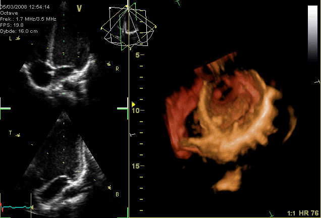
- The two atrioventricular (AV) valves, which are between the atria and the ventricles, are the mitral valveMitral valveThe mitral valve is a dual-flap valve in the heart that lies between the left atrium and the left ventricle...
and the tricuspid valveTricuspid valveThe tricuspid valve, or right atrioventricular valve, is on the right dorsal side of the mammalian heart, between the right atrium and the right ventricle. The normal tricuspid valve usually has three leaflets and three papillary muscles. They are connected to the papillary muscles by the chordae...
. - The two semilunar (SL) valves, which are in the arteries leaving the heart, are the aortic valveAortic valveThe aortic valve is one of the valves of the heart. It is normally tricuspid , although in 1% of the population it is found to be congenitally bicuspid . It lies between the left ventricle and the aorta....
and the pulmonary valvePulmonary valveThe pulmonary valve is the semilunar valve of the heart that lies between the right ventricle and the pulmonary artery and has three cusps. Similar to the aortic valve, the pulmonary valve opens in ventricular systole, when the pressure in the right ventricle rises above the pressure in the...
.
A form of heart disease occurs when a valve malfunctions and allows some blood to flow in the wrong direction. This is called regurgitation.
Atrioventricular or cuspid valves


Atrium (anatomy)
In anatomy, the atrium , sometimes called auricle , refers to a chamber or space. For example, the term is used for a portion of the lateral ventricle in the brain and the blood collection chamber of the heart...
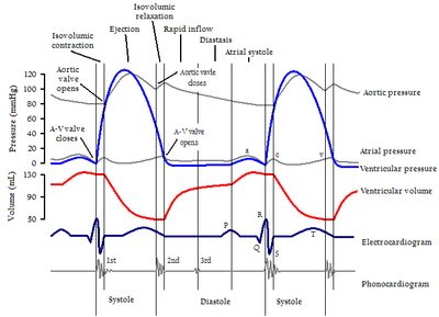
during systole
Systole (medicine)
Systole is the contraction of the heart. Used alone, it usually means the contraction of the left ventricle.In all mammals, the heart has 4 chambers. The left and right ventricles pump together. The atria and ventricles pump in sequence...
. They are anchored to the wall of the ventricle by chordae tendineae
Chordae tendineae
The chordae tendineae, or heart strings, are cord-like tendons that connect the papillary muscles to the tricuspid valve and the mitral valve in the heart....
, which prevent the valve from inverting.
The chordae tendineae
Chordae tendineae
The chordae tendineae, or heart strings, are cord-like tendons that connect the papillary muscles to the tricuspid valve and the mitral valve in the heart....
are attached to papillary muscle
Papillary muscle
In anatomy, the papillary muscles are muscles located in the ventricles of the heart. They attach to the cusps of the atrioventricular valves via the chordae tendinae and contract to prevent inversion or prolapse of these valves.- Action :There are five total papillary muscles in the heart, three...
s that cause tension to better hold the valve. Together, the papillary muscles and the chordae tendineae are known as the subvalvular apparatus. The function of the subvalvular apparatus is to keep the valves from prolapsing into the atria when they close. The subvalvular apparatus have no effect on the opening and closure of the valves, however. This is caused entirely by the pressure gradient across the valve. The peculiar insertion of chords on the leaflet free margin however provides systolic stress sharing between chords according to their different thickness.
The closure of the AV valves is heard as the first heart sound (S1).
Mitral valve (bicuspid)
Also known as the "bicuspid valve" because it contains two flaps, the mitral valve gets its name from the resemblance to a bishopBishop
A bishop is an ordained or consecrated member of the Christian clergy who is generally entrusted with a position of authority and oversight. Within the Catholic Church, Eastern Orthodox, Oriental Orthodox Churches, in the Assyrian Church of the East, in the Independent Catholic Churches, and in the...
's mitre
Mitre
The mitre , also spelled miter, is a type of headwear now known as the traditional, ceremonial head-dress of bishops and certain abbots in the Roman Catholic Church, as well as in the Anglican Communion, some Lutheran churches, and also bishops and certain other clergy in the Eastern Orthodox...
(a type of hat). It allows the blood to flow from the left atrium
Left atrium
The left atrium is one of the four chambers in the human heart. It receives oxygenated blood from the pulmonary veins, and pumps it into the left ventricle, via the mitral valve.-Foramen ovale:...
into the left ventricle
Left ventricle
The left ventricle is one of four chambers in the human heart. It receives oxygenated blood from the left atrium via the mitral valve, and pumps it into the aorta via the aortic valve.-Shape:...
. It is on the left side of the heart and has two cusps.
A common complication of rheumatic fever
Rheumatic fever
Rheumatic fever is an inflammatory disease that occurs following a Streptococcus pyogenes infection, such as strep throat or scarlet fever. Believed to be caused by antibody cross-reactivity that can involve the heart, joints, skin, and brain, the illness typically develops two to three weeks after...
is thickening and stenosis
Stenosis
A stenosis is an abnormal narrowing in a blood vessel or other tubular organ or structure.It is also sometimes called a stricture ....
of the mitral valve.
Tricuspid valve
The tricuspid valve is the three-flapped valve on the right side of the heart, between the right atriumRight atrium
The right atrium is one of four chambers in the hearts of mammals and archosaurs...
and the right ventricle
Right ventricle
The right ventricle is one of four chambers in the human heart. It receives deoxygenated blood from the right atrium via the tricuspid valve, and pumps it into the pulmonary artery via the pulmonary valve and pulmonary trunk....
which stops the backflow of blood between the two. It has three cusps.
Semilunar valves
Aortic valve
The aortic valve lies between the left ventricleLeft ventricle
The left ventricle is one of four chambers in the human heart. It receives oxygenated blood from the left atrium via the mitral valve, and pumps it into the aorta via the aortic valve.-Shape:...
and the aorta
Aorta
The aorta is the largest artery in the body, originating from the left ventricle of the heart and extending down to the abdomen, where it branches off into two smaller arteries...
. The aortic valve has three cusps. During ventricular systole
Systole (medicine)
Systole is the contraction of the heart. Used alone, it usually means the contraction of the left ventricle.In all mammals, the heart has 4 chambers. The left and right ventricles pump together. The atria and ventricles pump in sequence...
, pressure rises in the left ventricle. When the pressure in the left ventricle rises above the pressure in the aorta, the aortic valve opens, allowing blood to exit the left ventricle into the aorta. When ventricular systole ends, pressure in the left ventricle rapidly drops. When the pressure in the left ventricle decreases, the aortic pressure forces the aortic valve to close. The closure of the aortic valve contributes the A2 component of the second heart sound (S2).
The most common congenital abnormality of the heart is the bicuspid aortic valve
Bicuspid aortic valve
A bicuspid aortic valve is most commonly a congenital condition of the aortic valve where two of the aortic valvular leaflets fuse during development resulting in a valve that is bicuspid instead of the normal tricuspid configuration. Normally the only cardiac valve that is bicuspid is the mitral...
. In this condition, instead of three cusps, the aortic valve has two cusps. This condition is often undiagnosed until the person develops calcific aortic stenosis
Stenosis
A stenosis is an abnormal narrowing in a blood vessel or other tubular organ or structure.It is also sometimes called a stricture ....
. Aortic stenosis occurs in this condition usually in patients in their 40s or 50s, an average of over 10 years earlier than in people with normal aortic valves.
Pulmonary valve
The pulmonary valve (sometimes referred to as the pulmonic valve) is the semilunar valve of the heart that lies between the right ventricle and the pulmonary artery, and has three cusps. Similar to the aortic valve, the pulmonary valve opens in ventricular systole, when the pressure in the right ventricle rises above the pressure in the pulmonary artery. At the end of ventricular systole, when the pressure in the right ventricle falls rapidly, the pressure in the pulmonary artery will close the pulmonary valve.The closure of the pulmonary valve contributes the P2 component of the second heart sound (S2). The right heart is a low-pressure system, so the P2 component of the second heart sound is usually softer than the A2 component of the second heart sound. However, it is physiologically normal in some young people to hear both components separated during inhalation.
Heart valve dynamics
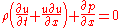
In general, motion of the heart valves is determined using the Navier-Stokes equation; using boundary conditions of the blood pressures, pericardial fluid, and external loading as the constraints.Motion of the heart valves is used as a boundary condition in the Navier-Stokes equation in determining the fluid dynamics of blood ejection from the left and right ventricles into the aorta and the lung.
Relationship between pressure and flow in open valves
The pressure drop, , across an open heart valve relates to the flow rate, Q, through the valve:
If:
-Inflow energy conserved
-Stagnant region behind leaflets
-Outflow momentum conserved
-Flat velocity profile
Valves with a single degree of freedom
Usually the aortic and mitral valves are incorporated in valve studies within a single degree of freedom. These relationships are based on the idea of the valve being a structure with a single degree of freedom. These relationships are based upon the Euler equations.Equations for the aortic valve in this case:

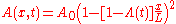

where:
u=axial velocity
p=pressure
A=cross sectional area of valve
L=axial length of valve
 (t)=single degree of freedom; when
See also
- Artificial heart valveArtificial heart valveAn artificial heart valve is a device implanted in the heart of a patient with heart valvular disease. When one of the four heart valves malfunctions, the medical choice may be to replace the natural valve with an artificial valve. This requires open-heart surgery.Valves are integral to the normal...
- Cardiac fibrosisCardiac fibrosisCardiac fibrosis refers to an abnormal thickening of the heart valves due to inappropriate proliferation of cardiac fibroblasts.Fibrocyte cells normally secrete collagen, and function to provide structural support for the heart...
- Congenital heart disease
- Disorders of the valves (Valvular heart diseaseValvular heart diseaseValvular heart disease is any disease process involving one or more of the valves of the heart . Valve problems may be congenital or acquired...
)- Aortic valve disorders:
- Aortic insufficiencyAortic insufficiencyAortic insufficiency , also known as aortic regurgitation , is the leaking of the aortic valve of the heart that causes blood to flow in the reverse direction during ventricular diastole, from the aorta into the left ventricle....
- Aortic stenosis
- Aortic valve repair
- Aortic valve replacementAortic valve replacementAortic valve replacement is a cardiac surgery procedure in which a patient's failing aortic valve is replaced with an alternate healthy valve. The aortic valve can be affected by a range of diseases; the valve can either become leaky or partially blocked...
- Aortic valvuloplastyAortic valvuloplastyAortic valvuloplasty is the repair of a stenotic aortic valve using a balloon catheter inside the valve. The balloon is placed into the aortic valve that has become stiff from calcium buildup...
- Aortic insufficiency
- Mitral valve disorders
- Mitral valve prolapseMitral valve prolapseMitral valve prolapse is a valvular heart disease characterized by the displacement of an abnormally thickened mitral valve leaflet into the left atrium during systole. There are various types of MVP, broadly classified as classic and nonclassic. In its nonclassic form, MVP carries a low risk of...
- Mitral valve repairMitral valve repairMitral valve repair is a cardiac surgery procedure performed by cardiac surgeons to treat stenosis or regurgitation of the mitral valve. The mitral valve is the "inflow valve" for the left side of the heart. Blood flows from the lungs, where it picks up oxygen, through the pulmonary veins, to the...
- Mitral valve replacementMitral valve replacementMitral valve replacement is a cardiac surgery procedure in which a patient’s mitral valve is replaced by a different valve. Mitral valve replacement is typically performed robotically or manually, when the valve becomes too tight for blood to flow into the left ventricle, or too loose in which...
- Mitral valvuloplasty
- Mitral stenosisMitral stenosisMitral stenosis is a valvular heart disease characterized by the narrowing of the orifice of the mitral valve of the heart.-Signs and symptoms:Symptoms of mitral stenosis include:...
- Mitral valve prolapse
- Pulmonary valve disorders
- Tricuspid valve disorders
- Aortic valve disorders:
- EndocarditisEndocarditisEndocarditis is an inflammation of the inner layer of the heart, the endocardium. It usually involves the heart valves . Other structures that may be involved include the interventricular septum, the chordae tendineae, the mural endocardium, or even on intracardiac devices...
- Heart soundsHeart soundsHeart sounds, or heartbeats, are the noises generated by the beating heart and the resultant flow of blood through it...
- Bjork–Shiley valve
External links
- Mitral Valve Repair at The Mount Sinai Hospital – "Mitral Valve Anatomy"]