

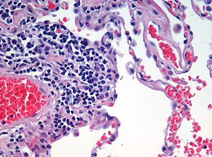
H&E stain
Encyclopedia
Histology
Histology is the study of the microscopic anatomy of cells and tissues of plants and animals. It is performed by examining cells and tissues commonly by sectioning and staining; followed by examination under a light microscope or electron microscope...
. It is the most widely used stain in medical diagnosis; for example when a pathologist looks at a biopsy
Biopsy
A biopsy is a medical test involving sampling of cells or tissues for examination. It is the medical removal of tissue from a living subject to determine the presence or extent of a disease. The tissue is generally examined under a microscope by a pathologist, and can also be analyzed chemically...
of a suspected cancer
Cancer
Cancer , known medically as a malignant neoplasm, is a large group of different diseases, all involving unregulated cell growth. In cancer, cells divide and grow uncontrollably, forming malignant tumors, and invade nearby parts of the body. The cancer may also spread to more distant parts of the...
, the histological section
Histological section
Histological section refers to thin slices of tissue applied to a microscopic slide, usually around 5 to 10 micrometres thick, which are viewed under a microscope...
is likely to be stained with H&E and termed H&E section, H+E section, or HE section.
The staining method involves application of hemalum, which is a complex formed from aluminium ions and oxidized haematoxylin
Haematoxylin
Haematoxylin, hematoxylin, Natural Black 1, or C.I. 75290 is extracted from the heartwood of the logwood tree. When oxidized it forms haematein, a compound that forms strongly coloured complexes with certain metal ions, the most notable ones being Fe and Al salts. Metal-haematein complexes are used...
. This colors nuclei of cells (and a few other objects, such as keratohyalin granules) blue. The nuclear staining is followed by counterstaining with an aqueous or alcoholic solution of eosin Y
Eosin Y
Eosin Y is a form of eosin....
, which colors other, eosinophilic
Eosinophilic
Eosinophilic refers to the staining of certain tissues, cells, or organelles after they have been washed with eosin, a dye.Eosin is an acidic dye; thus, the structure being stained is basic....
structures in various shades of red, pink and orange.
The staining of nuclei by hemalum does not require the presence of DNA and is probably due to binding of the dye-metal complex to arginine-rich basic nucleoproteins such as histones. The mechanism is different from that of nuclear staining by basic (cationic) dyes such as thionine or toluidine blue. Staining by basic dyes is prevented by chemical or enzymatic extraction of nucleic acids. Such extractions do not prevent staining of nuclei by hemalum.
The eosinophilic structures are generally composed of intracellular or extracellular protein
Protein
Proteins are biochemical compounds consisting of one or more polypeptides typically folded into a globular or fibrous form, facilitating a biological function. A polypeptide is a single linear polymer chain of amino acids bonded together by peptide bonds between the carboxyl and amino groups of...
. The Lewy bodies
Lewy body
Lewy bodies are abnormal aggregates of protein that develop inside nerve cells in Parkinson's disease , Lewy Body Dementia and some other disorders. They are identified under the microscope when histology is performed on the brain....
and Mallory bodies
Mallory body
In histopathology, a Mallory body, Mallory-Denk body, and Mallory's hyaline, is an inclusion found in the cytoplasm of liver cells.-Associated conditions:...
are examples of eosinophilic structures. Most of the cytoplasm
Cytoplasm
The cytoplasm is a small gel-like substance residing between the cell membrane holding all the cell's internal sub-structures , except for the nucleus. All the contents of the cells of prokaryote organisms are contained within the cytoplasm...
is eosinophilic. Red blood cell
Red blood cell
Red blood cells are the most common type of blood cell and the vertebrate organism's principal means of delivering oxygen to the body tissues via the blood flow through the circulatory system...
s are stained intensely red.
The structures do not have to be acidic or basic to be called basophilic and eosinophilic. The terminology is based on the affinity to the dyes.
Other colors, e.g. yellow and brown, can be present in the sample; they are caused by intrinsic pigments, e.g. melanin
Melanin
Melanin is a pigment that is ubiquitous in nature, being found in most organisms . In animals melanin pigments are derivatives of the amino acid tyrosine. The most common form of biological melanin is eumelanin, a brown-black polymer of dihydroxyindole carboxylic acids, and their reduced forms...
.
Some structures do not stain well. Basal lamina
Basal lamina
The basal lamina is a layer of extracellular matrix secreted by the epithelial cells, on which the epithelium sits. It is often confused with the basement membrane, and sometimes used inconsistently in the literature, see below....
e need to be stained by PAS stain or some silver stain
Silver stain
Silver staining is the use of silver to selectively alter the appearance of the target.-Use in medicine:It is used to stain histologic sections. This kind of staining is important especially to show proteins and DNA. It is used to show both substances inside and outside cells...
s, if they have to be well visible. Reticular fiber
Reticular fiber
Reticular fibers or reticulin is a histological term used to describe a type of fiber in connective tissue composed of type III collagen. Reticular fibers crosslink to form a fine meshwork...
s also require silver stain. Hydrophobic structures also tend to remain clear; these are usually rich in fats, e.g. adipocyte
Adipocyte
However, in some reports and textbooks, the number of fat cell increased in childhood and adolescence. The total number is constant in both obese and lean adult...
s, myelin
Myelin
Myelin is a dielectric material that forms a layer, the myelin sheath, usually around only the axon of a neuron. It is essential for the proper functioning of the nervous system. Myelin is an outgrowth of a type of glial cell. The production of the myelin sheath is called myelination...
around neuron
Neuron
A neuron is an electrically excitable cell that processes and transmits information by electrical and chemical signaling. Chemical signaling occurs via synapses, specialized connections with other cells. Neurons connect to each other to form networks. Neurons are the core components of the nervous...
axon
Axon
An axon is a long, slender projection of a nerve cell, or neuron, that conducts electrical impulses away from the neuron's cell body or soma....
s, and Golgi apparatus
Golgi apparatus
The Golgi apparatus is an organelle found in most eukaryotic cells. It was identified in 1898 by the Italian physician Camillo Golgi, after whom the Golgi apparatus is named....
membranes.
See also
- Papanicolaou stainPapanicolaou stainPapanicolaou stain is a multichromatic staining histological technique developed by George Papanikolaou, the father of cytopathology....
, other popular staining technique - Wright's stainWright's stainWright's stain is a histologic stain that facilitates the differentiation of blood cell types. It is used primarily to stain peripheral blood smears and bone marrow aspirates which are examined under a light microscope...
, used for cerebrospinal fluid and suspected lymphomaLymphomaLymphoma is a cancer in the lymphatic cells of the immune system. Typically, lymphomas present as a solid tumor of lymphoid cells. Treatment might involve chemotherapy and in some cases radiotherapy and/or bone marrow transplantation, and can be curable depending on the histology, type, and stage...
s - Van Gieson's stainVan Gieson's stainVan Gieson's Stain is a mixture of Picric Acid and Acid Fuchsin. It is the simplest method of differential staining of Collagen and other Connective Tissue...
- CytopathologyCytopathologyCytopathology is a branch of pathology that studies and diagnoses diseases on the cellular level. The discipline was founded by Rudolf Virchow in 1858. A common application of cytopathology is the Pap smear, used as a screening tool, to detect precancerous cervical lesions and prevent cervical...
- Staining
- HaematoxylinHaematoxylinHaematoxylin, hematoxylin, Natural Black 1, or C.I. 75290 is extracted from the heartwood of the logwood tree. When oxidized it forms haematein, a compound that forms strongly coloured complexes with certain metal ions, the most notable ones being Fe and Al salts. Metal-haematein complexes are used...
- EosinEosinEosin is a fluorescent red dye resulting from the action of bromine on fluorescein. It can be used to stain cytoplasm, collagen and muscle fibers for examination under the microscope. Structures that stain readily with eosin are termed eosinophilic....
- Acid-fastAcid-fastAcid-fastness is a physical property of certain bacteria, specifically their resistance to decolorization by acids during staining procedures.Acid-fast organisms are difficult to characterize using standard microbiological techniques Acid-fastness is a physical property of certain bacteria,...
- Gömöri methenamine silver stainGomori methenamine silver stainIn pathology, the Grocott's methenamine silver stain, abbreviated GMS, is a popular staining method in histology.It is used widely as a screen for fungal organisms...
External links
- SIGMA-ALDRICH H&E Informational Primer
- A video from the American Journal of Medical Videos showing H&E staining steps