

Foot
Encyclopedia
The foot is an anatomical structure found in many vertebrates. It is the terminal portion of a limb which bears weight and allows locomotion
. In many animals with feet, the foot is a separate organ at the terminal part of the leg
made up of one or more segments or bones, generally including claws or nails.
An anthropometric study of 1197 North American adult Caucasian males (mean age 35.5 years) found that a man's foot length was 26.3 cm with a standard deviation of 1.2 cm.
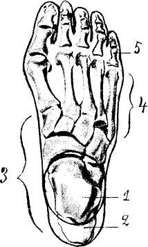
The foot can be subdivided into the hindfoot, the midfoot, and the forefoot:
The hindfoot is composed of the talus
or ankle bone and the calcaneus or heel bone. The two long bones of the lower leg, the tibia
and fibula, are connected to the top of the talus to form the ankle
. Connected to the talus at the subtalar joint
, the calcaneus, the largest bone of the foot, is cushioned inferiorly by a layer of fat.
The five irregular bones of the midfoot, the cuboid, navicular
, and three cuneiform
bones, form the arches of the foot
which serves as a shock absorber. The midfoot is connected to the hind- and fore-foot by muscles and the plantar fascia
.
The forefoot is composed of five toes and the corresponding five proximal long bones forming the metatarsus
. Similar to the fingers of the hand, the bones of the toes are called phalanges
and the big toe has two phalanges while the other four toes have three phalanges. The joints between the phalanges are called interphalangeal
and those between the metatarsus and phalanges are called metatarsophalangeal (MTP).
The instep is the arched part of the top of the foot between the toes and the ankle.
There can be many sesamoid bone
s near the metatarsophalangeal joints, although they are only regularly present in the distal portion of the first metatarsal bone
.
The human foot has two longitudinal
arches and a transverse arch maintained by the interlocking shapes of the foot bones, strong ligaments, and pulling muscles during activity. The slight mobility of these arches when weight is applied to and removed from the foot makes walking and running more economical in terms of energy. As can be examined in a footprint, the medial longitudinal arch curves above the ground. This arch stretches from the heel bone over the "keystone" ankle bone to the three medial metatarsals. In contrast, the lateral longitudinal arch is very low. With the cuboid serving as its keystone, it redistributes part of the weight to the calcaneus and the distal end of the fifth metatarsal. The two longitudinal arches serve as pillars for the transverse arch which run obliquely across the tarsometatarsal joints. Excessive strain on the tendons and ligaments of the feet can result in fallen arches or flat feet
.
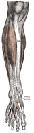 All muscles originating on the lower leg except the popliteus muscle
are attached to the bones of the foot. The tibia and fibula and the interosseous membrane separate these muscles into anterior and posterior groups, in their turn subdivided into subgroups and layers.
Anterior group
Extensor group: tibialis anterior
originates on the proximal half of the tibia and the interosseous membrane and is inserted near the tarsometatarsal joint of the first digit. In the non-weight-bearing leg tibialis anterior flexes the foot dorsally and lift its medial edge (supination
). In the weight-bearing leg it brings the leg towards the back of the foot, like in rapid walking. Extensor digitorum longus
arises on the lateral tibial condyle and along the fibula to be inserted on the second to fifth digits and proximally on the fifth metatarsal. The extensor digitorum longus acts similar to the tibialis anterior except that it also dorsiflexes the digits. Extensor hallucis longus
originates medially on the fibula and is inserted on the first digit. As the name implies it dorsiflexes the big toe and also acts on the ankle in the unstressed leg. In the weight-bearing leg it acts similar to the tibialis anterior.
Peroneal group: peroneus longus arises on the proximal aspect of the fibula and peroneus brevis below it on the same bone. Together, their tendons pass behind the lateral malleolus
. Distally, peroneus longus crosses the plantar side of the foot to reach its insertion on the first tarsometatarsal joint, while peroneus brevis reaches the proximal part of the fifth metatarsal. These two muscles are the strongest pronators and aid in plantar flexion. Longus also acts like a bowstring that braces the transverse arch of the foot.
Posterior group
The superficial layer of posterior leg muscles is formed by the triceps surae
and the plantaris
. The triceps surae consists of the soleus
and the two heads of the gastrocnemius
. The heads of gastrocnemius arise on the femur
, proximal to the condyles, and soleus arises on the proximal dorsal parts of the tibia and fibula. The tendons of these muscles merge to be inserted onto the calcaneus as the Achilles tendon
. Plantaris originates on the femur proximal to the lateral head of the gastrocnemius and its long tendon is embedded medially into the Achilles tendon. The triceps surae is the primary plantar flexor and its strength becomes most obvious during ballet dancing. It is fully activated only with the knee extended because the gastrocnemius is shortened during knee flexion. During walking it not only lifts the heel, but also flexes the knee, assisted by the plantaris.
In the deep layer of posterior muscles tibialis posterior
arises proximally on the back of the interosseous membrane and adjoining bones and divides into two parts in the sole of the foot to attach to the tarsus. In the non-weight-bearing leg, it produces plantar flexion and supination, and, in the weight-bearing leg, it proximates the heel to the calf. flexor hallucis longus
arises on the back of the fibula (i.e. on the lateral side), and its relatively thick muscle belly extends distally down to the flexor retinaculum where it passes over to the medial side to stretch across the sole to the distal phalanx of the first digit. The popliteus
is also part of this group, but, with its oblique course across the back of the knee, does not act on the foot.
On the back (top) of the foot, the tendons of extensor digitorum brevis
and extensor hallucis brevis
lie deep to the system of long extrinsic extensor tendons. They both arise on the calcaneus and extend into the dorsal aponeurosis
of digits one to four, just beyond the penultimate joints. They act to dorsiflex the digits.
Similar to the intrinsic muscles of the hand, there are three groups of muscles in the sole of foot, those of the first and last digits, and a central group:
Muscles of the big toe
: abductor hallucis
stretches medially along the border of the sole, from the calcaneus to the first digit. Below its tendon, the tendons of the long flexors pass through the tarsal canal
. It is an abductor and a weak flexor, and also helps maintain the arch of the foot. flexor hallucis brevis
arises on the medial cuneiform bone and related ligaments and tendons. An important plantar flexor, it is crucial for ballet dancing. Both these muscles are inserted with two heads proximally and distally to the first metatarsophalangeal joint. Adductor hallucis
is part of this group, though it originally formed a separate system (see contrahens
.) It has two heads, the oblique head originating obliquely across the central part of the midfoot, and the transverse head originating near the metatarsophalangeal joints of digits five to three. Both heads are inserted into the lateral sesamoid bone of the first digit. Adductor hallucis acts as a tensor of the plantar arches and also adducts the big toe and then might plantar flex the proximal phalanx.
Muscles of the little toe
: Stretching laterally from the calcaneus to the proximal phalanx of the fifth digit, abductor digiti minimi
form the lateral margin of the foot and is the largest of the muscles of the fifth digit. Arising from the base of the fifth metatarsal, flexor digiti minimi
is inserted together with abductor on the first phalanx. Often absent, opponens digiti minimi
originates near the cuboid bone and is inserted on the fifth metatarsal bone. These three muscles act to support the arch of the foot and to plantar flex the fifth digit.
Central muscle group: The four lumbricales
arise on the medial side of the tendons of flexor digitorum longus and are inserted on the medial margins of the proximal phalanges. Quadratus plantae
originates with two slips from the lateral and medial margins of the calcaneus and inserts into the lateral margin of the flexor digitorum tendon. It is also known as flexor accessorius. Flexor digitorum brevis
arise inferiorly on the calcaneus and its three tendons are inserted into the middle phalanges of digits two to four (sometimes also the fifth digit). These tendons divide before their insertions and the tendons of flexor digitorum longus pass through these divisions. Flexor digitorum brevis flexes the middle phalanges. It is occasionally absent. Between the toes, the dorsal and plantar interossei
stretch from the metatarsals to the proximal phalanges of digits two to five. The plantar interossei adducts and the dorsal interossei abducts these digits and are also plantar flexors at the metatarsophalangeal joints.
s and injuries, including athlete's foot
, bunion
s, ingrown toenails
, Morton's neuroma
, plantar fasciitis
, plantar wart
s and stress fracture
s. In addition, there are several genetic disorder
s that can affect the shape and function of the feet, including a club foot
or flat feet
.
This leaves humans more vulnerable to medical problems that are caused by poor leg and foot alignments. Also, the wearing of shoes, sneakers and boots can impede proper alignment and movement within the ankle and foot. For example, high heels are known to throw off the natural weight balance
(this can also affect the lower back). For the sake of posture, flat soles and heels are advised.
A doctor
who specializes in the treatment of the feet practices podiatry
and is called a podiatrist. A pedorthist
specializes in the use and modification of footwear to treat problems related to the lower limbs.
Fracture
s of the foot include:
Foot sweat
is the major cause of foot odor
. Sweat itself is odorless, but it creates a beneficial environment for certain bacteria to grow and produce bad-smelling substances.
is the soft foot of a mammal, generally a quadruped, that has claws or nails. A hard foot is called a hoof
.
Depending on style of locomotion, animals can be classified as plantigrade
(sole walking), digitigrade
(toe walking), or unguligrade (nail walking).
The metatarsals are the bones that make up the main part of the foot in humans, and part of the leg in large animals or paw in smaller animals. The number of metatarsals are directly related to the mode of locomotion with many larger animals having their digits reduced to two (elk
, cow, sheep) or one (horse
). The metatarsal bones of feet and paws are tightly grouped compared to, most notably, the human hand where the thumb metacarpal diverges from the rest of the metacarpus.
Animal locomotion
Animal locomotion, which is the act of self-propulsion by an animal, has many manifestations, including running, swimming, jumping and flying. Animals move for a variety of reasons, such as to find food, a mate, or a suitable microhabitat, and to escape predators...
. In many animals with feet, the foot is a separate organ at the terminal part of the leg
Leg
Łęg may refer to the following places in Poland:*A former name for the town of Ełk *Part of the Czyżyny district of Kraków*Łęg, Pleszew County in Greater Poland Voivodeship...
made up of one or more segments or bones, generally including claws or nails.
Anatomy
The human foot and ankle is a strong and complex mechanical structure containing more than 26 bones, 33 joints (20 of which are actively articulated), and more than a hundred muscles, tendons, and ligaments.An anthropometric study of 1197 North American adult Caucasian males (mean age 35.5 years) found that a man's foot length was 26.3 cm with a standard deviation of 1.2 cm.
The foot can be subdivided into the hindfoot, the midfoot, and the forefoot:
The hindfoot is composed of the talus
Talus bone
-External links:* *...
or ankle bone and the calcaneus or heel bone. The two long bones of the lower leg, the tibia
Tibia
The tibia , shinbone, or shankbone is the larger and stronger of the two bones in the leg below the knee in vertebrates , and connects the knee with the ankle bones....
and fibula, are connected to the top of the talus to form the ankle
Ankle
The ankle joint is formed where the foot and the leg meet. The ankle, or talocrural joint, is a synovial hinge joint that connects the distal ends of the tibia and fibula in the lower limb with the proximal end of the talus bone in the foot...
. Connected to the talus at the subtalar joint
Subtalar joint
In human anatomy, the subtalar joint, also known as thetalocalcaneal joint, is a joint of the foot. It occurs at the meeting point of the talus and the calcaneus.-Motion:...
, the calcaneus, the largest bone of the foot, is cushioned inferiorly by a layer of fat.
The five irregular bones of the midfoot, the cuboid, navicular
Navicular bone
The navicular bone is a small bone found in the feet of both humans and horses.- Human anatomy :The navicular bone is one of the tarsal bones, found in the foot. Its name derives from the bone's resemblance to a small boat, caused by the strongly concave proximal articular surface...
, and three cuneiform
Cuneiform (anatomy)
There are three cuneiform bones in the human foot:* the medial cuneiform* the intermediate cuneiform also known as the middle* the lateral cuneiform...
bones, form the arches of the foot
Arches of the foot
-External links:* * *...
which serves as a shock absorber. The midfoot is connected to the hind- and fore-foot by muscles and the plantar fascia
Plantar fascia
The plantar fascia is the thick connective tissue which supports the arch on the bottom of the foot. It runs from the tuberosity of the calcaneus forward to the heads of the metatarsal bones .The often painful condition plantar fasciitis is an inflammatory condition of the plantar...
.
The forefoot is composed of five toes and the corresponding five proximal long bones forming the metatarsus
Metatarsus
The metatarsus or metatarsal bones are a group of five long bones in the foot located between the tarsal bones of the hind- and mid-foot and the phalanges of the toes. Lacking individual names, the metatarsal bones are numbered from the medial side : the first, second, third, fourth, and fifth...
. Similar to the fingers of the hand, the bones of the toes are called phalanges
Phalanx bones
In anatomy, phalanx bones are those that form the fingers and toes. In primates such as humans and monkeys, the thumb and big toe have two phalanges, while the other fingers and toes consist of three. Phalanges are classified as long bones.The phalanges do not have individual names...
and the big toe has two phalanges while the other four toes have three phalanges. The joints between the phalanges are called interphalangeal
Interphalangeal articulations of foot
The interphalangeal articulations of the foot are the joints between the phalanges of the toes. They are ginglymoid joints, and each has a plantar and two collateral ligaments...
and those between the metatarsus and phalanges are called metatarsophalangeal (MTP).
The instep is the arched part of the top of the foot between the toes and the ankle.
Skeleton
- tibiaTibiaThe tibia , shinbone, or shankbone is the larger and stronger of the two bones in the leg below the knee in vertebrates , and connects the knee with the ankle bones....
, fibula - tarsusTarsus (skeleton)In tetrapods, the tarsus is a cluster of articulating bones in each foot situated between the lower end of tibia and fibula of the lower leg and the metatarsus. In the foot the tarsus articulates with the bones of the metatarsus, which in turn articulate with the bones of the individual toes...
: talusTalus bone-External links:* *...
, calcaneus, cuneiformesCuneiform (anatomy)There are three cuneiform bones in the human foot:* the medial cuneiform* the intermediate cuneiform also known as the middle* the lateral cuneiform...
, cuboid, and navicularNavicular boneThe navicular bone is a small bone found in the feet of both humans and horses.- Human anatomy :The navicular bone is one of the tarsal bones, found in the foot. Its name derives from the bone's resemblance to a small boat, caused by the strongly concave proximal articular surface... - metatarsusMetatarsusThe metatarsus or metatarsal bones are a group of five long bones in the foot located between the tarsal bones of the hind- and mid-foot and the phalanges of the toes. Lacking individual names, the metatarsal bones are numbered from the medial side : the first, second, third, fourth, and fifth...
: firstFirst metatarsal boneThe first metatarsal bone is the bone in the body of the foot just behind the big toe.It is remarkable for its great thickness, and is the shortest of the metatarsal bones.The body is strong, and of well-marked prismoid form....
, secondSecond metatarsal boneThe second metatarsal bone is the longest of the metatarsal bones, being prolonged backward into the recess formed by the three cuneiform bones.Its base is broad above, narrow and rough below....
, thirdThird metatarsal boneThe third metatarsal bone articulates proximally, by means of a triangular smooth surface, with the third cuneiform; medially, by two facets, with the second metatarsal; and laterally, by a single facet, with the fourth metatarsal....
, fourthFourth metatarsal boneThe fourth metatarsal bone is smaller in size than the third; its base presents an oblique quadrilateral surface for articulation with the cuboid; a smooth facet on the medial side, divided by a ridge into an anterior portion for articulation with the third metatarsal, and a posterior portion for...
, and fifth metatarsal boneFifth metatarsal boneThe fifth metatarsal bone is recognized by a rough eminence, the tuberosity, on the lateral side of its base.The base articulates behind, by a triangular surface cut obliquely in a transverse direction, with the cuboid; and medially, with the fourth metatarsal.On the medial part of its dorsal... - phalangesPhalanges of the footThe Olphalanges of the foot are the bones in the toes. They correspond, in number and general arrangement, with those of the hand; there are two in the big toe, and three in each of the other toes...
There can be many sesamoid bone
Sesamoid bone
In anatomy, a sesamoid bone is a bone embedded within a tendon.Sesamoids are found in locations where a tendon passes over a joint, such as the hand, knee, and foot. Functionally, they act to protect the tendon and to increase its mechanical effect. The presence of the sesamoid bone holds the...
s near the metatarsophalangeal joints, although they are only regularly present in the distal portion of the first metatarsal bone
First metatarsal bone
The first metatarsal bone is the bone in the body of the foot just behind the big toe.It is remarkable for its great thickness, and is the shortest of the metatarsal bones.The body is strong, and of well-marked prismoid form....
.
Arches
The human foot has two longitudinal
Anatomical terms of location
Standard anatomical terms of location are designations employed in science that deal with the anatomy of animals to avoid ambiguities that might otherwise arise. They are not language-specific, and thus require no translation...
arches and a transverse arch maintained by the interlocking shapes of the foot bones, strong ligaments, and pulling muscles during activity. The slight mobility of these arches when weight is applied to and removed from the foot makes walking and running more economical in terms of energy. As can be examined in a footprint, the medial longitudinal arch curves above the ground. This arch stretches from the heel bone over the "keystone" ankle bone to the three medial metatarsals. In contrast, the lateral longitudinal arch is very low. With the cuboid serving as its keystone, it redistributes part of the weight to the calcaneus and the distal end of the fifth metatarsal. The two longitudinal arches serve as pillars for the transverse arch which run obliquely across the tarsometatarsal joints. Excessive strain on the tendons and ligaments of the feet can result in fallen arches or flat feet
Flat feet
Flat feet is a formal reference to a medical condition in which the arch of the foot collapses, with the entire sole of the foot coming into complete or near-complete contact with the ground...
.
Muscles
The muscles acting on the foot can be classified into extrinsic muscles, those originating on the anterior or posterior aspect of the lower leg, and intrinsic muscles, originating on the dorsal or plantar aspects of the foot.Extrinsic
Popliteus muscle
The popliteus muscle in the leg is used to unlock the knee during walking/running by laterally rotating the femur on the tibia during a closed chain movement ....
are attached to the bones of the foot. The tibia and fibula and the interosseous membrane separate these muscles into anterior and posterior groups, in their turn subdivided into subgroups and layers.
Anterior group
Extensor group: tibialis anterior
Tibialis anterior muscle
In human anatomy, the tibialis anterior is a muscle that originates in the upper two-thirds of the lateral surface of the tibia and inserts into the medial cuneiform and first metatarsal bones of the foot. Its acts to dorsiflex and invert the foot. This muscle is mostly located near the shin.It is...
originates on the proximal half of the tibia and the interosseous membrane and is inserted near the tarsometatarsal joint of the first digit. In the non-weight-bearing leg tibialis anterior flexes the foot dorsally and lift its medial edge (supination
Supination
Supination is a position of either the forearm or foot; in the forearm when the palm faces anteriorly, or faces up . Supination in the foot occurs when a person appears "bow-legged" with their weight supported primarily on the anterior of their feet.The hand is supine in the anatomical position...
). In the weight-bearing leg it brings the leg towards the back of the foot, like in rapid walking. Extensor digitorum longus
Extensor digitorum longus muscle
The Extensor digitorum longus is a pennate muscle, situated at the lateral part of the front of the leg.-Origin and insertion:It arises from the lateral condyle of the tibia; from the upper three-fourths of the anterior surface of the body of the fibula; from the upper part of the interosseous...
arises on the lateral tibial condyle and along the fibula to be inserted on the second to fifth digits and proximally on the fifth metatarsal. The extensor digitorum longus acts similar to the tibialis anterior except that it also dorsiflexes the digits. Extensor hallucis longus
Extensor hallucis longus muscle
The Extensor hallucis longus is a thin muscle, situated between the Tibialis anterior and the Extensor digitorum longus that functions to extend the big toe, dorsiflex the foot, and assists with foot inversion....
originates medially on the fibula and is inserted on the first digit. As the name implies it dorsiflexes the big toe and also acts on the ankle in the unstressed leg. In the weight-bearing leg it acts similar to the tibialis anterior.
Peroneal group: peroneus longus arises on the proximal aspect of the fibula and peroneus brevis below it on the same bone. Together, their tendons pass behind the lateral malleolus
Malleolus
Each leg is supported by two bones, the tibia on the inner side of the leg and the fibula on the outer side of the leg.The medial malleolus is the prominence on the inner side of the ankle, formed by the lower end of the tibia....
. Distally, peroneus longus crosses the plantar side of the foot to reach its insertion on the first tarsometatarsal joint, while peroneus brevis reaches the proximal part of the fifth metatarsal. These two muscles are the strongest pronators and aid in plantar flexion. Longus also acts like a bowstring that braces the transverse arch of the foot.
Posterior group
The superficial layer of posterior leg muscles is formed by the triceps surae
Triceps surae
The triceps surae is a pair of muscles located at the calf - the gastrocnemius and the soleus...
and the plantaris
Plantaris muscle
Plantaris is a vestigial structure and one of the superficial muscles of the posterior crural compartment of the leg.It is innervated by the tibial nerve ....
. The triceps surae consists of the soleus
Soleus muscle
- References :* Gray, Henry. Pick, T. Pickering, & Howden, Robert . Gray's Anatomy . New York: Barnes & Noble Books.- External links :...
and the two heads of the gastrocnemius
Gastrocnemius muscle
In humans, the gastrocnemius muscle is a very powerful superficial pennate muscle that is in the back part of the lower leg. It runs from its two heads just above the knee to the heel, and is involved in standing, walking, running and jumping. Along with the soleus muscle it forms the calf muscle...
. The heads of gastrocnemius arise on the femur
Femur
The femur , or thigh bone, is the most proximal bone of the leg in tetrapod vertebrates capable of walking or jumping, such as most land mammals, birds, many reptiles such as lizards, and amphibians such as frogs. In vertebrates with four legs such as dogs and horses, the femur is found only in...
, proximal to the condyles, and soleus arises on the proximal dorsal parts of the tibia and fibula. The tendons of these muscles merge to be inserted onto the calcaneus as the Achilles tendon
Achilles tendon
The Achilles tendon , also known as the calcaneal tendon or the tendo calcaneus, is a tendon of the posterior leg. It serves to attach the plantaris, gastrocnemius and soleus muscles to the calcaneus bone.- Anatomy :The Achilles is the tendonous extension of 3 muscles in the lower leg:...
. Plantaris originates on the femur proximal to the lateral head of the gastrocnemius and its long tendon is embedded medially into the Achilles tendon. The triceps surae is the primary plantar flexor and its strength becomes most obvious during ballet dancing. It is fully activated only with the knee extended because the gastrocnemius is shortened during knee flexion. During walking it not only lifts the heel, but also flexes the knee, assisted by the plantaris.
In the deep layer of posterior muscles tibialis posterior
Tibialis posterior muscle
The Tibialis posterior is the most central of all the leg muscles, and is located in the posterior compartment of the leg.It is the key stabilizing muscle of the lower leg....
arises proximally on the back of the interosseous membrane and adjoining bones and divides into two parts in the sole of the foot to attach to the tarsus. In the non-weight-bearing leg, it produces plantar flexion and supination, and, in the weight-bearing leg, it proximates the heel to the calf. flexor hallucis longus
Flexor hallucis longus muscle
The Flexor hallucis longus muscle is a muscle of the leg.It is one of the deep muscles of the posterior compartment of the leg. The other deep muscles of the leg are flexor digitorum longus and tibialis posterior. Tibialis posterior is most powerful of these deep muscles.The Flexor hallucis...
arises on the back of the fibula (i.e. on the lateral side), and its relatively thick muscle belly extends distally down to the flexor retinaculum where it passes over to the medial side to stretch across the sole to the distal phalanx of the first digit. The popliteus
Popliteus muscle
The popliteus muscle in the leg is used to unlock the knee during walking/running by laterally rotating the femur on the tibia during a closed chain movement ....
is also part of this group, but, with its oblique course across the back of the knee, does not act on the foot.
Intrinsic
On the back (top) of the foot, the tendons of extensor digitorum brevis
Extensor digitorum brevis muscle
The extensor digitorum brevis muscle is a muscle on the upper surface of the foot that helps extend digits 2 through 4.-Structure:...
and extensor hallucis brevis
Extensor hallucis brevis muscle
The extensor hallucis brevis is a muscle on the top of the foot that helps to extend the big toe.-Structure:The extensor hallucis brevis is essentially the medial part of the extensor digitorum brevis muscle...
lie deep to the system of long extrinsic extensor tendons. They both arise on the calcaneus and extend into the dorsal aponeurosis
Aponeurosis
Aponeuroses are layers of flat broad tendons. They have a shiny, whitish-silvery color, are histologically similar to tendons, and are very sparingly supplied with blood vessels and nerves. When dissected, aponeuroses are papery, and peel off by sections...
of digits one to four, just beyond the penultimate joints. They act to dorsiflex the digits.
Similar to the intrinsic muscles of the hand, there are three groups of muscles in the sole of foot, those of the first and last digits, and a central group:
Muscles of the big toe
Hallux
In tetrapods, the hallux is the innermost toe of the foot. Despite its name it may not be the longest toe on the foot of some individuals...
: abductor hallucis
Abductor hallucis muscle
The Abductor hallucis lies along the medial border of the foot and covers the origins of the plantar vessels and nerves.It arises from the medial process of the tuberosity of the calcaneus, from the laciniate ligament, from the plantar aponeurosis, and from the intermuscular septum between it and...
stretches medially along the border of the sole, from the calcaneus to the first digit. Below its tendon, the tendons of the long flexors pass through the tarsal canal
Tarsal tunnel
The tarsal tunnel is found along the inner leg behind the medial malleolus.The tarsal tunnel is made up of bone on the inside and the flexor retinaculum on the outside.-Nerve distribution:...
. It is an abductor and a weak flexor, and also helps maintain the arch of the foot. flexor hallucis brevis
Flexor hallucis brevis muscle
The Flexor hallucis brevis arises, by a pointed tendinous process, from the medial part of the under surface of the cuboid bone, from the contiguous portion of the third cuneiform, and from the prolongation of the tendon of the Tibialis posterior which is attached to that bone.It divides in front...
arises on the medial cuneiform bone and related ligaments and tendons. An important plantar flexor, it is crucial for ballet dancing. Both these muscles are inserted with two heads proximally and distally to the first metatarsophalangeal joint. Adductor hallucis
Adductor hallucis muscle
The Adductor hallucis arises by two heads—oblique and transverse and is responsible for adducting the big toe...
is part of this group, though it originally formed a separate system (see contrahens
Contrahens
The contrahentes are muscles widely present in the hands of mammals, including monkeys and gorillas. They are on the palmar/plantar side. There is one each for digits I II IV V but not III...
.) It has two heads, the oblique head originating obliquely across the central part of the midfoot, and the transverse head originating near the metatarsophalangeal joints of digits five to three. Both heads are inserted into the lateral sesamoid bone of the first digit. Adductor hallucis acts as a tensor of the plantar arches and also adducts the big toe and then might plantar flex the proximal phalanx.
Muscles of the little toe
Fifth toe
The fifth toe is the outermost and usually the smallest toe of the foot.It is associated with many medical conditions, largely due to the use of shoes....
: Stretching laterally from the calcaneus to the proximal phalanx of the fifth digit, abductor digiti minimi
Abductor digiti minimi muscle (foot)
The Abductor digiti minimi is a muscle which lies along the lateral border of the foot, and is in relation by its medial margin with the lateral plantar vessels and nerves....
form the lateral margin of the foot and is the largest of the muscles of the fifth digit. Arising from the base of the fifth metatarsal, flexor digiti minimi
Flexor digiti minimi brevis muscle (foot)
The Flexor digiti minimi brevis lies under the metatarsal bone of the little toe, and resembles one of the Interossei....
is inserted together with abductor on the first phalanx. Often absent, opponens digiti minimi
Opponens digiti minimi muscle
The opponens digiti minimi is a muscle in the hand. It is of a triangular form, and placed immediately beneath the palmaris brevis, abductor minimi digiti, and flexor brevis minimi digiti...
originates near the cuboid bone and is inserted on the fifth metatarsal bone. These three muscles act to support the arch of the foot and to plantar flex the fifth digit.
Central muscle group: The four lumbricales
Lumbrical muscle (foot)
The lumbricals are four small skeletal muscles, accessory to the tendons of the flexor digitorum longus and numbered from the medial side of the foot; they arise from these tendons, as far back as their angles of division, each springing from two tendons, except the first.The muscles end in...
arise on the medial side of the tendons of flexor digitorum longus and are inserted on the medial margins of the proximal phalanges. Quadratus plantae
Quadratus plantae muscle
The Quadratus plantæ is separated from the muscles of the first layer by the lateral plantar vessels and nerve. It acts to aid in flexing the 2nd to 5th toes and is one of the few muscles in the foot with no homolog in the hand.It arises by two heads, which are separated from each other by the long...
originates with two slips from the lateral and medial margins of the calcaneus and inserts into the lateral margin of the flexor digitorum tendon. It is also known as flexor accessorius. Flexor digitorum brevis
Flexor digitorum brevis muscle
The flexor digitorum brevis lies in the middle of the sole of the foot, immediately above the central part of the plantar aponeurosis, with which it is firmly united....
arise inferiorly on the calcaneus and its three tendons are inserted into the middle phalanges of digits two to four (sometimes also the fifth digit). These tendons divide before their insertions and the tendons of flexor digitorum longus pass through these divisions. Flexor digitorum brevis flexes the middle phalanges. It is occasionally absent. Between the toes, the dorsal and plantar interossei
Plantar interossei muscles
The plantar interossei, three in number, lie beneath rather than between the metatarsal bones, and each is connected with but one metatarsal bone....
stretch from the metatarsals to the proximal phalanges of digits two to five. The plantar interossei adducts and the dorsal interossei abducts these digits and are also plantar flexors at the metatarsophalangeal joints.
Medical aspects
Due to their position and function, feet are exposed to a variety of potential infectionInfection
An infection is the colonization of a host organism by parasite species. Infecting parasites seek to use the host's resources to reproduce, often resulting in disease...
s and injuries, including athlete's foot
Athlete's foot
Athlete's foot is a fungal infection of the skin that causes scaling, flaking, and itch of affected areas. It is caused by fungi in the genus Trichophyton and is typically transmitted in moist areas where people walk barefoot, such as showers or bathhouses...
, bunion
Bunion
A bunion is a deformity characterized by lateral deviation of the great toe, often erroneously described as an enlargement of bone or tissue around the joint at the head of the big toe...
s, ingrown toenails
Ingrown nail
Onychocryptosis , also known as an ingrown toenail, or unguis incarnatus is a common form of nail disease. It is an often painful condition in which the nail grows so that it cuts into one or both sides of the paronychium or nail bed...
, Morton's neuroma
Morton's neuroma
Morton's neuroma is a benign neuroma of an intermetatarsal plantar nerve, most commonly of the third and fourth intermetatarsal spaces.This problem is characterised by pain and/or numbness, sometimes relieved by removing footwear.Despite the name, the...
, plantar fasciitis
Plantar fasciitis
Plantar fasciitis is a painful inflammatory process of the plantar fascia, the connective tissue on the sole of the foot.Longstanding cases of plantar fasciitis often demonstrate more degenerative changes than inflammatory changes, in which case they are termed plantar fasciosis. The suffix...
, plantar wart
Plantar wart
-External links:* at the Mayo Clinic website* at The Merck Manual* at dermnet.com...
s and stress fracture
Stress fracture
A stress fracture is one type of incomplete fracture in bones. It is caused by "unusual or repeated stress" and also heavy continuous weight on the ankle or leg...
s. In addition, there are several genetic disorder
Genetic disorder
A genetic disorder is an illness caused by abnormalities in genes or chromosomes, especially a condition that is present from before birth. Most genetic disorders are quite rare and affect one person in every several thousands or millions....
s that can affect the shape and function of the feet, including a club foot
Club foot
A club foot, or congenital talipes equinovarus , is a congenital deformity involving one foot or both. The affected foot appears rotated internally at the ankle. TEV is classified into 2 groups: Postural TEV or Structural TEV....
or flat feet
Flat feet
Flat feet is a formal reference to a medical condition in which the arch of the foot collapses, with the entire sole of the foot coming into complete or near-complete contact with the ground...
.
This leaves humans more vulnerable to medical problems that are caused by poor leg and foot alignments. Also, the wearing of shoes, sneakers and boots can impede proper alignment and movement within the ankle and foot. For example, high heels are known to throw off the natural weight balance
Neutral spine
A neutral spine or good posture refers to the "three natural curves [that] are present in a healthy spine."- Posture :The word "posture" comes from the Latin verb "ponere" which is defined as "to put or place." The general concept of human posture refers to "the carriage of the body as a whole, the...
(this can also affect the lower back). For the sake of posture, flat soles and heels are advised.
A doctor
Physician
A physician is a health care provider who practices the profession of medicine, which is concerned with promoting, maintaining or restoring human health through the study, diagnosis, and treatment of disease, injury and other physical and mental impairments...
who specializes in the treatment of the feet practices podiatry
Podiatry
Podiatry is a branch of medicine devoted to the study, diagnosis, and treatment of disorders of the foot, ankle, and lower leg. The term podiatry came into use first in the early 20th century United States, where it now denotes a Doctor of Podiatric Medicine , a specialist who is qualified by their...
and is called a podiatrist. A pedorthist
Pedorthist
Pedorthist is the title of a health care professional who specializes in the use of footwear and supportive devices to address conditions which affect the feet and lower limbs. They are trained in the assessment of lower limb anatomy and biomechanics, and the appropriate use of corrective footware...
specializes in the use and modification of footwear to treat problems related to the lower limbs.
Fracture
Bone fracture
A bone fracture is a medical condition in which there is a break in the continuity of the bone...
s of the foot include:
- Lisfranc fractureLisfranc fractureThe Lisfranc fracture is a fracture of the foot in which one or all of the metatarsals are displaced from the tarsus.It is named after 18th- and 19th-century surgeon and gynecologist Jacques Lisfranc de St. Martin.-Causes:...
- in which one or all of the metatarsals are displaced from the tarsusTarsus (skeleton)In tetrapods, the tarsus is a cluster of articulating bones in each foot situated between the lower end of tibia and fibula of the lower leg and the metatarsus. In the foot the tarsus articulates with the bones of the metatarsus, which in turn articulate with the bones of the individual toes... - Jones fractureJones fractureA Jones fracture is a fracture of the diaphysis of the fifth metatarsal of the foot. The fifth metatarsal is at the base of the small toe. The proximal end, where the Jones fracture occurs, is in the midportion of the foot. Patients who sustain a Jones fracture have pain over this area, swelling,...
- a fracture of the fifth metatarsal - March fractureMarch fractureMarch fracture, also known as fatigue fracture or stress fracture of metatarsal bone, is the fracture of the distal third of one of the metatarsals occurring because of recurrent stress. It is more common in soldiers, but also occurs in hikers, organists, and even those, like hospital doctors,...
- a fracture of the distal third of one of the metatarsals occurring because of recurrent stress - Calcaneal fractureCalcaneal fractureCalcaneal fracture, also known as Lover's fracture and Don Juan fracture, is a fracture of the calcaneus. It is usually caused by a fall from height when one lands on his or her feet. These fractures represent approximately 2% of all fractures but 60% of tarsal bone fractures...
Foot sweat
SWEAT
SWEAT is an OLN/TSN show hosted by Julie Zwillich that aired in 2003-2004.Each of the 13 half-hour episodes of SWEAT features a different outdoor sport: kayaking, mountain biking, ice hockey, beach volleyball, soccer, windsurfing, rowing, Ultimate, triathlon, wakeboarding, snowboarding, telemark...
is the major cause of foot odor
Foot odor
Foot odor is a type of body odor that affects the feet of humans and is generally considered to be an unpleasant smell.-Cause:...
. Sweat itself is odorless, but it creates a beneficial environment for certain bacteria to grow and produce bad-smelling substances.
Evolutionary variations
A pawPaw
A paw is the soft foot of a mammal, generally a quadruped, that has claws or nails. A hard foot is called a hoof. Paws are used to pad feet for walking and increase friction.- Common characteristics :...
is the soft foot of a mammal, generally a quadruped, that has claws or nails. A hard foot is called a hoof
Hoof
A hoof , plural hooves or hoofs , is the tip of a toe of an ungulate mammal, strengthened by a thick horny covering. The hoof consists of a hard or rubbery sole, and a hard wall formed by a thick nail rolled around the tip of the toe. The weight of the animal is normally borne by both the sole...
.
Depending on style of locomotion, animals can be classified as plantigrade
Plantigrade
right|151px|thumb|Human skeleton, showing plantigrade habitIn terrestrial animals, plantigrade locomotion means walking with the podials and metatarsals flat on the ground. It is one of three forms of locomotion adopted by mammals...
(sole walking), digitigrade
Digitigrade
A digitigrade is an animal that stands or walks on its digits, or toes. Digitigrades include walking birds , cats, dogs, and many other mammals, but not plantigrades or unguligrades...
(toe walking), or unguligrade (nail walking).
The metatarsals are the bones that make up the main part of the foot in humans, and part of the leg in large animals or paw in smaller animals. The number of metatarsals are directly related to the mode of locomotion with many larger animals having their digits reduced to two (elk
Elk
The Elk is the large deer, also called Cervus canadensis or wapiti, of North America and eastern Asia.Elk may also refer to:Other antlered mammals:...
, cow, sheep) or one (horse
Horse
The horse is one of two extant subspecies of Equus ferus, or the wild horse. It is a single-hooved mammal belonging to the taxonomic family Equidae. The horse has evolved over the past 45 to 55 million years from a small multi-toed creature into the large, single-toed animal of today...
). The metatarsal bones of feet and paws are tightly grouped compared to, most notably, the human hand where the thumb metacarpal diverges from the rest of the metacarpus.
See also
- Flat feetFlat feetFlat feet is a formal reference to a medical condition in which the arch of the foot collapses, with the entire sole of the foot coming into complete or near-complete contact with the ground...
- Foot bindingFoot bindingFoot binding was the custom of binding the feet of young girls painfully tight to prevent further growth. The practice probably originated among court dancers in the early Song dynasty, but spread to upper class families and eventually became common among all classes. The tiny narrow feet were...
- Foot fetishismFoot fetishismFoot fetishism, foot partialism, foot worship, or podophilia is a pronounced sexual interest in feet. It is the most common form of sexual preference for otherwise non-sexual objects or body parts.-Characteristics:...
- Foot gymnasticsFoot gymnasticsFoot gymnastics are games and exercises intended to strengthen the muscles of legs and feet, improve the motion sequences of walking and sports, support therapy of varicose veins and dorsal pain...
- Foot pressure
- Foot washing
- Gait analysisGait analysisGait analysis is the systematic study of animal locomotion, more specific as a study of human motion, using the eye and the brain of observers, augmented by instrumentation for measuring body movements, body mechanics, and the activity of the muscles. Gait analysis is used to assess, plan, and...
- Pes cavusPes cavusHigh arch is a human foot type in which the sole of the foot is distinctly hollow when bearing weight. That is, there is a fixed plantar flexion of the foot...
- Sole (foot)Sole (foot)The sole is the bottom of the foot.In humans the sole of the foot is anatomically referred to as the plantar aspect. The equivalent surface in ungulates is the hoof.- Human sole :...
- Runner's toe, repetitive injury seen in runners
- Ball (anatomy)Ball (anatomy)The ball of the foot is where the toes join with the rest of the foot.Source:...