

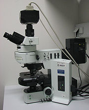
Fluorescence microscope
Encyclopedia
Optical microscope
The optical microscope, often referred to as the "light microscope", is a type of microscope which uses visible light and a system of lenses to magnify images of small samples. Optical microscopes are the oldest design of microscope and were possibly designed in their present compound form in the...
used to study properties of organic or inorganic substances using the phenomena of fluorescence
Fluorescence
Fluorescence is the emission of light by a substance that has absorbed light or other electromagnetic radiation of a different wavelength. It is a form of luminescence. In most cases, emitted light has a longer wavelength, and therefore lower energy, than the absorbed radiation...
and phosphorescence
Phosphorescence
Phosphorescence is a specific type of photoluminescence related to fluorescence. Unlike fluorescence, a phosphorescent material does not immediately re-emit the radiation it absorbs. The slower time scales of the re-emission are associated with "forbidden" energy state transitions in quantum...
instead of, or in addition to, reflection
Reflection (physics)
Reflection is the change in direction of a wavefront at an interface between two differentmedia so that the wavefront returns into the medium from which it originated. Common examples include the reflection of light, sound and water waves...
and absorption. The "fluorescence microscope" refers to any microscope that uses fluorescence to generate an image, whether it is a more simple set up like an epifluorescent microscope, or a more complicated design such as a confocal microscope, which uses optical sectioning to get better resolution of the fluorescent image.
All fluorescence microscopy methods share the same principle. A sample is illuminated with light of a wavelength which causes fluorescence in the sample. The light emitted by fluorescence, which is at a different, longer, wavelength than the illumination, is then detected through a microscope objective. Two filters are normally used in this technique; an illumination (or excitation) filter which ensures the illumination is near monochromatic and at the correct wavelength, and a second emission (or detection) filter which ensures none of the excitation light source reaches the detector. Fluorescence microscopy takes a fundamentally different approach to generating a light microscope image compared to transmitted or reflected white light techniques such as phase contrast and differential interference contrast microscopy
Differential interference contrast microscopy
Differential interference contrast microscopy , also known as Nomarski Interference Contrast or Nomarski microscopy, is an optical microscopy illumination technique used to enhance the contrast in unstained, transparent samples...
. These two contrasting optical microscopy methods give very different but complementary data.
Principle
The specimen is illuminated with light of a specific wavelengthWavelength
In physics, the wavelength of a sinusoidal wave is the spatial period of the wave—the distance over which the wave's shape repeats.It is usually determined by considering the distance between consecutive corresponding points of the same phase, such as crests, troughs, or zero crossings, and is a...
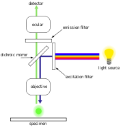
(or wavelengths) which is absorbed by the fluorophores, causing them to emit light of longer wavelengths (i.e., of a different color than the absorbed light). The illumination light is separated from the much weaker emitted fluorescence through the use of a spectral emission filter. Typical components of a fluorescence microscope are a light source (xenon arc lamp
Xenon arc lamp
A xenon arc lamp is a specialized type of gas discharge lamp, an electric light that produces light by passing electricity through ionized xenon gas at high pressure to produce a bright white light that closely mimics natural sunlight...
or mercury-vapor lamp
Mercury-vapor lamp
A mercury-vapor lamp is a gas discharge lamp that uses an electric arc through vaporized mercury to produce light. The arc discharge is generally confined to a small fused quartz arc tube mounted within a larger borosilicate glass bulb...
), the excitation filter
Excitation filter
An excitation filter is a high quality optical-glass filter commonly used in fluorescence microscopy and spectroscopic applications for selection of the excitation wavelength of light from a light source...
, the dichroic mirror (or dichroic beamsplitter
Dichroic filter
A dichroic filter, thin-film filter, or interference filter is a very accurate color filter used to selectively pass light of a small range of colors while reflecting other colors. By comparison, dichroic mirrors and dichroic reflectors tend to be characterized by the color of light that they...
), and the emission filter (see figure below). The filters and the dichroic are chosen to match the spectral excitation and emission characteristics of the fluorophore used to label the specimen. In this manner, the distribution of a single fluorophore (color) is imaged at a time. Multi-color images of several types of fluorophores must be composed by combining several single-color images.
Most fluorescence microscopes in use are epifluorescence microscopes (i.e., excitation and observation of the fluorescence are from above (epi–) the specimen). These microscopes have become an important part in the field of biology, opening the doors for more advanced microscope designs, such as the confocal microscope
Confocal microscopy
Confocal microscopy is an optical imaging technique used to increase optical resolution and contrast of a micrograph by using point illumination and a spatial pinhole to eliminate out-of-focus light in specimens that are thicker than the focal plane. It enables the reconstruction of...
and the total internal reflection fluorescence microscope
Total internal reflection fluorescence microscope
A total internal reflection fluorescence microscope is a type of microscope with which a thin region of a specimen, usually less than 200 nm, can be observed.-Background:...
(TIRF).
Epifluorescence microscopy
Life sciences
The life sciences comprise the fields of science that involve the scientific study of living organisms, like plants, animals, and human beings. While biology remains the centerpiece of the life sciences, technological advances in molecular biology and biotechnology have led to a burgeoning of...
, is epifluorescence microscopy. The excitatory light is passed from above (or, for inverted microscopes, from below), through the objective
Objective (optics)
In an optical instrument, the objective is the optical element that gathers light from the object being observed and focuses the light rays to produce a real image. Objectives can be single lenses or mirrors, or combinations of several optical elements. They are used in microscopes, telescopes,...
lens and then onto the specimen instead of passing it first through the specimen. The fluorescence
Fluorescence
Fluorescence is the emission of light by a substance that has absorbed light or other electromagnetic radiation of a different wavelength. It is a form of luminescence. In most cases, emitted light has a longer wavelength, and therefore lower energy, than the absorbed radiation...
in the specimen gives rise to emitted light which is focused to the detector by the same objective that is used for the excitation. Since most of the excitatory light is transmitted through the specimen, only reflected excitatory light reaches the objective together with the emitted light and this method therefore gives an improved signal to noise ratio. An additional dichroic filter
Dichroic filter
A dichroic filter, thin-film filter, or interference filter is a very accurate color filter used to selectively pass light of a small range of colors while reflecting other colors. By comparison, dichroic mirrors and dichroic reflectors tend to be characterized by the color of light that they...
between the objective and the detector can filter out the remaining excitation light from fluorescent light.
Light sources
Fluorescence microscopy requires intense, near-monochromatic, illumination which some widespread light sources, like halogen lampHalogen lamp
A halogen lamp, also known as a tungsten halogen lamp, is an incandescent lamp with a tungsten filament contained within an inert gas and a small amount of a halogen such as iodine or bromine. The chemical halogen cycle redeposits evaporated tungsten back on to the filament, extending the life of...
s cannot provide. There are two main types of light source used; xenon arc lamp
Xenon arc lamp
A xenon arc lamp is a specialized type of gas discharge lamp, an electric light that produces light by passing electricity through ionized xenon gas at high pressure to produce a bright white light that closely mimics natural sunlight...
or mercury-vapor lamp
Mercury-vapor lamp
A mercury-vapor lamp is a gas discharge lamp that uses an electric arc through vaporized mercury to produce light. The arc discharge is generally confined to a small fused quartz arc tube mounted within a larger borosilicate glass bulb...
s with an excitation filter
Excitation filter
An excitation filter is a high quality optical-glass filter commonly used in fluorescence microscopy and spectroscopic applications for selection of the excitation wavelength of light from a light source...
and laser
Laser
A laser is a device that emits light through a process of optical amplification based on the stimulated emission of photons. The term "laser" originated as an acronym for Light Amplification by Stimulated Emission of Radiation...
s. Lasers are most widely used for more complex fluorescence microscopy techniques like confocal microscopy
Confocal microscopy
Confocal microscopy is an optical imaging technique used to increase optical resolution and contrast of a micrograph by using point illumination and a spatial pinhole to eliminate out-of-focus light in specimens that are thicker than the focal plane. It enables the reconstruction of...
and total internal reflection fluorescence microscopy
Total internal reflection fluorescence microscope
A total internal reflection fluorescence microscope is a type of microscope with which a thin region of a specimen, usually less than 200 nm, can be observed.-Background:...
while xenon and mercury lamps with an excitation filter are commonly used for widefield epifluorescence microscopes.
Sample preparation
In order for a sample to be suitable for fluorescence microscopy it must be fluorescent. There are several methods of creating a fluorescent sample; the main techniques are labelling with fluorescent stains or, in the case of biological samples, expression of a fluorescent protein. Alternatively the intrinsic fluorescence of a sample (i.e., autofluorescenceAutofluorescence
Autofluorescence is the natural emission of light by biological entities such as mitochondria and lysosomes, and is used to distinguish the light originating from artificially added fluorescent markers...
) can be used. In the life sciences fluorescence microscopy is a powerful tool which allows the specific and sensitive staining of a specimen in order to detect the distribution of protein
Protein
Proteins are biochemical compounds consisting of one or more polypeptides typically folded into a globular or fibrous form, facilitating a biological function. A polypeptide is a single linear polymer chain of amino acids bonded together by peptide bonds between the carboxyl and amino groups of...
s or other molecules of interest. As a result there is a diverse range of techniques for fluorescent staining of biological samples.
Biological fluorescent stains
Many fluorescent stains have been designed for a range of biological molecules. Some of these are small molecules which are intrinsically fluorescent and bind a biological molecule of interest. Major examples of these are nucleic acidNucleic acid
Nucleic acids are biological molecules essential for life, and include DNA and RNA . Together with proteins, nucleic acids make up the most important macromolecules; each is found in abundance in all living things, where they function in encoding, transmitting and expressing genetic information...
stains like DAPI
DAPI
DAPI or 4',6-diamidino-2-phenylindole is a fluorescent stain that binds strongly to A-T rich regions in DNA. It is used extensively in fluorescence microscopy...
and Hoechst
Hoechst stain
Hoechst stains are part of a family of blue fluorescent dyes used to stain DNA. These Bis-benzimides were originally developed by the Hoechst AG, which numbered all their compounds so that the dye Hoechst 33342 is the 33342nd compound made by the company. There are three related Hoechst stains:...
which bind the minor groove of DNA
DNA
Deoxyribonucleic acid is a nucleic acid that contains the genetic instructions used in the development and functioning of all known living organisms . The DNA segments that carry this genetic information are called genes, but other DNA sequences have structural purposes, or are involved in...
, thus labelling the nuclei of cells. Others are drugs or toxins which bind specific cellular structures and have been derivatised with a fluorescent reporter. A major example of this class of fluorescent stain is fluorescently labelled-phalloidin
Phalloidin
Phalloidin is one of a group of toxins from the death cap known as phallotoxins.-Background:Pioneering work on this toxin was done by the Nobel laureate Heinrich Wieland in the 1930s...
which is used to stain actin
Actin
Actin is a globular, roughly 42-kDa moonlighting protein found in all eukaryotic cells where it may be present at concentrations of over 100 μM. It is also one of the most highly-conserved proteins, differing by no more than 20% in species as diverse as algae and humans...
fibres in mammal
Mammal
Mammals are members of a class of air-breathing vertebrate animals characterised by the possession of endothermy, hair, three middle ear bones, and mammary glands functional in mothers with young...
ian cells.
There are many fluorescent reported molecules, called fluorophore
Fluorophore
A fluorophore, in analogy to a chromophore, is a component of a molecule which causes a molecule to be fluorescent. It is a functional group in a molecule which will absorb energy of a specific wavelength and re-emit energy at a different wavelength...
s or fluorochromes such as fluorescein
Fluorescein
Fluorescein is a synthetic organic compound available as a dark orange/red powder soluble in water and alcohol. It is widely used as a fluorescent tracer for many applications....
, Alexa Fluors or DyLight 488
DyLight Fluor
The DyLight Fluor family of fluorescent dyes are produced by Dyomics in collaboration with Thermo Fisher Scientific. DyLight dyes are typically used in biotechnology and research applications as biomolecule, cell and tissue labels for fluorescence microscopy, cell biology or molecular...
, which can be chemically linked to a different molecule which binds the target of interest within the sample.
Immunofluorescence
Immuofluorescence is an antibodyAntibody
An antibody, also known as an immunoglobulin, is a large Y-shaped protein used by the immune system to identify and neutralize foreign objects such as bacteria and viruses. The antibody recognizes a unique part of the foreign target, termed an antigen...
based on technique which uses the highly specific binding of an antibody to its antigen
Antigen
An antigen is a foreign molecule that, when introduced into the body, triggers the production of an antibody by the immune system. The immune system will then kill or neutralize the antigen that is recognized as a foreign and potentially harmful invader. These invaders can be molecules such as...
in order to label specific proteins or other molecules within the cell. A sample is treated with a primary antibody specific for the molecule of interest A fluorophore can be directly conjugated to the primary antibody. Alternatively a secondary antibody, conjugated to a fluorophore, which binds specifically to the first antibody can be used. For example a primary antibody raised in a mouse which recognises tubulin
Tubulin
Tubulin is one of several members of a small family of globular proteins. The most common members of the tubulin family are α-tubulin and β-tubulin, the proteins that make up microtubules. Each has a molecular weight of approximately 55 kiloDaltons. Microtubules are assembled from dimers of α- and...
combined with a secondary anti-mouse antibody derivatised with a fluorophore could be used to label microtubules in a cell.
Fluorescent proteins
The modern understanding of geneticsGenetics
Genetics , a discipline of biology, is the science of genes, heredity, and variation in living organisms....
and the techniques available for modifying DNA allows scientists to genetically modify proteins to also carry a fluorescent protein reporter. In biological samples this allows a scientist to directly make a protein of interest fluorescent. The protein location can then be directly tracked, including in live cells.
Limitations
Fluorophores lose their ability to fluoresce as they are illuminated in a process called photobleachingPhotobleaching
Photobleaching is the photochemical destruction of a fluorophore. In microscopy, photobleaching may complicate the observation of fluorescent molecules, since they will eventually be destroyed by the light exposure necessary to stimulate them into fluorescing...
. Photobleaching occurs as the fluorescent molecules accumulate chemical damage from the electrons excited during fluorescence. Photobleaching can severely limit the time over which a sample can be observed by fluorescent microscopy. Several techniques exist to reduce photobleaching such as the use of more robust fluorophores, by minimizing illumination, or by using photoprotective scavenger
Scavenger (chemistry)
A scavenger in chemistry is a chemical substance added to a mixture in order to remove or inactivate impurities or unwanted reaction products. Their use is wide-ranged:...
chemicals.
Fluorescence microscopy with fluorescent reporter proteins has enabled analysis of live cells by fluorescence microscopy, however cells are susceptible to phototoxicity, particularly with short wavelength light. Furthermore fluorescent molecules have a tendency to generate reactive chemical species when under illumination which enhances the phototoxic effect.
Unlike transmitted and reflected light microscopy techniques fluorescence microscopy only allows observation of the specific structures which have been fluorescently labeled. For example observing a tissue sample prepared with a fluorescent DNA stain by fluorescent microscopy only reveals the organisation of the DNA within the cells and reveals nothing else about the cell morphologies.
Improvements and sub-diffraction techniques
The wave nature of light limits the size of the spot to which light can be focused due to the diffraction limit. This limitation was described in the 19th century by Ernst Abbe and limits an optical microscope's resolution to approximately half of the wavelength of the light used. Fluorescence microscopy is central to many techniques which aim to reach past this limit by specialised optical configurations.Several improvements in microscopy techniques have been invented in the 20th century and have resulted in increased resolution and contrast to some extent. However they did not overcome the diffraction limit. In 1978 first theoretical ideas have been developed to break this barrier by using a 4Pi microscope as a confocal laser scanning fluorescence microscope where the light is focused ideally from all sides to a common focus which is used to scan the object by 'point-by-point' excitation combined with 'point-by-point' detection.
However, the first experimental demonstration of the 4pi microscope took place in 1994. 4Pi microscopy maximizes the amount of available focusing directions by using two opposing objective lenses or Multi-photon microscopy using redshifted light and multi-photon excitation.
The first technique to really achieve a sub-diffraction resolution was STED microscopy
STED microscopy
Stimulated Emission Depletion microscopy, or STED microscopy, is a fluorescence microscopy technique that uses the non-linear de-excitation of fluorescent dyes to overcome the resolution limit imposed by diffraction with standard confocal laser scanning microscopes and conventional far-field...
, proposed in 1994. This method and all techniques following the RESOLFT
RESOLFT
RESOLFT, an acronym for REversible Saturable OpticaL Fluorescence Transitions, denotes a group of optical microscopy techniques with very high resolution...
concept rely on a strong non-linear interaction between light and fluorescing molecules. The molecules are driven strongly between distinguishable molecular states at each specific location, so that finally light can be emitted at only a small fraction of space, hence an increased resolution.
As well in the 1990s another super resolution microscopy method based on wide field microscopy has been developed. Substantially improved size resolution of cellular nanostructure
Nanostructure
A nanostructure is an object of intermediate size between molecular and microscopic structures.In describing nanostructures it is necessary to differentiate between the number of dimensions on the nanoscale. Nanotextured surfaces have one dimension on the nanoscale, i.e., only the thickness of the...
s stained with a fluorescent marker was achieved by development of SPDM localization microscopy and the structured laser illumination (spatially modulated illumination, SMI). Combining the principle of SPDM with SMI resulted in the development of the Vertico SMI
Vertico SMI
Vertico-SMI is currently the fastest light microscope for the 3D analysis of complete cells in the nanometer range. It is based on two technologies developed in 1996, SMI and SPDM...
microscope. Single molecule detection of normal blinking
Fluorescence intermittency
Fluorescence intermittency, or blinking, is the phenomenon of random switching between ON and OFF states of the emitter under its continuous excitation...
fluorescent dyes like Green fluorescent protein
Green fluorescent protein
The green fluorescent protein is a protein composed of 238 amino acid residues that exhibits bright green fluorescence when exposed to blue light. Although many other marine organisms have similar green fluorescent proteins, GFP traditionally refers to the protein first isolated from the...
(GFP) can be achieved by using a further development of SPDM the so-called SPDMphymod technology which makes it possible to detect and count two different fluorescent molecule types at the molecular level (this technology is referred to as 2CLM, 2 Color Localization Microscopy).
Alternatively, the advent of photoactivated localization microscopy could achieve similar results by relying on blinking or switching of single molecules, where the fraction of fluorescing molecules is very small at each time. This stochastic response of molecules on the applied light corresponds also to a highly nonlinear interaction, leading to subdiffraction resolution.
External Links
- Fluorophores.org, the database of fluorescent dyes
- MicroscopyU
- "Fluorescence Microscopy" lecture by Nico Stuurman (UCSF)