
Fetus
Encyclopedia
A fetus (ˈfiːtəs; also spelled foetus, fœtus, faetus, or fætus, see below) is a developing mammal
or other viviparous vertebrate
after the embryo
nic stage and before birth
.
In humans, the fetal stage of prenatal development starts at the beginning of the 11th week in gestational age
, which is the 9th week after fertilization.
fētus (“offspring”, “bringing forth”, “hatching of young”). It has Indo-European
roots related to sucking or suckling, from the Aryan
prefix bheu-, meaning "To come into being".
Fœtus or foetus is the British, Irish and Commonwealth spelling, which has been in use since at least 1594. It arose as a hypercorrection
based on an incorrect etymology (i.e. due to insufficient knowledge of Latin) that may have originated with an error by Saint Isidore of Seville, in AD 620. This spelling is the most common in most Commonwealth nations (except in medical literature, where its use is barred). The etymologically correct original spelling, fetus is used in Canada and the United States. In addition, fetus is now the standard English spelling throughout the world in medical journals. The spelling "faetus" was used historically.
The spelling in the Oxford Encyclopedic English Dictionary, Third Edition (1996), page 537, is 'foetus' with 'foetuses' as the plural; 'fetus' (page 514) is given as the 'US variant of foetus.' However, later editions of the OED clarify the etymology behind the Commonwealth spelling.
Fetuses are not capable of feeling pain at the beginning of the fetal stage, and may not be able to feel pain until the third trimester. At this point in development, uncontrolled movements and twitches occur as muscles, the brain and pathways begin to develop.
 The amount of body fat rapidly increases. Lungs are not fully mature. Thalamic
brain connections, which mediate sensory input, form. Bones are fully developed, but are still soft and pliable. Iron
, calcium
, and phosphorus
become more abundant. Fingernails reach the end of the fingertips. The lanugo
begins to disappear, until it is gone except on the upper arms and shoulders. Small breast buds are present on both sexes. Head hair becomes coarse and thicker. Birth is imminent and occurs around the 40th week. The fetus is considered full-term between weeks 37 and 40, which means that the fetus is considered sufficiently developed for life outside the uterus. It may be 48 to 53 cm (19 to 21 inches) in length, when born. Control of movement is limited at birth, and purposeful voluntary movements develop all the way until puberty.
l, or fetal.
Maternal factors include maternal weight
, body mass index
, nutritional state, emotional stress
, toxin exposure (including tobacco
, alcohol
, heroin, and other drugs which can also harm the fetus in other ways), and uterine
blood
flow.
Placental factors include size, microstructure (densities and architecture), umbilical
blood flow, transporters and binding proteins, nutrient utilization and nutrient production.
Fetal factors include the fetus genome, nutrient production, and hormone
output. Also, female fetuses tend to weigh less than males, at full term.
Fetal growth is often classified as follows: small for gestational age (SGA), appropriate for gestational age (AGA), and large for gestational age (LGA). SGA can result in low birth weight
, although premature birth can also result in low birth weight. Low birth weight increases risk for perinatal mortality (death
shortly after birth), asphyxia
, hypothermia
, polycythemia
, hypocalcemia, immune dysfunction, neurologic abnormalities, and other long-term health problems. SGA may be associated with growth delay, or it may instead be associated with absolute stunting of growth.
 Viability refers to a point in fetal development at which the fetus may survive outside the womb. The lower limit of viability is approximately five months gestational age
, and usually later.
 There is no sharp limit of development, age, or weight at which a fetus automatically becomes viable. According to data years 2003-2005, 20 to 35 percent of babies born at 23 weeks of gestation survive, while 50 to 70 percent of babies born at 24 to 25 weeks, and more than 90 percent born at 26 to 27 weeks, survive. It is rare for a baby weighing less than 500 gm to survive.
When such babies are born, the main causes of perinatal mortality
is that the respiratory system and the central nervous system are not completely differentiated. If given expert postnatal care, some fetuses weighing less than 500 gm may survive, and are referred to as extremely low birth weight or immature infants. Preterm birth is the most common cause of perinatal mortality, causing almost 30 percent of neonatal deaths.
, its existence, and its implications are debated politically and academically. According to the conclusions of a review published in 2005, "Evidence regarding the capacity for fetal pain is limited but indicates that fetal perception of pain is unlikely before the third trimester." However, there may be an emerging consensus among developmental neurobiologists that the establishment of thalamocortical connections" (at about 26 weeks) is a critical event with regard to fetal perception of pain. Nevertheless, because pain can involve sensory, emotional and cognitive factors, it is "impossible to know" when painful experiences may become possible, even if it is known when thalamocortical connections are established.
Whether a fetus has the ability to feel pain
and to suffer
is part of the abortion debate
. For example, in the USA legislation has been proposed by pro-life
advocates that abortion providers should be required to tell a woman that the fetus may feel pain during the abortion procedure, and require her to accept or decline anesthesia for the fetus.
 The circulatory system
of a human fetus works differently from that of born humans, mainly because the lungs are not in use: the fetus obtains oxygen
and nutrients from the woman through the placenta
and the umbilical cord
.
Blood from the placenta is carried to the fetus by the umbilical vein
. About half of this enters the fetal ductus venosus
and is carried to the inferior vena cava
, while the other half enters the liver
proper from the inferior border of the liver. The branch of the umbilical vein that supplies the right lobe of the liver first joins with the portal vein. The blood then moves to the right atrium of the heart
. In the fetus, there is an opening between the right and left atrium (the foramen ovale
), and most of the blood flows from the right into the left atrium, thus bypassing pulmonary circulation
. The majority of blood flow is into the left ventricle from where it is pumped through the aorta
into the body. Some of the blood moves from the aorta through the internal iliac arteries to the umbilical arteries, and re-enters the placenta, where carbon dioxide
and other waste products from the fetus are taken up and enter the woman's circulation.
Some of the blood from the right atrium does not enter the left atrium, but enters the right ventricle and is pumped into the pulmonary artery
. In the fetus, there is a special connection between the pulmonary artery and the aorta, called the ductus arteriosus
, which directs most of this blood away from the lungs (which aren't being used for respiration at this point as the fetus is suspended in amniotic fluid
).
"). More blood moves from the right atrium to the right ventricle and into the pulmonary arteries, and less flows through the foramen ovale
to the left atrium. The blood from the lungs travels through the pulmonary veins to the left atrium, increasing the pressure there. The decreased right atrial pressure and the increased left atrial pressure pushes the septum primum against the septum secundum, closing the foramen ovale, which now becomes the fossa ovalis. This completes the separation of the circulatory system into two halves, the left and the right.
The ductus arteriosus normally closes off within one or two days of birth, leaving behind the ligamentum arteriosum. The umbilical vein and the ductus venosus closes off within two to five days after birth, leaving behind the ligamentum teres and the ligamentum venosus of the liver respectively.
In addition to differences in circulation, the developing fetus also employs a different type of oxygen transport molecule
than adults (adults use adult hemoglobin
). Fetal hemoglobin
enhances the fetus' ability to draw oxygen from the placenta. Its dissociation curve to oxygen is shifted to the left, meaning that it will take up oxygen at a lower concentration than adult hemoglobin will. This enables fetal hemoglobin to absorb oxygen from adult hemoglobin in the placenta, which has a lower pressure of oxygen than at the lungs.
s to permit sufficient time for the surgical correction of the anomalies. Conversely, in cases of patent ductus arteriosus
, where the ductus does not properly close, drugs that inhibit prostaglandin synthesis can be used to encourage its closure, so that surgery can be avoided.
A developing fetus is highly susceptible to anomalies in its growth and metabolism, increasing the risk of birth defects. One area of concern is the pregnant woman's lifestyle choices made during pregnancy. Diet is especially important in the early stages of development. Studies show that supplementation of the woman's diet with folic acid
reduces the risk of spina bifida
and other neural tube
defects. Another dietary concern is whether the woman eats breakfast. Skipping breakfast could lead to extended periods of lower than normal nutrients in the woman's blood, leading to a higher risk of prematurity, or other birth defects in the fetus. During this time alcohol consumption may increase the risk of the development of Fetal alcohol syndrome
, a condition leading to mental retardation
in some infants.
Smoking during pregnancy
may also lead to reduced birth weight. Low birth weight is defined as 2500 grams (5.5 lb). Low birth weight is a concern for medical providers due to the tendency of these infants, described as premature by weight, to have a higher risk of secondary medical problems.
and/or tolerated due to facets of the physician-patient relationship in many countries such as Australia, India, Canada, most European countries, and the United States. Many of those countries that allow abortion during the fetal stage have gestational time limits, so that late-term abortions are not normally allowed.
Mammal
Mammals are members of a class of air-breathing vertebrate animals characterised by the possession of endothermy, hair, three middle ear bones, and mammary glands functional in mothers with young...
or other viviparous vertebrate
Vertebrate
Vertebrates are animals that are members of the subphylum Vertebrata . Vertebrates are the largest group of chordates, with currently about 58,000 species described. Vertebrates include the jawless fishes, bony fishes, sharks and rays, amphibians, reptiles, mammals, and birds...
after the embryo
Embryo
An embryo is a multicellular diploid eukaryote in its earliest stage of development, from the time of first cell division until birth, hatching, or germination...
nic stage and before birth
Childbirth
Childbirth is the culmination of a human pregnancy or gestation period with the birth of one or more newborn infants from a woman's uterus...
.
In humans, the fetal stage of prenatal development starts at the beginning of the 11th week in gestational age
Gestational age
Gestational age relates to the age of an embryo or fetus . There is some ambiguity in how it is defined:*In embryology, gestational age is the time elapsed since conception. This interval is also termed fertilisation age....
, which is the 9th week after fertilization.
Etymology and spelling variations
The word fetus (plural fetuses) is from the LatinLatin
Latin is an Italic language originally spoken in Latium and Ancient Rome. It, along with most European languages, is a descendant of the ancient Proto-Indo-European language. Although it is considered a dead language, a number of scholars and members of the Christian clergy speak it fluently, and...
fētus (“offspring”, “bringing forth”, “hatching of young”). It has Indo-European
Proto-Indo-European language
The Proto-Indo-European language is the reconstructed common ancestor of the Indo-European languages, spoken by the Proto-Indo-Europeans...
roots related to sucking or suckling, from the Aryan
Aryan
Aryan is an English language loanword derived from Sanskrit ārya and denoting variously*In scholarly usage:**Indo-Iranian languages *in dated usage:**the Indo-European languages more generally and their speakers...
prefix bheu-, meaning "To come into being".
Fœtus or foetus is the British, Irish and Commonwealth spelling, which has been in use since at least 1594. It arose as a hypercorrection
Hypercorrection
In linguistics or usage, hypercorrection is a non-standard usage that results from the over-application of a perceived rule of grammar or a usage prescription...
based on an incorrect etymology (i.e. due to insufficient knowledge of Latin) that may have originated with an error by Saint Isidore of Seville, in AD 620. This spelling is the most common in most Commonwealth nations (except in medical literature, where its use is barred). The etymologically correct original spelling, fetus is used in Canada and the United States. In addition, fetus is now the standard English spelling throughout the world in medical journals. The spelling "faetus" was used historically.
The spelling in the Oxford Encyclopedic English Dictionary, Third Edition (1996), page 537, is 'foetus' with 'foetuses' as the plural; 'fetus' (page 514) is given as the 'US variant of foetus.' However, later editions of the OED clarify the etymology behind the Commonwealth spelling.
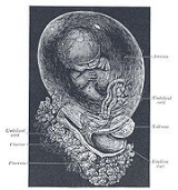
Week 9 to 16
The fetal stage commences at the beginning of the 9th week. At the start of the fetal stage, the fetus is typically about 30 millimetres (1.2 in) in length from crown to rump, and weighs about 8 grams. The head makes up nearly half of the fetus' size. Breathing-like movement of the fetus is necessary for stimulation of lung development, rather than for obtaining oxygen. The heart, hands, feet, brain and other organs are present, but are only at the beginning of development and have minimal operation.Fetuses are not capable of feeling pain at the beginning of the fetal stage, and may not be able to feel pain until the third trimester. At this point in development, uncontrolled movements and twitches occur as muscles, the brain and pathways begin to develop.
Week 16 to 25
A woman pregnant for the first time (i.e. a primiparous woman) typically feels fetal movements at about 21 weeks, whereas a woman who has already given birth at least two times (i.e. a multiparous woman) will typically feel movements by 20 weeks. By the end of the fifth month, the fetus is about 20 cm (8 inches).Week 26 to 40
Thalamus
The thalamus is a midline paired symmetrical structure within the brains of vertebrates, including humans. It is situated between the cerebral cortex and midbrain, both in terms of location and neurological connections...
brain connections, which mediate sensory input, form. Bones are fully developed, but are still soft and pliable. Iron
Iron
Iron is a chemical element with the symbol Fe and atomic number 26. It is a metal in the first transition series. It is the most common element forming the planet Earth as a whole, forming much of Earth's outer and inner core. It is the fourth most common element in the Earth's crust...
, calcium
Calcium
Calcium is the chemical element with the symbol Ca and atomic number 20. It has an atomic mass of 40.078 amu. Calcium is a soft gray alkaline earth metal, and is the fifth-most-abundant element by mass in the Earth's crust...
, and phosphorus
Phosphorus
Phosphorus is the chemical element that has the symbol P and atomic number 15. A multivalent nonmetal of the nitrogen group, phosphorus as a mineral is almost always present in its maximally oxidized state, as inorganic phosphate rocks...
become more abundant. Fingernails reach the end of the fingertips. The lanugo
Lanugo
Lanugo is fine, downy hair as a type of fur. It is often found in teratomas .-Fetal development:Lanugo grows on fetuses as a normal part of gestation, but is usually shed and replaced by vellus hair at about 33 to 36 weeks of gestational age...
begins to disappear, until it is gone except on the upper arms and shoulders. Small breast buds are present on both sexes. Head hair becomes coarse and thicker. Birth is imminent and occurs around the 40th week. The fetus is considered full-term between weeks 37 and 40, which means that the fetus is considered sufficiently developed for life outside the uterus. It may be 48 to 53 cm (19 to 21 inches) in length, when born. Control of movement is limited at birth, and purposeful voluntary movements develop all the way until puberty.
Variation in growth
There is much variation in the growth of the fetus. When fetal size is less than expected, that condition is known as intrauterine growth restriction (IUGR) also called fetal growth restriction (FGR); factors affecting fetal growth can be maternal, placentaPlacenta
The placenta is an organ that connects the developing fetus to the uterine wall to allow nutrient uptake, waste elimination, and gas exchange via the mother's blood supply. "True" placentas are a defining characteristic of eutherian or "placental" mammals, but are also found in some snakes and...
l, or fetal.
Maternal factors include maternal weight
Weight
In science and engineering, the weight of an object is the force on the object due to gravity. Its magnitude , often denoted by an italic letter W, is the product of the mass m of the object and the magnitude of the local gravitational acceleration g; thus:...
, body mass index
Body mass index
The body mass index , or Quetelet index, is a heuristic proxy for human body fat based on an individual's weight and height. BMI does not actually measure the percentage of body fat. It was invented between 1830 and 1850 by the Belgian polymath Adolphe Quetelet during the course of developing...
, nutritional state, emotional stress
Stress (medicine)
Stress is a term in psychology and biology, borrowed from physics and engineering and first used in the biological context in the 1930s, which has in more recent decades become commonly used in popular parlance...
, toxin exposure (including tobacco
Tobacco
Tobacco is an agricultural product processed from the leaves of plants in the genus Nicotiana. It can be consumed, used as a pesticide and, in the form of nicotine tartrate, used in some medicines...
, alcohol
Alcohol
In chemistry, an alcohol is an organic compound in which the hydroxy functional group is bound to a carbon atom. In particular, this carbon center should be saturated, having single bonds to three other atoms....
, heroin, and other drugs which can also harm the fetus in other ways), and uterine
Uterus
The uterus or womb is a major female hormone-responsive reproductive sex organ of most mammals including humans. One end, the cervix, opens into the vagina, while the other is connected to one or both fallopian tubes, depending on the species...
blood
Blood
Blood is a specialized bodily fluid in animals that delivers necessary substances such as nutrients and oxygen to the cells and transports metabolic waste products away from those same cells....
flow.
Placental factors include size, microstructure (densities and architecture), umbilical
Umbilical cord
In placental mammals, the umbilical cord is the connecting cord from the developing embryo or fetus to the placenta...
blood flow, transporters and binding proteins, nutrient utilization and nutrient production.
Fetal factors include the fetus genome, nutrient production, and hormone
Hormone
A hormone is a chemical released by a cell or a gland in one part of the body that sends out messages that affect cells in other parts of the organism. Only a small amount of hormone is required to alter cell metabolism. In essence, it is a chemical messenger that transports a signal from one...
output. Also, female fetuses tend to weigh less than males, at full term.
Fetal growth is often classified as follows: small for gestational age (SGA), appropriate for gestational age (AGA), and large for gestational age (LGA). SGA can result in low birth weight
Low birth weight
Low birth weight is defined as a birth weight of a liveborn infant of less than 2,500 g. regardless of gestational age-Causes:LBW is either the result of preterm birth or of the infant being small for gestational age , or a combination of...
, although premature birth can also result in low birth weight. Low birth weight increases risk for perinatal mortality (death
Death
Death is the permanent termination of the biological functions that sustain a living organism. Phenomena which commonly bring about death include old age, predation, malnutrition, disease, and accidents or trauma resulting in terminal injury....
shortly after birth), asphyxia
Asphyxia
Asphyxia or asphyxiation is a condition of severely deficient supply of oxygen to the body that arises from being unable to breathe normally. An example of asphyxia is choking. Asphyxia causes generalized hypoxia, which primarily affects the tissues and organs...
, hypothermia
Hypothermia
Hypothermia is a condition in which core temperature drops below the required temperature for normal metabolism and body functions which is defined as . Body temperature is usually maintained near a constant level of through biologic homeostasis or thermoregulation...
, polycythemia
Polycythemia
Polycythemia is a disease state in which the proportion of blood volume that is occupied by red blood cells increases...
, hypocalcemia, immune dysfunction, neurologic abnormalities, and other long-term health problems. SGA may be associated with growth delay, or it may instead be associated with absolute stunting of growth.
Viability
Gestational age
Gestational age relates to the age of an embryo or fetus . There is some ambiguity in how it is defined:*In embryology, gestational age is the time elapsed since conception. This interval is also termed fertilisation age....
, and usually later.
When such babies are born, the main causes of perinatal mortality
Perinatal mortality
Perinatal mortality , also perinatal death, refers to the death of a fetus or neonate and is the basis to calculate the perinatal mortality rate. Variations in the precise definition of the perinatal mortality exist specifically concerning the issue of inclusion or exclusion of early fetal and...
is that the respiratory system and the central nervous system are not completely differentiated. If given expert postnatal care, some fetuses weighing less than 500 gm may survive, and are referred to as extremely low birth weight or immature infants. Preterm birth is the most common cause of perinatal mortality, causing almost 30 percent of neonatal deaths.
Fetal pain
Fetal painPain
Pain is an unpleasant sensation often caused by intense or damaging stimuli such as stubbing a toe, burning a finger, putting iodine on a cut, and bumping the "funny bone."...
, its existence, and its implications are debated politically and academically. According to the conclusions of a review published in 2005, "Evidence regarding the capacity for fetal pain is limited but indicates that fetal perception of pain is unlikely before the third trimester." However, there may be an emerging consensus among developmental neurobiologists that the establishment of thalamocortical connections" (at about 26 weeks) is a critical event with regard to fetal perception of pain. Nevertheless, because pain can involve sensory, emotional and cognitive factors, it is "impossible to know" when painful experiences may become possible, even if it is known when thalamocortical connections are established.
Whether a fetus has the ability to feel pain
Pain
Pain is an unpleasant sensation often caused by intense or damaging stimuli such as stubbing a toe, burning a finger, putting iodine on a cut, and bumping the "funny bone."...
and to suffer
Suffering
Suffering, or pain in a broad sense, is an individual's basic affective experience of unpleasantness and aversion associated with harm or threat of harm. Suffering may be qualified as physical or mental. It may come in all degrees of intensity, from mild to intolerable. Factors of duration and...
is part of the abortion debate
Abortion debate
The abortion debate refers to discussion and controversy surrounding the moral and legal status of abortion. The two main groups involved in the abortion debate are the self-described "pro-choice" movement and the "pro-life" movement...
. For example, in the USA legislation has been proposed by pro-life
Pro-life
Opposition to the legalization of abortion is centered around the pro-life, or anti-abortion, movement, a social and political movement opposing elective abortion on moral grounds and supporting its legal prohibition or restriction...
advocates that abortion providers should be required to tell a woman that the fetus may feel pain during the abortion procedure, and require her to accept or decline anesthesia for the fetus.
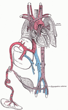
Circulatory system
Circulatory system
The circulatory system is an organ system that passes nutrients , gases, hormones, blood cells, etc...
of a human fetus works differently from that of born humans, mainly because the lungs are not in use: the fetus obtains oxygen
Oxygen
Oxygen is the element with atomic number 8 and represented by the symbol O. Its name derives from the Greek roots ὀξύς and -γενής , because at the time of naming, it was mistakenly thought that all acids required oxygen in their composition...
and nutrients from the woman through the placenta
Placenta
The placenta is an organ that connects the developing fetus to the uterine wall to allow nutrient uptake, waste elimination, and gas exchange via the mother's blood supply. "True" placentas are a defining characteristic of eutherian or "placental" mammals, but are also found in some snakes and...
and the umbilical cord
Umbilical cord
In placental mammals, the umbilical cord is the connecting cord from the developing embryo or fetus to the placenta...
.
Blood from the placenta is carried to the fetus by the umbilical vein
Umbilical vein
The umbilical vein is a vein present during fetal development that carries oxygenated blood from the placenta to the growing fetus.The blood pressure inside the umbilical vein is approximately 20 mmHg.-Development:...
. About half of this enters the fetal ductus venosus
Ductus venosus
In the fetus, the ductus venosus shunts approximately half of the blood flow of the umbilical vein directly to the inferior vena cava. Thus, it allows oxygenated blood from the placenta to bypass the liver. In conjunction with the other fetal shunts, the foramen ovale and ductus arteriosus, it...
and is carried to the inferior vena cava
Inferior vena cava
The inferior vena cava , also known as the posterior vena cava, is the large vein that carries de-oxygenated blood from the lower half of the body into the right atrium of the heart....
, while the other half enters the liver
Liver
The liver is a vital organ present in vertebrates and some other animals. It has a wide range of functions, including detoxification, protein synthesis, and production of biochemicals necessary for digestion...
proper from the inferior border of the liver. The branch of the umbilical vein that supplies the right lobe of the liver first joins with the portal vein. The blood then moves to the right atrium of the heart
Heart
The heart is a myogenic muscular organ found in all animals with a circulatory system , that is responsible for pumping blood throughout the blood vessels by repeated, rhythmic contractions...
. In the fetus, there is an opening between the right and left atrium (the foramen ovale
Foramen ovale (heart)
In the fetal heart, the foramen ovale , also ostium secundum of Born or falx septi, allows blood to enter the left atrium from the right atrium. It is one of two fetal cardiac shunts, the other being the ductus arteriosus...
), and most of the blood flows from the right into the left atrium, thus bypassing pulmonary circulation
Pulmonary circulation
Pulmonary circulation is the half portion of the cardiovascular system which carries Oxygen-depleted Blood away from the heart, to the Lungs, and returns oxygenated blood back to the heart. Encyclopedic description and discovery of the pulmonary circulation is widely attributed to Doctor Ibn...
. The majority of blood flow is into the left ventricle from where it is pumped through the aorta
Aorta
The aorta is the largest artery in the body, originating from the left ventricle of the heart and extending down to the abdomen, where it branches off into two smaller arteries...
into the body. Some of the blood moves from the aorta through the internal iliac arteries to the umbilical arteries, and re-enters the placenta, where carbon dioxide
Carbon dioxide
Carbon dioxide is a naturally occurring chemical compound composed of two oxygen atoms covalently bonded to a single carbon atom...
and other waste products from the fetus are taken up and enter the woman's circulation.
Some of the blood from the right atrium does not enter the left atrium, but enters the right ventricle and is pumped into the pulmonary artery
Pulmonary artery
The pulmonary arteries carry deoxygenated blood from the heart to the lungs. They are the only arteries that carry deoxygenated blood....
. In the fetus, there is a special connection between the pulmonary artery and the aorta, called the ductus arteriosus
Ductus arteriosus
In the developing fetus, the ductus arteriosus , also called the ductus Botalli, is a shunt connecting the pulmonary artery to the aortic arch. It allows most of the blood from the right ventricle to bypass the fetus's fluid-filled lungs. Upon closure at birth, it becomes the ligamentum arteriosum...
, which directs most of this blood away from the lungs (which aren't being used for respiration at this point as the fetus is suspended in amniotic fluid
Amniotic fluid
Amniotic fluid or liquor amnii is the nourishing and protecting liquid contained by the amniotic sac of a pregnant woman.- Development of amniotic fluid :...
).
Postnatal development
With the first breath after birth, the system changes suddenly. The pulmonary resistance is dramatically reduced ("pulmo" is from the Latin for "lungLung
The lung is the essential respiration organ in many air-breathing animals, including most tetrapods, a few fish and a few snails. In mammals and the more complex life forms, the two lungs are located near the backbone on either side of the heart...
"). More blood moves from the right atrium to the right ventricle and into the pulmonary arteries, and less flows through the foramen ovale
Foramen ovale (heart)
In the fetal heart, the foramen ovale , also ostium secundum of Born or falx septi, allows blood to enter the left atrium from the right atrium. It is one of two fetal cardiac shunts, the other being the ductus arteriosus...
to the left atrium. The blood from the lungs travels through the pulmonary veins to the left atrium, increasing the pressure there. The decreased right atrial pressure and the increased left atrial pressure pushes the septum primum against the septum secundum, closing the foramen ovale, which now becomes the fossa ovalis. This completes the separation of the circulatory system into two halves, the left and the right.
The ductus arteriosus normally closes off within one or two days of birth, leaving behind the ligamentum arteriosum. The umbilical vein and the ductus venosus closes off within two to five days after birth, leaving behind the ligamentum teres and the ligamentum venosus of the liver respectively.
Differences from the adult circulatory system
Remnants of the fetal circulation can be found in adults:| Fetal | Adult Adult An adult is a human being or living organism that is of relatively mature age, typically associated with sexual maturity and the attainment of reproductive age.... |
|---|---|
| foramen ovale Foramen ovale There are multiple structures in the human body with the name foramen ovale :* In the fetal heart, the foramen ovale is a shunt from the right atrium to left atrium.... |
fossa ovalis Fossa ovalis (heart) Found in the right atrium of the heart, the fossa ovalis is an embryonic remnant of the foramen ovale, which normally closes shortly after birth.In a heart specimen of a neonate, the fossa ovalis is translucent, but later in life the membrane thickens... |
| ductus arteriosus Ductus arteriosus In the developing fetus, the ductus arteriosus , also called the ductus Botalli, is a shunt connecting the pulmonary artery to the aortic arch. It allows most of the blood from the right ventricle to bypass the fetus's fluid-filled lungs. Upon closure at birth, it becomes the ligamentum arteriosum... |
ligamentum arteriosum Ligamentum arteriosum The ligamentum arteriosum is a small ligament attached to the superior surface of the pulmonary trunk and the inferior surface of the aortic arch... |
| extra-hepatic portion of the fetal left umbilical vein Umbilical vein The umbilical vein is a vein present during fetal development that carries oxygenated blood from the placenta to the growing fetus.The blood pressure inside the umbilical vein is approximately 20 mmHg.-Development:... |
ligamentum teres hepatis (the "round ligament of the liver"). |
| intra-hepatic portion of the fetal left umbilical vein (the ductus venosus Ductus venosus In the fetus, the ductus venosus shunts approximately half of the blood flow of the umbilical vein directly to the inferior vena cava. Thus, it allows oxygenated blood from the placenta to bypass the liver. In conjunction with the other fetal shunts, the foramen ovale and ductus arteriosus, it... ) |
ligamentum venosum Ligamentum venosum The ligamentum venosum is the fibrous remnant of the ductus venosus of the fetal circulation. Usually, it is attached to the left branch of the portal vein within the porta hepatis... |
| proximal portions of the fetal left and right umbilical arteries | umbilical branches of the internal iliac arteries |
| distal portions of the fetal left and right umbilical arteries | medial umbilical ligaments (urachus Urachus The urachus is a fibrous remnant of the allantois, a canal that drains the urinary bladder of the fetus that joins and runs within the umbilical cord... ) |
In addition to differences in circulation, the developing fetus also employs a different type of oxygen transport molecule
Transport protein
A membrane transport protein is a membrane protein involved in the movement of ions, small molecules, or macromolecules, such as another protein across a biological membrane. Transport proteins are integral membrane proteins; that is they exist within and span the membrane across which they...
than adults (adults use adult hemoglobin
Hemoglobin
Hemoglobin is the iron-containing oxygen-transport metalloprotein in the red blood cells of all vertebrates, with the exception of the fish family Channichthyidae, as well as the tissues of some invertebrates...
). Fetal hemoglobin
Fetal hemoglobin
Fetal hemoglobin, or foetal haemoglobin, is the main oxygen transport protein in the fetus during the last seven months of development in the uterus and in the newborn until roughly 6 months old...
enhances the fetus' ability to draw oxygen from the placenta. Its dissociation curve to oxygen is shifted to the left, meaning that it will take up oxygen at a lower concentration than adult hemoglobin will. This enables fetal hemoglobin to absorb oxygen from adult hemoglobin in the placenta, which has a lower pressure of oxygen than at the lungs.
Developmental problems
Congenital anomalies are anomalies that are acquired before birth. Infants with certain congenital anomalies of the heart can survive only as long as the ductus remains open: in such cases the closure of the ductus can be delayed by the administration of prostaglandinProstaglandin
A prostaglandin is any member of a group of lipid compounds that are derived enzymatically from fatty acids and have important functions in the animal body. Every prostaglandin contains 20 carbon atoms, including a 5-carbon ring....
s to permit sufficient time for the surgical correction of the anomalies. Conversely, in cases of patent ductus arteriosus
Patent ductus arteriosus
Patent ductus arteriosus is a congenital disorder in the heart wherein a neonate's ductus arteriosus fails to close after birth. Early symptoms are uncommon, but in the first year of life include increased work of breathing and poor weight gain...
, where the ductus does not properly close, drugs that inhibit prostaglandin synthesis can be used to encourage its closure, so that surgery can be avoided.
A developing fetus is highly susceptible to anomalies in its growth and metabolism, increasing the risk of birth defects. One area of concern is the pregnant woman's lifestyle choices made during pregnancy. Diet is especially important in the early stages of development. Studies show that supplementation of the woman's diet with folic acid
Folic acid
Folic acid and folate , as well as pteroyl-L-glutamic acid, pteroyl-L-glutamate, and pteroylmonoglutamic acid are forms of the water-soluble vitamin B9...
reduces the risk of spina bifida
Spina bifida
Spina bifida is a developmental congenital disorder caused by the incomplete closing of the embryonic neural tube. Some vertebrae overlying the spinal cord are not fully formed and remain unfused and open. If the opening is large enough, this allows a portion of the spinal cord to protrude through...
and other neural tube
Neural tube
In the developing vertebrate, the neural tube is the embryo's precursor to the central nervous system, which comprises the brain and spinal cord...
defects. Another dietary concern is whether the woman eats breakfast. Skipping breakfast could lead to extended periods of lower than normal nutrients in the woman's blood, leading to a higher risk of prematurity, or other birth defects in the fetus. During this time alcohol consumption may increase the risk of the development of Fetal alcohol syndrome
Fetal alcohol syndrome
Fetal alcohol syndrome is a pattern of mental and physical defects that can develop in a fetus in association with high levels of alcohol consumption during pregnancy. Current research also implicates other lifestyle choices made by the prospective mother...
, a condition leading to mental retardation
Mental retardation
Mental retardation is a generalized disorder appearing before adulthood, characterized by significantly impaired cognitive functioning and deficits in two or more adaptive behaviors...
in some infants.
Smoking during pregnancy
Smoking and pregnancy
Tobacco smoking and pregnancy is related to many effects on health and reproduction, in addition to the general health effects of tobacco. A number of studies have shown that tobacco use is a significant factor in miscarriages among pregnant smokers, and that it contributes to a number of other...
may also lead to reduced birth weight. Low birth weight is defined as 2500 grams (5.5 lb). Low birth weight is a concern for medical providers due to the tendency of these infants, described as premature by weight, to have a higher risk of secondary medical problems.
Legal issues
Abortion of a pregnancy is legalAbortion law
Abortion law is legislation and common law which pertains to the provision of abortion. Abortion has been a controversial subject in many societies through history because of the moral, ethical, practical, and political power issues that surround it. It has been banned frequently and otherwise...
and/or tolerated due to facets of the physician-patient relationship in many countries such as Australia, India, Canada, most European countries, and the United States. Many of those countries that allow abortion during the fetal stage have gestational time limits, so that late-term abortions are not normally allowed.
See also
- AbortionAbortionAbortion is defined as the termination of pregnancy by the removal or expulsion from the uterus of a fetus or embryo prior to viability. An abortion can occur spontaneously, in which case it is usually called a miscarriage, or it can be purposely induced...
- ChildChildBiologically, a child is generally a human between the stages of birth and puberty. Some vernacular definitions of a child include the fetus, as being an unborn child. The legal definition of "child" generally refers to a minor, otherwise known as a person younger than the age of majority...
- Fetal positionFetal positionFetal position is a medical term used to describe the positioning of the body of a prenatal fetus as it develops...
- Fetal rightsFetal rightsFetal rights is a term used in some countries in reference to legislation that grants legal rights to fetuses. The term is used most often in the context of the abortion debate, as the basis for an argument in support of the pro-life stance....
- FetoscopyFetoscopyFetoscopy is an endoscopic procedure during pregnancy to allow access to the fetus, the amniotic cavity, the umbilical cord, and the fetal side of the placenta. A small incision is made in the abdomen, and an endoscope is inserted through the abdominal wall and uterus into the amniotic cavity...
- Neural developmentNeural developmentNeural development comprises the processes that generate, shape, and reshape the nervous system, from the earliest stages of embryogenesis to the final years of life. The study of neural development aims to describe the cellular basis of brain development and to address the underlying mechanisms...
- PregnancyPregnancyPregnancy refers to the fertilization and development of one or more offspring, known as a fetus or embryo, in a woman's uterus. In a pregnancy, there can be multiple gestations, as in the case of twins or triplets...
- SuperfetationSuperfetationSuperfetation is the simultaneous occurrence of more than one stage of developing embryo in the same animal. In mammals it manifests as the formation of a fetus from a different menstrual cycle while another embryo is already present in the uterus...
- Women's rightsWomen's rightsWomen's rights are entitlements and freedoms claimed for women and girls of all ages in many societies.In some places these rights are institutionalized or supported by law, local custom, and behaviour, whereas in others they may be ignored or suppressed...
External links
- "Prenatal Image Gallery Index" from The Endowment for Human Development (providing numerous motion pictures of human fetal movement that can be viewed online).
- "In the Womb," video from National Geographic.