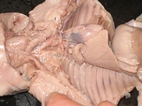
Fetal pigs are unborn pigs used in elementary as well as advanced
biologyBiology is the natural science concerned with the study of life and living organisms, including their structure, function, growth, origin, evolution, distribution, and taxonomy...
classes as objects for
dissectionDissection is usually the process of disassembling and observing something to determine its internal structure and as an aid to discerning the functions and relationships of its components...
.
PigPigs are a genus of even-toed ungulates within the family Suidae. The name hog most commonly refers to the domestic pig in everyday parlance, but technically encompasses several distinct species, including the wild boar...
s, as a mammalian
speciesIn biology, a species is:* a taxonomic rank or* a unit at that rank ....
, provide a good specimen for the study of physiological systems and processes.
Use in Biology Labs
Along with frogs and earthworms, fetal pigs are among the most common animals used in classroom dissection. There are several reasons for this, the biggest being that pigs, like humans, are mammals. Shared traits include common hair, mammary glands, live birth, similar organ systems, metabolic levels, and basic body form. They also allow for the study of
fetalA fetus is a developing mammal or other viviparous vertebrate after the embryonic stage and before birth. The plural is fetuses....
circulation, which differs from that of an adult. Secondly, fetal pigs are easy to obtain because they are by-products of the pork industry. Fetal pigs are the unborn piglets of sows that were killed by the meat packing industry. These pigs are not bred and killed for this purpose, but are extracted from the deceased sow’s uterus. Fetal pigs not used in classroom dissections are often used in fertilizer or simply discarded. Thirdly, fetal pigs are cheap, which is an essential component for dissection use by schools. They can be ordered for a little more than $25 at biological product companies. Fourthly, fetal pigs are easy to dissect because of their soft tissue and incompletely developed bones that are still made of cartilage. In addition, they are relatively large with well-developed organs that are easily visible. As long as the pork industry exists, fetal pigs will be relatively abundant, making them the prime choice for classroom dissections.
Development
The size of the fetal pig depends on the age it was allowed for the mother to
gestateGestation is the carrying of an embryo or fetus inside a female viviparous animal. Mammals duringpregnancy can have one or more gestations at the same time ....
:
| Size |
Time |
| 40 mm |
54 days |
| 80 mm |
68 days |
| 100 mm |
75 days |
| 158 mm |
86 days |
| 220 mm |
100 days |
| 300 mm |
114 days |
Nutrition
No studies have found significant data regarding the mother swine’s diet and fetal pig survival rate. However, there is a correlation between a mother pig having a nutritious diet containing proteins, vitamins and minerals during gestation period and the survival rate of piglets. The correlation, however, is not statistically different. Weight is also not a factor of survival rate due to the fact that a healthier diet does not lead to a heavier child or a greater chance of live birth. ..
Placental Development
The
placentaThe placenta is an organ unique to mammals that connects the developing fetus to the uterine wall. The placenta supplies the fetus with oxygen and food, and allows fetal waste to be disposed of via the maternal kidneys...
is used as a means of transferring nutrients from the mother to the fetus. The efficiency at which nutrients are transferred dictates the health and growth of the fetus. FRP, or fetal weight: placental weight ratio, was commonly used to determine placental efficiency. However, increasing FPR does not prove to increase litter size. Instead, a more accurate way of determining fetus growth is through certain characteristics of the placental lining. The placenta is made of a folded
trophoblastTrophoblasts are cells forming the outer layer of a blastocyst, which provide nutrients to the embryo and develop into a large part of the placenta...
/endometrial epithelial bilayer. The width and length of the placenta folds are positively related and increase as gestation progresses.
The width of the placental folds decreases until day 85 of gestation. From here, the width increases with gestation and is at its largest around day 105. The rate at which these folds increase is negatively related to fetus size. Thus, greater fold widths will be seen in smaller fetuses. Although increasing placental fold width does increase the interaction between fetus and mother, nutrient exchange is not most efficient in smaller fetal pigs, as would be expected. Many other factors, including depth of placental folds, are also responsible for these interactions.
Prenatal Development
The prenatal development of the fetus includes all the tissue and organ development. Within hours of mating, the sperm and egg undergo fertilization in the oviduct and three days later the egg moves into the uterus. The cells begin to specialize by day six, and attach themselves to the uterus lining by day eleven. From fertilization to day 18, the
endodermEndoderm, is one of the germ layers formed during animal embryogenesis. Cells migrating inward along the archenteron form the inner layer of the gastrula, which develops into the endoderm....
,
ectodermThe ectoderm is the start of a tissue that covers the body surfaces. It emerges first and forms from the outermost of the germ layers.Generally speaking, the ectoderm differentiates to form the nervous system, and the epidermis .In vertebrates, the ectoderm has three parts: external ectoderm , the ...
and
mesodermIn humans, the mesoderm is one of the three primary germ cell layers - the other two are the ectoderm and endoderm - in the very early embryo. The mesoderm is the middle layer. It differentiates to gives rise to a number of tissues and structures including bone, muscle, connective tissue, and the...
have been forming inside the embryo, and are completely formed by day 18, the same day the placenta forms. The
endodermEndoderm, is one of the germ layers formed during animal embryogenesis. Cells migrating inward along the archenteron form the inner layer of the gastrula, which develops into the endoderm....
transforms into the lungs, trachea, thyroid gland, and digestive tract of the fetus. The
ectodermThe ectoderm is the start of a tissue that covers the body surfaces. It emerges first and forms from the outermost of the germ layers.Generally speaking, the ectoderm differentiates to form the nervous system, and the epidermis .In vertebrates, the ectoderm has three parts: external ectoderm , the ...
has a greater role in the development of the fetus. It forms into the skin, nervous system, enamel of the teeth, lining of the intestine, mammary and sweat glands, hoofs, and hair. The
mesodermIn humans, the mesoderm is one of the three primary germ cell layers - the other two are the ectoderm and endoderm - in the very early embryo. The mesoderm is the middle layer. It differentiates to gives rise to a number of tissues and structures including bone, muscle, connective tissue, and the...
forms the major organ components that help keep the fetus alive. It forms the muscles and connective tissues of the body, blood vessels and cells, the skeleton, kidneys, adrenal glands, heart, and the reproductive organs. By day 20, most of the major organs are visible, and the last half of
gestationGestation is the carrying of an embryo or fetus inside a female viviparous animal. Mammals duringpregnancy can have one or more gestations at the same time ....
focuses greatly on increasing the size of the fetuses..
Development of Lymphoid and HaematopoiesisHaematopoiesis is the formation of blood cellular components. All cellular blood components are derived from haematopoietic stem cells...
tissues
The development of the lymphatic system and the formation of blood circulation occur at different stages of fetal pig development. The first lymphatic organ to become present is the thymus.
LymphocyteA lymphocyte is a type of white blood cell in the vertebrate immune system.By their appearance under the light microscope, there are two broad categories of lymphocytes, namely the large granular lymphocytes and the small lymphocytes. Functionally distinct subsets of lymphocytes correlate with...
builds up in the spleen on the 70th day. By day 77, the thymus is already completely developed and is distinguishable from other organs. Also, follicles are present on the tongue and intestines on day 77. On the 84th day,
Periarteriolar lymphoid sheathsPeriarteriolar lymphoid sheaths are a portion of the white pulp of the spleen. They are populated largely by T cells....
appear in the fetal pig. By this time, the liver and bone marrow are active and functional..
Environmental Effects on Swine Reproductive Performance
Studies have shown that litter size, the amount of floor space during the growing period, and the number of pigs the gilt, or female pig, is placed with while growing affect the reproduction rates of the gilts. Data from a study in 1976 by Nelson and Robinson showed that gilts from a small litter size ovulated more than the gilts from the larger litters. The study suggests stress plays a role in impacting the reproduction. The amount of floor space has been shown to impact the time it takes gilts to reach puberty. An adequate amount of floor space allowed the higher percentage of gilts to reach puberty sooner than those gilts who had less floor space. The gilts placed in smaller groups bore one more pig per litter than gilts in larger groups. Still, the environment in which the fetal gilt develops is significant to the reproductive and physiological development..
Preparation of Species =
Fetal pigs are often preserved in
formaldehydeFormaldehyde is a chemical compound with the formula CH2O. It is the simplest aldehyde. Formaldehyde also exists as the cyclic trimer trioxane and the polymer paraformaldehyde. It exists in water as the hydrate H2C2. Aqueous solutions of formaldehyde are referred...
, a carcinogenic substance. A 1980 study found that exposure to formaldehyde could possibly cause nasal cancer in rats, leading to research on whether this was possible in humans or not. . In 1995 it was concluded by the International Agency for Research on Cancer (IARC) that formaldehyde could be a probable carcinogen for humans. .
Your dissection kit most likely includes the following instruments:
~
Dissecting scissors—sharp straight points for cutting
~
Dissecting scalpel—sharp cutting edge for cutting
~
Dissecting student forceps—serrated tips for firm gripping action
~
Glass dropper with rubber bulb—for extracting liquids
~
Plastic Ruler—for measuring
~
Dissecting Mall probe—for separating and tracking veins, nerves, and organs from connective tissue. Angular point at one end only.
~
Dissecting teasing needle straight with metal chuck—for separating and manipulating tissue without tearing
~
Dissecting teasing needle angular with metal chuck—for separating and manipulating tissue without tearing
Choosing your fetal pig
If possible, choose the largest fetal pig available: It will be easier to see the organ systems because the pig will be further developed. The fetal pig is wrapped in a plastic bag containing formalin, which preserves the animal. Pour out excess liquid before placing the specimen on the dissection pan.
Tie specimen to dissecting pan
Place pig with its ventral side facing upwards (belly up, back on dissecting pan). Cut two pieces of rope about two feet each in length. Using the first rope, securely tie the first front leg, then bring rope underneath the dissecting pan and tie the other front leg. Repeat with the back legs using the second piece of rope. The specimen will have its back on the dissecting pan, with its legs spread open to make the dissection possible.
Helpful hints for a successful dissection
- Read all directions prior to beginning the dissection. Follow the directions.
- Ask your instructor if you do not understand the directions or how to follow the incision guidelines.
- Choose a designated reader and dissector at any given time. These roles can rotate so that everyone in the group gets a chance to participate. Wear gloves when handling any part of the pig.
- Use scissors for most incisions.
- Use the scalpel only when absolutely necessary. Be careful to not cut too deep, as this may puncture the organs.
Making the cut
Make an incision just above the umbilical cord. Feel inside with your finger for the diaphragm, and make a cut across the crest, anterior to the diaphragm. Extend the initial cut (above the umbilical cord) up to the chest (just under the neck) and across the top of the shoulders. Make a small incision caudal to the diaphragm and extend it laterally, and extend the cut to the base of the umbilical cord. Cut around the umbilical cord; do not cut the umbilical cord! Make two cuts just above the hind-limbs.
Anatomy of Fetal Pig =
The anatomy of a fetal pig is similar to that of the adult pig in various aspects. Systems that are similar include the nervous, skeletal, respiratory (neglecting the under developed diaphragm), and muscular. Other important body systems have significant differences from the adult pig that need to be addressed.
Circulatory System
Pigs, like all mammals, have a four chambered heart. Blood enters the right atrium via the superior and
inferior vena cavaThe inferior vena cava is the large vein that carries de-oxygenated blood from the lower half of the body into the right atrium of the heart....
. The blood is then pumped into the right ventricle from where it is pumped to the lungs to be oxygenated via the pulmonary arteries. Oxygen-rich blood is then pumped through the left atrium and into the left ventricle. Location of the fetal heart will show that the walls of the left ventricle are thicker than those of the other chambers. This is due to fact that the muscle of the left ventricle must be strong enough to pump oxygen-rich blood throughout the body.
The aortic arch of a fetal pig has two arteries attached to it, the
brachiocephalic arteryThe brachiocephalic artery is an artery of the mediastinum that supplies blood to the right arm and the head and neck....
and the
subclavian arteryIn human anatomy, the subclavian artery is a major artery of the upper thorax that mainly supplies blood to the head and arms. It is located below the clavicle, hence the name...
. As the
aortaThe aorta is the largest artery in the body, originating from the left ventricle of the heart and bringing oxygenated blood to all parts of the body in the systemic circulation....
descends, it splits into two large iliac arteries. An umbilical artery branches near the base of each iliac artery. The umbilical arteries run through the umbilical cord, carrying blood to the maternal placenta where it becomes oxygenated, nutrient-rich, and free of waste. This oxygenated, nutrient-rich blood is then returned to the liver of the fetus via the umbilical vein.
There are only a few differences between the circulatory system of an adult pig and a fetal pig, besides from the umbilical arteries and vein. There is a shunt between the wall of the right and left atrium called the
foramen ovaleThere are multiple structures in the human body with the name foramen ovale :* In the fetal heart, the foramen ovale is a shunt from the right atrium to left atrium....
. This allows blood to pass directly from the right to left atrium. There is also the ductus arterius which allows blood from the right atrium to be diverted to the aortic arch. Both of these shunts close a few minutes after birth.
Digestive
The
monogastricA monogastric organism has a simple single-chambered stomach, whereas ruminants have a four-chambered complex stomach. Examples of monogastric animals include humans, pigs, dogs, and cats....
digestive system of the fetal pig harbors many similarities with many other mammals. The fetal pig's digestive organs are well developed before birth, although it does not ingest food. These organs include the
esophagusThe esophagus or oesophagus , sometimes known as the gullet, is an organ in vertebrates which consists of a muscular tube through which food passes from the pharynx to the stomach. The word esophagus is derived from the Latin œsophagus, which derives from the Greek word oisophagos , lit...
,
stomachIn most mammals, the stomach is a hollow, muscular organ of the gastrointestinal tract , between the esophagus and the small intestine. It is involved in the second phase of digestion, following mastication . The word stomach is derived from the Latin stomachus, which derives from the Greek word...
, small and large intestines. Mesenteries serve to connect the organs of the fetal pig together. In order for digestion to occur, the fetal pig would have to ingest food. Instead, it gains much needed nutrition from the mother pig via the umbilical cord. In the adult pig, food will follow the general flow through the esophagus, which can be located behind the tracheae. From the oral cavity, the esophagus leads to the stomach, small intestine, and large intestine. Other organs developing during fetal pig development such as the
gallbladderThe gallbladder is a small non-vital organ that aids in the digestive process and stores bile produced in the liver.-Human anatomy:...
,
pancreasThe pancreas is a gland organ in the digestive and endocrine system of vertebrates. It is both an endocrine gland producing several important hormones, including insulin, glucagon, and somatostatin, as well as an exocrine gland, secreting pancreatic juice containing digestive enzymes that pass to...
and
spleenThe spleen is an organ found in virtually all vertebrate animals with important roles in regard to red blood cells and the immune system In humans, it is located in the left upper quadrant of the abdomen. It removes old red blood cells, holds a reserve in case of hemorrhagic shock, especially in...
are all critical in contributing to the overall flow of the digestive system. After being digested and absorbed, the food follows through the large intestine and is excreted through the rectum and anus. In the fetal pig however, the metabolic wastes are sent back to the mother through the umbilical cord where the mother excretes the wastes. Other remaining wastes remain in the fetal pig until birth.
The oral cavity of the fetal pig begins developing before birth. The tongue's taste buds, located in the enlarged papillae, facilitate food handling after birth. These taste buds develop during fetal development. Adult pigs have up to 15,000 taste buds, a much larger number than the average human tongue, which has 9,000.
http://faculty.washington.edu/chudler/amaze.html. The dental anatomy of the fetal pig shows differences from adult pigs. The fetal pig develops primary teeth (which are later replaced with permanent teeth). Some may erupt during fetal stage, which is why some of the pigs that are/will be dissected show evidence of teeth. Depending on the age of the fetal pig, it is natural to see eruptions of third
incisorIncisors are the first kind of tooth in heterodont mammals. They are located in the premaxilla above and mandible below.-Function:...
and canine in the fetal pig. Because the fetal pigs were still in the mother’s uterus, teeth will still form which supports reasons for hollow unerupted teeth that may be seen during the dissection. Similar to human dental anatomy, the overall dental anatomy of the pig consists of incisors, canines, pre-molars, and molars. Exploring the dental anatomy even further, piglets can have 28th teeth total and adult pigs can have 44 teeth total. If you would like to compare this to the
dental anatomyDental anatomy or anatomy of teeth is a field of anatomy dedicated to the study of human tooth structures. The development, appearance, and classification of teeth fall within its purvue. Tooth formation begins before birth, and teeth's eventual morphology is dictated during this time...
of a human, there are 20 primary teeth and 28-30 permanent teeth.
Urogenital
The fetal pig urogenital system is similar to the adult pig's system with the exception of the reproductive organs. The fetal pig urinary track is relatively developed and easy to locate during dissection. The kidneys are located behind the abdominal organs and are partially embedded into the dorsal body wall by the spine. The ureters carry the urine to the
urinary bladderIn anatomy, the urinary bladder is the organ that collects urine excreted by the kidneys prior to disposal by urination. A hollow muscular, and distensible organ, the bladder sits on the pelvic floor...
, the large sack-like organ by the umbilical artery and vein, to the
urethraIn anatomy, the urethra is a tube which connects the urinary bladder to the outside of the body. The urethra has an excretory function in both sexes to pass urine to the outside, and also a reproductive function in the male, as a passage for semen.The external urethral sphincter is a striated...
. From there, the urine can be excreted.
Female:
To externally determine if the fetal pig is a female, there will be a fleshy protrusion ventral near the anus called the
genital papillaThe genital papilla is a part of female external genitalia not present in humans, which apperars as a small, fleshy flab of tissue. The papilla covers the opening of the vagina....
. The female's internal reproductive system is located below the kidneys. The two sac-like organs attached to the coil-like fallopian tubes are the ovaries.. The uterus, which becomes the vagina, is located where the fallopian tubes meet. This system can be difficult to find as it is small as well as extremely dorsal and posterior to the other systems.
Male:
To externally determine if the fetal pig is male, look for the urogenital opening located behind the umbilical cord. Also note the swelling behind the hind legs of the fetal pig . This will be the
scrotumIn some male mammals the scrotum is a protuberance of skin and muscle containing the testicles. It is an extension of the abdomen, and is located between the penis and anus. In humans and some other mammals, the base of the scrotum becomes covered with curly pubic hairs at puberty...
. The male's internal reproductive system has two scrotal sacs, which depending on the age of the fetal pig may or may not have developed testes.. The
epididymisThe epididymis is part of the male reproductive system and is present in all male mammals. It is a narrow, tightly-coiled tube connecting the efferent ducts from the rear of each testicle to its vas deferens.-Regions:...
coil on the testes connects to the vas deferens. The vas deferens crosses over the ureter and enters the urethra, which then connects to the penis located just posterior to the skin. Similar to the female system, the male system may also be difficult to identify all parts. If the fetal pig is indeed male, take caution to not cut very deep into the scrotum when dissecting.