
Electrophysiology
Encyclopedia
Electrophysiology is the study of the electrical properties of biological cell
s and tissues. It involves measurements of voltage
change or electric current
on a wide variety of scales from single ion channel
protein
s to whole organs like the heart
. In neuroscience
, it includes measurements of the electrical activity of neurons, and particularly action potential
activity. Recordings of large-scale electric signals from the nervous system such as electroencephalography
, may also be referred to as electrophysiological recordings.
solution or another electrolyte solution. The principal preparations include 1) living organisms, 2) excised tissue (acute or cultured), 3) dissociated cells from excised tissue (acute or cultured), 4) artificially grown cells or tissues, or 5) hybrids of the above.
If an electrode is small enough (micrometers) in diameter, then the electro-physiologist may choose to insert the tip into a single cell. Such a configuration allows direct observation and recording of the intracellular electrical activity of a single cell. However, at the same time such invasive setup reduces the life of the cell and causes a leak of substances across the cell membrane. Intracellular activity may also be observed using a specially formed (hollow) glass pipette containing an electrolyte. In this technique, the microscopic pipette tip is pressed against the cell membrane, to which it tightly adheres by an interaction between glass and lipids of the cell membrane. The electrolyte within the pipette may be brought into fluid continuity with the cytoplasm by delivering a pulse of pressure to the electrolyte in order to rupture the small patch of membrane encircled by the pipette rim (whole-cell recording). Alternatively, ionic continuity may be established by "perforating" the patch by allowing exogenous pore-forming agent within the electrolyte to insert themselves into the membrane patch (perforated patch recording). Finally, the patch may be left intact (patch recording
).
The electrophysiologist may choose not to insert the tip into a single cell. Instead, the electrode tip may be left in continuity with the extracellular space. If the tip is small enough, such a configuration may allow indirect observation and recording of action potential
s from a single cell, and is termed single-unit recording
. Depending on the preparation and precise placement, an extracellular configuration may pick up the activity of several nearby cells simultaneously, and this is termed multi-unit recording.
As electrode size increases, the resolving power decreases. Larger electrodes are sensitive only to the net activity of many cells, termed local field potentials. Still larger electrodes, such as uninsulated needles and surface electrodes used by clinical and surgical neurophysiologists, are sensitive only to certain types of synchronous activity within populations of cells numbering in the millions.
Other classical electrophysiological techniques include single channel recording and amperometry
.
s and fluorescing proteins.
After introducing one or more such compounds into tissue via perfusion, injection or gene expression, the 1 or 2-dimensional distribution of electrical activity may be observed and recorded.
Many particular electrophysiological readings have specific names:
can be measured. Typically, the resting membrane potential of a healthy cell will be -60 to -80 mV, and during an action potential the membrane potential might reach +40 mV.
In 1963, Alan Lloyd Hodgkin
and Andrew Fielding Huxley won the Nobel Prize in Physiology or Medicine for their contribution to understanding the mechanisms underlying the generation of action potentials in neurons. Their experiments involved intracellular recordings from the giant axon
of Atlantic squid (Loligo pealei), and were among the first applications of the "voltage clamp" technique.
Today, most microelectrodes used for intracellular recording are glass micropipettes, with a tip diameter of < 1 micrometre, and a resistance of several megaohms. The micropipettes are filled with a solution that has a similar ionic composition to the intracellular fluid of the cell. A chlorided silver wire inserted in to the pipet connects the electrolyte electrically to the amplifier and signal processing circuit. The voltage measured by the electrode is compared to the voltage of a reference electrode, usually a silver chloride-coated silver wire in contact with the extracellular fluid around the cell. In general, the smaller the electrode tip, the higher its electrical resistance
, so an electrode is a compromise between size (small enough to penetrate a single cell with minimum damage to the cell) and resistance (low enough so that small neuronal signals can be discerned from thermal noise in the electrode tip).
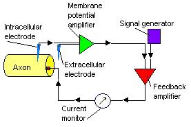 The voltage clamp technique allows an experimenter to "clamp" the cell potential at a chosen value. This makes it possible to measure how much ionic current crosses a cell's membrane at any given voltage. This is important because many of the ion channel
s in the membrane of a neuron are voltage-gated ion channel
s, which open only when the membrane voltage is within a certain range. Voltage clamp measurements of current are made possible by the near-simultaneous digital subtraction of transient capacitive currents that pass as the recording electrode and cell membrane are charged to alter the cell's potential.
by injecting current into a cell through the recording electrode. Unlike in the voltage clamp mode, where the membrane potential is held at a level determined by the experimenter, in "current clamp" mode the membrane potential is free to vary, and the amplifier records whatever voltage the cell generates on its own or as a result of stimulation. This technique is used to study how a cell responds when electric current enters a cell; this is important for instance for understanding how neurons respond to neurotransmitter
s that act by opening membrane ion channels.
Most current-clamp amplifiers provide little or no amplification of the voltage changes recorded from the cell. The "amplifier" is actually an electrometer
, sometimes referred to as a "unity gain amplifier"; its main job is to change the nature of small signals (in the mV range) produced by cells so that they can be accurately recorded by low-impedance
electronics. The amplifier increases the current behind the signal while decreasing the resistance over which that current passes. Consider this example based on Ohm's law: A voltage of 10 mV is generated by passing 10 nanoamperes of current across 1 MΩ of resistance. The electrometer changes this "high impedance signal" to a "low impedance signal" by using a voltage follower circuit. A voltage follower reads the voltage on the input (caused by a small current through a big resistor
). It then instructs a parallel circuit that has a large current source behind it (the electrical mains) and adjusts the resistance of that parallel circuit to give the same output voltage, but across a lower resistance.
This technique was developed by Erwin Neher
and Bert Sakmann
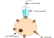
who received the Nobel Prize in 1991. Conventional intracellular recording involves impaling a cell with a fine electrode; patch-clamp recording takes a different approach. A patch-clamp microelectrode is a micropipette with a relatively large tip diameter. The microelectrode is placed next to a cell, and gentle suction is applied through the microelectrode to draw a piece of the cell membrane (the 'patch') into the microelectrode tip; the glass tip forms a high resistance 'seal' with the cell membrane. This configuration is the "cell-attached" mode, and it can be used for studying the activity of the ion channels that are present in the patch of membrane.
If more suction is now applied, the small patch of membrane in the electrode tip can be displaced, leaving the electrode sealed to the rest of the cell. This "whole-cell" mode allows very stable intracellular recording. A disadvantage (compared to conventional intracellular recording with sharp electrodes) is that the intracellular fluid of the cell mixes with the solution inside the recording electrode, and so some important components of the intracellular fluid can be diluted. A variant of this technique, the "perforated patch" technique, tries to minimise these problems.
Instead of applying suction to displace the membrane patch from the electrode tip, it is also possible to make small holes on the patch with pore-forming agents so that large molecules such as proteins can stay inside the cell and ions can pass through the holes freely. Also the patch of membrane can be pulled away from the rest of the cell. This approach enables the membrane properties of the patch to be analysed pharmacologically.
to fill the cells recorded from, for later confirmation of their morphology under a microscope. The dyes are injected by applying a positive or negative, DC or pulsed voltage to the electrodes depending on the polarity of the dye.
recorded the activity of single neurons in the primary visual cortex
of the anesthetized cat, and showed how single neurons in this area respond to very specific features of a visual stimulus. Hubel and Wiesel were awarded the Nobel Prize in Physiology or Medicine in 1981.
If the electrode tip is slightly larger, then the electrode might record the activity generated by several neurons. This type of recording is often called "multi-unit recording", and is often used in conscious animals to record changes in the activity in a discrete brain area during normal activity. Recordings from one or more such electrodes that are closely spaced can be used to identify the number of cells around it as well as which of the spikes come from which cell. This process is called spike sorting
and is suitable in areas where there are identified types of cells with well defined spike characteristics.
If the electrode tip is bigger still, in general the activity of individual neurons cannot be distinguished but the electrode will still be able to record a field potential generated by the activity of many cells.
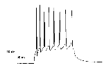
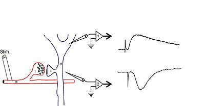 Extracellular field potential
s are local current sinks or sources that are generated by the collective activity of many cells. Usually, a field potential is generated by the simultaneous activation of many neurons by synaptic transmission. The diagram to the right shows hippocampal synaptic field potentials. At the right, the lower trace shows a negative wave that corresponds to a current sink caused by positive charges entering cells through postsynaptic glutamate receptor
s, while the upper trace shows a positive wave that is generated by the current that leaves the cell (at the cell body) to complete the circuit. For more information, see local field potential
.
uses a carbon electrode to record changes in the chemical composition of the oxidized components of a biological solution. Oxidation and reduction is accomplished by changing the voltage at the active surface of the recording electrode in a process known as "scanning". Because certain brain chemicals lose or gain electrons at characteristic voltages, individual species can be identified. Amperometry has been used for studying exocytosis in the neural and endocrine systems. Many monoamine neurotransmitters; e.g., norepinephrine
(noradrenalin), dopamine
, and serotonin
(5-HT) are oxidizable. The method can also be used with cells that do not secrete oxidizable neurotransmitters by "loading" them with 5-HT or dopamine.
Instead of positioning a pipette on an adherent cell, cell suspension is pipetted on a chip
containing a microstructured aperture.

A single cell is then positioned on the hole by suction and a tight connection (Gigaseal) is formed.
The planar geometry offers a variety of advantages compared to the classical experiment:
- it allows for integration of microfluidics
, which enables automatic compound application for ion channel
screening.
- the system is accessible for optical or scanning probe
techniques
- perfusion
of the intracellular
side can be performed.
s, membrane vesicle
s, or membrane fragments containing the channel or transporter of interest are adsorbed to a lipid monolayer painted over a functionalized electrode. This electrode consists of a glass support, a chromium
layer, a gold
layer, and an octadecyl mercaptane monolayer. Because the painted membrane is supported by the electrode, it is called a solid-supported membrane. It is important to note that mechanical perturbations, which usually destroy a biological lipid membrane, do not influence the life-time of an SSM. The capacitive
electrode (composed of the SSM and the absorbed vesicles) is so mechanically stable that solutions may be rapidly exchanged at its surface. This property allows the application of rapid substrate/ligand concentration jumps to investigate the electrogenic activity of the protein of interest, measured via capacitive coupling between the vesicles and the electrode.
by changing the cell membrane potential. In this way, when a positive sample is added to the sensor, a characteristic, ‘signature-like’ change in electrical potential occurs. BERA has been used for the detection for human viruses (Hepatitis B and C viruses, herpes viruses) and veterinary disease agents (foot and mouth disease virus, prions, blue tongue virus) and plants (tobacco and cucumber viruses) in a highly specific, rapid (1–2 minutes), reproducible and cost-efficient fashion. The method has also been used for the detection of environmental toxins, such as herbicides and the determination of very low concentrations of superoxide anion in clinical samples.
A recent advance in the evolution of the BERA technology is the development of a technique called molecular identification through membrane engineering (MIME). This technique allows for building cells with absolutely defined specificity for virtually any molecule of interest, by embedding thousand of artificial receptors into the cell membrane.
or reporting guidelines specify the minimum amount of meta data (information) and data required to meet a specific aims or aims. Usually the aim is to provide enough meta data and data to enable the unambiguous reproduction and interpretation of an experiment. MI guidelines are normally informal human readable specifications that inform the development of formal data models (e.g. XML
or UML
), data exchange formats (e.g. FuGE, MAGE-ML, MAGE-TAB) or knowledge models such as an ontology
(e.g. OBI
, MGED-Ontology).
The Minimum Information about a Neuroscience investigation (MINI) family of reporting guideline documents, produced by community consultation and continually available for public comment aims to provide a consistent set of guidelines in order to report an electrophysiology experiment. A MINI module represents the minimum information that should be reported about a dataset to facilitate computational access and analysis to allow a reader to interpret and critically evaluate the processes performed and the conclusions reached, and to support their experimental corroboration. In practice a MINI module comprises a checklist of information that should be provided (for example about the protocols employed) whena data set is described for publication. The full specification of the MINI module can be found here.
Cell (biology)
The cell is the basic structural and functional unit of all known living organisms. It is the smallest unit of life that is classified as a living thing, and is often called the building block of life. The Alberts text discusses how the "cellular building blocks" move to shape developing embryos....
s and tissues. It involves measurements of voltage
Voltage
Voltage, otherwise known as electrical potential difference or electric tension is the difference in electric potential between two points — or the difference in electric potential energy per unit charge between two points...
change or electric current
Electric current
Electric current is a flow of electric charge through a medium.This charge is typically carried by moving electrons in a conductor such as wire...
on a wide variety of scales from single ion channel
Ion channel
Ion channels are pore-forming proteins that help establish and control the small voltage gradient across the plasma membrane of cells by allowing the flow of ions down their electrochemical gradient. They are present in the membranes that surround all biological cells...
protein
Protein
Proteins are biochemical compounds consisting of one or more polypeptides typically folded into a globular or fibrous form, facilitating a biological function. A polypeptide is a single linear polymer chain of amino acids bonded together by peptide bonds between the carboxyl and amino groups of...
s to whole organs like the heart
Heart
The heart is a myogenic muscular organ found in all animals with a circulatory system , that is responsible for pumping blood throughout the blood vessels by repeated, rhythmic contractions...
. In neuroscience
Neuroscience
Neuroscience is the scientific study of the nervous system. Traditionally, neuroscience has been seen as a branch of biology. However, it is currently an interdisciplinary science that collaborates with other fields such as chemistry, computer science, engineering, linguistics, mathematics,...
, it includes measurements of the electrical activity of neurons, and particularly action potential
Action potential
In physiology, an action potential is a short-lasting event in which the electrical membrane potential of a cell rapidly rises and falls, following a consistent trajectory. Action potentials occur in several types of animal cells, called excitable cells, which include neurons, muscle cells, and...
activity. Recordings of large-scale electric signals from the nervous system such as electroencephalography
Electroencephalography
Electroencephalography is the recording of electrical activity along the scalp. EEG measures voltage fluctuations resulting from ionic current flows within the neurons of the brain...
, may also be referred to as electrophysiological recordings.
Classical electrophysiological techniques
Electrophysiology is the science and branch of physiology that pertains to the flow of ions in biological tissues and, in particular, to the electrical recording techniques that enable the measurement of this flow. Classical electrophysiology techniques involve placing electrodes into various preparations of biological tissue. The principal types of electrodes are: 1) simple solid conductors, such as discs and needles (singles or arrays, often insulated except for the tip), 2) tracings on printed circuit boards, also insulated except for the tip, and 3) hollow tubes filled with an electrolyte, such as glass pipettes filled with potassium chloridePotassium chloride
The chemical compound potassium chloride is a metal halide salt composed of potassium and chlorine. In its pure state, it is odorless and has a white or colorless vitreous crystal appearance, with a crystal structure that cleaves easily in three directions. Potassium chloride crystals are...
solution or another electrolyte solution. The principal preparations include 1) living organisms, 2) excised tissue (acute or cultured), 3) dissociated cells from excised tissue (acute or cultured), 4) artificially grown cells or tissues, or 5) hybrids of the above.
If an electrode is small enough (micrometers) in diameter, then the electro-physiologist may choose to insert the tip into a single cell. Such a configuration allows direct observation and recording of the intracellular electrical activity of a single cell. However, at the same time such invasive setup reduces the life of the cell and causes a leak of substances across the cell membrane. Intracellular activity may also be observed using a specially formed (hollow) glass pipette containing an electrolyte. In this technique, the microscopic pipette tip is pressed against the cell membrane, to which it tightly adheres by an interaction between glass and lipids of the cell membrane. The electrolyte within the pipette may be brought into fluid continuity with the cytoplasm by delivering a pulse of pressure to the electrolyte in order to rupture the small patch of membrane encircled by the pipette rim (whole-cell recording). Alternatively, ionic continuity may be established by "perforating" the patch by allowing exogenous pore-forming agent within the electrolyte to insert themselves into the membrane patch (perforated patch recording). Finally, the patch may be left intact (patch recording
Patch clamp
The patch clamp technique is a laboratory technique in electrophysiology that allows the study of single or multiple ion channels in cells. The technique can be applied to a wide variety of cells, but is especially useful in the study of excitable cells such as neurons, cardiomyocytes, muscle...
).
The electrophysiologist may choose not to insert the tip into a single cell. Instead, the electrode tip may be left in continuity with the extracellular space. If the tip is small enough, such a configuration may allow indirect observation and recording of action potential
Action potential
In physiology, an action potential is a short-lasting event in which the electrical membrane potential of a cell rapidly rises and falls, following a consistent trajectory. Action potentials occur in several types of animal cells, called excitable cells, which include neurons, muscle cells, and...
s from a single cell, and is termed single-unit recording
Single-unit recording
In neurophysiology and neurology, single-unit recording is the use of an electrode to record the electrophysiological activity from a single neuron.-History:...
. Depending on the preparation and precise placement, an extracellular configuration may pick up the activity of several nearby cells simultaneously, and this is termed multi-unit recording.
As electrode size increases, the resolving power decreases. Larger electrodes are sensitive only to the net activity of many cells, termed local field potentials. Still larger electrodes, such as uninsulated needles and surface electrodes used by clinical and surgical neurophysiologists, are sensitive only to certain types of synchronous activity within populations of cells numbering in the millions.
Other classical electrophysiological techniques include single channel recording and amperometry
Amperometry
Amperometry in chemistry and biochemistry is detection of ions in a solution based on electric current or changes in electric current.Amperometry is used in electrophysiology to study vesicle release events using a carbon fiber electrode...
.
Optical electrophysiological techniques
Optical electrophysiological techniques were created by scientists and engineers to overcome one of the main limitations of classical techniques. Classical techniques allow observation of electrical activity at approximately a single point within a volume of tissue. Essentially, classical techniques singularize a distributed phenomenon. Interest in the spatial distribution of bioelectric activity prompted development of molecules capable of emitting light in response to their electrical or chemical environment. Examples are voltage sensitive dyeVoltage sensitive dye
Voltage-sensitive dyes, also known as potentiometric dyes, are dyes which change their spectral properties in response to voltage changes. They are able to provide linear measurements of firing activity of single neurons, large neuronal populations or activity of myocytes...
s and fluorescing proteins.
After introducing one or more such compounds into tissue via perfusion, injection or gene expression, the 1 or 2-dimensional distribution of electrical activity may be observed and recorded.
Many particular electrophysiological readings have specific names:
- Electrocardiography - for the heartHuman heartThe human heart is a muscular organ that provides a continuous blood circulation through the cardiac cycle and is one of the most vital organs in the human body...
- ElectroencephalographyElectroencephalographyElectroencephalography is the recording of electrical activity along the scalp. EEG measures voltage fluctuations resulting from ionic current flows within the neurons of the brain...
- for the brainHuman brainThe human brain has the same general structure as the brains of other mammals, but is over three times larger than the brain of a typical mammal with an equivalent body size. Estimates for the number of neurons in the human brain range from 80 to 120 billion... - ElectrocorticographyElectrocorticographyElectrocorticography is the practice of using electrodes placed directly on the exposed surface of the brain to record electrical activity from the cerebral cortex. ECoG may be performed either in the operating room during surgery or outside of surgery...
- from the cerebral cortexCerebral cortexThe cerebral cortex is a sheet of neural tissue that is outermost to the cerebrum of the mammalian brain. It plays a key role in memory, attention, perceptual awareness, thought, language, and consciousness. It is constituted of up to six horizontal layers, each of which has a different... - ElectromyographyElectromyographyElectromyography is a technique for evaluating and recording the electrical activity produced by skeletal muscles. EMG is performed using an instrument called an electromyograph, to produce a record called an electromyogram. An electromyograph detects the electrical potential generated by muscle...
- for the muscleMuscleMuscle is a contractile tissue of animals and is derived from the mesodermal layer of embryonic germ cells. Muscle cells contain contractile filaments that move past each other and change the size of the cell. They are classified as skeletal, cardiac, or smooth muscles. Their function is to...
s - ElectrooculographyElectrooculographyElectrooculography is a technique for measuring the resting potential of the retina. The resulting signal is called the electrooculogram. The main applications are in ophthalmological diagnosis and in recording eye movements...
- for the eyeHuman eyeThe human eye is an organ which reacts to light for several purposes. As a conscious sense organ, the eye allows vision. Rod and cone cells in the retina allow conscious light perception and vision including color differentiation and the perception of depth...
s - ElectroretinographyElectroretinographyElectroretinography measures the electrical responses of various cell types in the retina, including the photoreceptors , inner retinal cells , and the ganglion cells. Electrodes are usually placed on the cornea and the skin near the eye, although it is possible to record the ERG from skin electrodes...
- for the retinaRetinaThe vertebrate retina is a light-sensitive tissue lining the inner surface of the eye. The optics of the eye create an image of the visual world on the retina, which serves much the same function as the film in a camera. Light striking the retina initiates a cascade of chemical and electrical... - ElectroantennographyElectroantennographyElectroantennogram or EAG is a technique by which we measure the average output of the antenna to the brain for a given odor. It is commonly used in the electrophysiology while studying the function of olfactory pathway in insects...
- for the olfactory receptors in arthropods - AudiologyAudiologyAudiology is the branch of science that studies hearing, balance, and related disorders. Its practitioners, who treat those with hearing loss and proactively prevent related damage are audiologists. Employing various testing strategies Audiology (from Latin , "to hear"; and from Greek , -logia) is...
- for the auditory systemAuditory systemThe auditory system is the sensory system for the sense of hearing.- Outer ear :The folds of cartilage surrounding the ear canal are called the pinna...
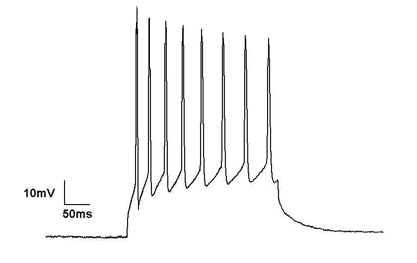
Intracellular recording
Intracellular recording involves measuring voltage and/or current across the membrane of a cell. To make an intracellular recording, the tip of a fine (sharp) microelectrode must be inserted inside the cell, so that the membrane potentialMembrane potential
Membrane potential is the difference in electrical potential between the interior and exterior of a biological cell. All animal cells are surrounded by a plasma membrane composed of a lipid bilayer with a variety of types of proteins embedded in it...
can be measured. Typically, the resting membrane potential of a healthy cell will be -60 to -80 mV, and during an action potential the membrane potential might reach +40 mV.
In 1963, Alan Lloyd Hodgkin
Alan Lloyd Hodgkin
Sir Alan Lloyd Hodgkin, OM, KBE, PRS was a British physiologist and biophysicist, who shared the 1963 Nobel Prize in Physiology or Medicine with Andrew Huxley and John Eccles....
and Andrew Fielding Huxley won the Nobel Prize in Physiology or Medicine for their contribution to understanding the mechanisms underlying the generation of action potentials in neurons. Their experiments involved intracellular recordings from the giant axon
Squid giant axon
The squid giant axon is the very large axon that controls part of the water jet propulsion system in squid. It was discovered by English zoologist and neurophysiologist John Zachary Young in 1936...
of Atlantic squid (Loligo pealei), and were among the first applications of the "voltage clamp" technique.
Today, most microelectrodes used for intracellular recording are glass micropipettes, with a tip diameter of < 1 micrometre, and a resistance of several megaohms. The micropipettes are filled with a solution that has a similar ionic composition to the intracellular fluid of the cell. A chlorided silver wire inserted in to the pipet connects the electrolyte electrically to the amplifier and signal processing circuit. The voltage measured by the electrode is compared to the voltage of a reference electrode, usually a silver chloride-coated silver wire in contact with the extracellular fluid around the cell. In general, the smaller the electrode tip, the higher its electrical resistance
Electrical resistance
The electrical resistance of an electrical element is the opposition to the passage of an electric current through that element; the inverse quantity is electrical conductance, the ease at which an electric current passes. Electrical resistance shares some conceptual parallels with the mechanical...
, so an electrode is a compromise between size (small enough to penetrate a single cell with minimum damage to the cell) and resistance (low enough so that small neuronal signals can be discerned from thermal noise in the electrode tip).
Voltage clamp
Ion channel
Ion channels are pore-forming proteins that help establish and control the small voltage gradient across the plasma membrane of cells by allowing the flow of ions down their electrochemical gradient. They are present in the membranes that surround all biological cells...
s in the membrane of a neuron are voltage-gated ion channel
Voltage-gated ion channel
Voltage-gated ion channels are a class of transmembrane ion channels that are activated by changes in electrical potential difference near the channel; these types of ion channels are especially critical in neurons, but are common in many types of cells....
s, which open only when the membrane voltage is within a certain range. Voltage clamp measurements of current are made possible by the near-simultaneous digital subtraction of transient capacitive currents that pass as the recording electrode and cell membrane are charged to alter the cell's potential.
Current clamp
The current clamp technique records the membrane potentialMembrane potential
Membrane potential is the difference in electrical potential between the interior and exterior of a biological cell. All animal cells are surrounded by a plasma membrane composed of a lipid bilayer with a variety of types of proteins embedded in it...
by injecting current into a cell through the recording electrode. Unlike in the voltage clamp mode, where the membrane potential is held at a level determined by the experimenter, in "current clamp" mode the membrane potential is free to vary, and the amplifier records whatever voltage the cell generates on its own or as a result of stimulation. This technique is used to study how a cell responds when electric current enters a cell; this is important for instance for understanding how neurons respond to neurotransmitter
Neurotransmitter
Neurotransmitters are endogenous chemicals that transmit signals from a neuron to a target cell across a synapse. Neurotransmitters are packaged into synaptic vesicles clustered beneath the membrane on the presynaptic side of a synapse, and are released into the synaptic cleft, where they bind to...
s that act by opening membrane ion channels.
Most current-clamp amplifiers provide little or no amplification of the voltage changes recorded from the cell. The "amplifier" is actually an electrometer
Electrometer
An electrometer is an electrical instrument for measuring electric charge or electrical potential difference. There are many different types, ranging from historical hand-made mechanical instruments to high-precision electronic devices...
, sometimes referred to as a "unity gain amplifier"; its main job is to change the nature of small signals (in the mV range) produced by cells so that they can be accurately recorded by low-impedance
Electrical impedance
Electrical impedance, or simply impedance, is the measure of the opposition that an electrical circuit presents to the passage of a current when a voltage is applied. In quantitative terms, it is the complex ratio of the voltage to the current in an alternating current circuit...
electronics. The amplifier increases the current behind the signal while decreasing the resistance over which that current passes. Consider this example based on Ohm's law: A voltage of 10 mV is generated by passing 10 nanoamperes of current across 1 MΩ of resistance. The electrometer changes this "high impedance signal" to a "low impedance signal" by using a voltage follower circuit. A voltage follower reads the voltage on the input (caused by a small current through a big resistor
Resistor
A linear resistor is a linear, passive two-terminal electrical component that implements electrical resistance as a circuit element.The current through a resistor is in direct proportion to the voltage across the resistor's terminals. Thus, the ratio of the voltage applied across a resistor's...
). It then instructs a parallel circuit that has a large current source behind it (the electrical mains) and adjusts the resistance of that parallel circuit to give the same output voltage, but across a lower resistance.
The patch-clamp technique
This technique was developed by Erwin Neher
Erwin Neher
Erwin Neher is a German biophysicist.Erwin Neher studied physics at the Technical University of Munich from 1963 to 1966. In 1966, He was awarded a Fulbright Scholarship to study in the US...
and Bert Sakmann
Bert Sakmann
-External links:*...
who received the Nobel Prize in 1991. Conventional intracellular recording involves impaling a cell with a fine electrode; patch-clamp recording takes a different approach. A patch-clamp microelectrode is a micropipette with a relatively large tip diameter. The microelectrode is placed next to a cell, and gentle suction is applied through the microelectrode to draw a piece of the cell membrane (the 'patch') into the microelectrode tip; the glass tip forms a high resistance 'seal' with the cell membrane. This configuration is the "cell-attached" mode, and it can be used for studying the activity of the ion channels that are present in the patch of membrane.
If more suction is now applied, the small patch of membrane in the electrode tip can be displaced, leaving the electrode sealed to the rest of the cell. This "whole-cell" mode allows very stable intracellular recording. A disadvantage (compared to conventional intracellular recording with sharp electrodes) is that the intracellular fluid of the cell mixes with the solution inside the recording electrode, and so some important components of the intracellular fluid can be diluted. A variant of this technique, the "perforated patch" technique, tries to minimise these problems.
Instead of applying suction to displace the membrane patch from the electrode tip, it is also possible to make small holes on the patch with pore-forming agents so that large molecules such as proteins can stay inside the cell and ions can pass through the holes freely. Also the patch of membrane can be pulled away from the rest of the cell. This approach enables the membrane properties of the patch to be analysed pharmacologically.
Sharp electrode technique
In situations where one wants to record the potential inside the cell membrane with minimal effect on the ionic constitution of the intracellular fluid a sharp electrode can be used. These micropipettes (electrodes) are again like those for patch clamp pulled from glass capillaries, but the pore is much smaller so that there is very little ion exchange between the intracellular fluid and the electrolyte in the pipette. The resistance of the micropipette electrode is tens or hundreds of MΩ. Often the tip of the electrode is filled with various kinds of dyes like Lucifer yellowLucifer yellow
Lucifer yellow is a fluorescent dye used in cell biology. The key property of Lucifer yellow is that it can readily visualized in both living and fixed cells using a fluorescence microscope. Lucifer yellow was engineered by Walter W. Stewart at NIH and patented in 1978.- Preparations :For common...
to fill the cells recorded from, for later confirmation of their morphology under a microscope. The dyes are injected by applying a positive or negative, DC or pulsed voltage to the electrodes depending on the polarity of the dye.
Single-unit recording
An electrode introduced into the brain of a living animal will detect electrical activity that is generated by the neurons adjacent to the electrode tip. If the electrode is a microelectrode, with a tip size of about 1 micrometre, the electrode will usually detect the activity of at most one neuron. Recording in this way is in general called "single-unit" recording. The action potentials recorded are very much like the action potentials that are recorded intracellularly, but the signals are very much smaller (typically about 1 mV). Most recordings of the activity of single neurons in anesthetized animals are made in this way, and all recordings of single neurons in conscious animals. Recordings of single neurons in living animals have provided important insights into how the brain processes information. For example, David Hubel and Torsten WieselTorsten Wiesel
Torsten Nils Wiesel was a Swedish co-recipient with David H. Hubel of the 1981 Nobel Prize in Physiology or Medicine, for their discoveries concerning information processing in the visual system; the prize was shared with Roger W...
recorded the activity of single neurons in the primary visual cortex
Visual cortex
The visual cortex of the brain is the part of the cerebral cortex responsible for processing visual information. It is located in the occipital lobe, in the back of the brain....
of the anesthetized cat, and showed how single neurons in this area respond to very specific features of a visual stimulus. Hubel and Wiesel were awarded the Nobel Prize in Physiology or Medicine in 1981.
If the electrode tip is slightly larger, then the electrode might record the activity generated by several neurons. This type of recording is often called "multi-unit recording", and is often used in conscious animals to record changes in the activity in a discrete brain area during normal activity. Recordings from one or more such electrodes that are closely spaced can be used to identify the number of cells around it as well as which of the spikes come from which cell. This process is called spike sorting
Spike sorting
Spike sorting is a class of techniques used in the analysis of electrophysiological data. Spike sorting algorithms use the shape of waveforms collected with one or more electrodes in the brain to distinguish the activity of one or more neurons from background electrical noise.Neurons produce action...
and is suitable in areas where there are identified types of cells with well defined spike characteristics.
If the electrode tip is bigger still, in general the activity of individual neurons cannot be distinguished but the electrode will still be able to record a field potential generated by the activity of many cells.
Field potentials
Extracellular field potential
The extracellular field potential is the electrical potential produced by cells, e.g. nerve or muscle cells, outside of the cell. Electrophysiological studies investigate these potentials using extracellular microelectrodes...
s are local current sinks or sources that are generated by the collective activity of many cells. Usually, a field potential is generated by the simultaneous activation of many neurons by synaptic transmission. The diagram to the right shows hippocampal synaptic field potentials. At the right, the lower trace shows a negative wave that corresponds to a current sink caused by positive charges entering cells through postsynaptic glutamate receptor
Glutamate receptor
Glutamate receptors are synaptic receptors located primarily on the membranes of neuronal cells. Glutamate is one of the 20 amino acids used to assemble proteins and as a result is abundant in many areas of the body, but it also functions as a neurotransmitter and is particularly abundant in the...
s, while the upper trace shows a positive wave that is generated by the current that leaves the cell (at the cell body) to complete the circuit. For more information, see local field potential
Local field potential
A local field potential is a particular class of electrophysiological signals, which is dominated by the electrical current flowing from all nearby dendritic synaptic activity within a volume of tissue. A voltage is produced by the summed synaptic current flowing across the resistance of the local...
.
Amperometry
AmperometryAmperometry
Amperometry in chemistry and biochemistry is detection of ions in a solution based on electric current or changes in electric current.Amperometry is used in electrophysiology to study vesicle release events using a carbon fiber electrode...
uses a carbon electrode to record changes in the chemical composition of the oxidized components of a biological solution. Oxidation and reduction is accomplished by changing the voltage at the active surface of the recording electrode in a process known as "scanning". Because certain brain chemicals lose or gain electrons at characteristic voltages, individual species can be identified. Amperometry has been used for studying exocytosis in the neural and endocrine systems. Many monoamine neurotransmitters; e.g., norepinephrine
Norepinephrine
Norepinephrine is the US name for noradrenaline , a catecholamine with multiple roles including as a hormone and a neurotransmitter...
(noradrenalin), dopamine
Dopamine
Dopamine is a catecholamine neurotransmitter present in a wide variety of animals, including both vertebrates and invertebrates. In the brain, this substituted phenethylamine functions as a neurotransmitter, activating the five known types of dopamine receptors—D1, D2, D3, D4, and D5—and their...
, and serotonin
Serotonin
Serotonin or 5-hydroxytryptamine is a monoamine neurotransmitter. Biochemically derived from tryptophan, serotonin is primarily found in the gastrointestinal tract, platelets, and in the central nervous system of animals including humans...
(5-HT) are oxidizable. The method can also be used with cells that do not secrete oxidizable neurotransmitters by "loading" them with 5-HT or dopamine.
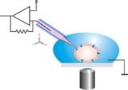
Planar patch clamp
Planar patch clamp is a novel method developed for high throughput electrophysiology.Instead of positioning a pipette on an adherent cell, cell suspension is pipetted on a chip
Biochip
The development of biochips is a major thrust of the rapidly growing biotechnology industry, which encompasses a very diverse range ofresearch efforts including genomics, proteomics, and pharmaceuticals, among other activities...
containing a microstructured aperture.
A single cell is then positioned on the hole by suction and a tight connection (Gigaseal) is formed.
The planar geometry offers a variety of advantages compared to the classical experiment:
- it allows for integration of microfluidics
Microfluidics
Microfluidics deals with the behavior, precise control and manipulation of fluids that are geometrically constrained to a small, typically sub-millimeter, scale.Typically, micro means one of the following features:* small volumes...
, which enables automatic compound application for ion channel
Ion channel
Ion channels are pore-forming proteins that help establish and control the small voltage gradient across the plasma membrane of cells by allowing the flow of ions down their electrochemical gradient. They are present in the membranes that surround all biological cells...
screening.
- the system is accessible for optical or scanning probe
Scanning probe microscopy
Scanning Probe Microscopy is a branch of microscopy that forms images of surfaces using a physical probe that scans the specimen. An image of the surface is obtained by mechanically moving the probe in a raster scan of the specimen, line by line, and recording the probe-surface interaction as a...
techniques
- perfusion
Perfusion
In physiology, perfusion is the process of nutritive delivery of arterial blood to a capillary bed in the biological tissue. The word is derived from the French verb "perfuser" meaning to "pour over or through."...
of the intracellular
Intracellular
Not to be confused with intercellular, meaning "between cells".In cell biology, molecular biology and related fields, the word intracellular means "inside the cell".It is used in contrast to extracellular...
side can be performed.
Solid-supported membrane (SSM) based
With this electrophysiological approach, proteoliposomeLiposome
Liposomes are artificially prepared vesicles made of lipid bilayer. Liposomes can be filled with drugs, and used to deliver drugs for cancer and other diseases. Liposomes are composite structures made of phospholipids and may contain small amounts of other molecules...
s, membrane vesicle
Vesicle (biology)
A vesicle is a bubble of liquid within another liquid, a supramolecular assembly made up of many different molecules. More technically, a vesicle is a small membrane-enclosed sack that can store or transport substances. Vesicles can form naturally because of the properties of lipid membranes , or...
s, or membrane fragments containing the channel or transporter of interest are adsorbed to a lipid monolayer painted over a functionalized electrode. This electrode consists of a glass support, a chromium
Chromium
Chromium is a chemical element which has the symbol Cr and atomic number 24. It is the first element in Group 6. It is a steely-gray, lustrous, hard metal that takes a high polish and has a high melting point. It is also odorless, tasteless, and malleable...
layer, a gold
Gold
Gold is a chemical element with the symbol Au and an atomic number of 79. Gold is a dense, soft, shiny, malleable and ductile metal. Pure gold has a bright yellow color and luster traditionally considered attractive, which it maintains without oxidizing in air or water. Chemically, gold is a...
layer, and an octadecyl mercaptane monolayer. Because the painted membrane is supported by the electrode, it is called a solid-supported membrane. It is important to note that mechanical perturbations, which usually destroy a biological lipid membrane, do not influence the life-time of an SSM. The capacitive
Capacitance
In electromagnetism and electronics, capacitance is the ability of a capacitor to store energy in an electric field. Capacitance is also a measure of the amount of electric potential energy stored for a given electric potential. A common form of energy storage device is a parallel-plate capacitor...
electrode (composed of the SSM and the absorbed vesicles) is so mechanically stable that solutions may be rapidly exchanged at its surface. This property allows the application of rapid substrate/ligand concentration jumps to investigate the electrogenic activity of the protein of interest, measured via capacitive coupling between the vesicles and the electrode.
Bioelectric recognition assay (BERA)
The bioelectric recognition assay (BERA) is a novel method for determination of various chemical and biological molecules by measuring changes in the membrane potential of cells immobilized in a gel matrix. Apart from the increased stability of the electrode-cell interface, immobilization preserves the viability and physiological functions of the cells. BERA is used primarily in biosensor applications in order to assay analytes that can interact with the immobilized cellsImmobilized whole cell
The immobilized whole cell system is an alternative to enzyme immobilization. Unlike enzyme immobilization, where the enzyme is attached to a substrate , in immobilized whole cell systems, the target cell is immobilized. Such methods may be implemented when the enzymes required are difficult or...
by changing the cell membrane potential. In this way, when a positive sample is added to the sensor, a characteristic, ‘signature-like’ change in electrical potential occurs. BERA has been used for the detection for human viruses (Hepatitis B and C viruses, herpes viruses) and veterinary disease agents (foot and mouth disease virus, prions, blue tongue virus) and plants (tobacco and cucumber viruses) in a highly specific, rapid (1–2 minutes), reproducible and cost-efficient fashion. The method has also been used for the detection of environmental toxins, such as herbicides and the determination of very low concentrations of superoxide anion in clinical samples.
A recent advance in the evolution of the BERA technology is the development of a technique called molecular identification through membrane engineering (MIME). This technique allows for building cells with absolutely defined specificity for virtually any molecule of interest, by embedding thousand of artificial receptors into the cell membrane.
Reporting guidelines for electrophysiology experiments
Minimum Information (MI) standardsMinimum Information Standards
Minimum Information standards or reporting guidelines specify the minimum amount of meta data and data required to meet a specific aim or aims. Usually the aim is to provide enough meta data and data to enable the unambiguous reproduction and interpretation of an experiment...
or reporting guidelines specify the minimum amount of meta data (information) and data required to meet a specific aims or aims. Usually the aim is to provide enough meta data and data to enable the unambiguous reproduction and interpretation of an experiment. MI guidelines are normally informal human readable specifications that inform the development of formal data models (e.g. XML
XML
Extensible Markup Language is a set of rules for encoding documents in machine-readable form. It is defined in the XML 1.0 Specification produced by the W3C, and several other related specifications, all gratis open standards....
or UML
Unified Modeling Language
Unified Modeling Language is a standardized general-purpose modeling language in the field of object-oriented software engineering. The standard is managed, and was created, by the Object Management Group...
), data exchange formats (e.g. FuGE, MAGE-ML, MAGE-TAB) or knowledge models such as an ontology
Ontology
Ontology is the philosophical study of the nature of being, existence or reality as such, as well as the basic categories of being and their relations...
(e.g. OBI
Ontology for Biomedical Investigations
The Ontology for Biomedical Investigations is an open access, integrated ontology for the description of biological and clinical investigations. OBI provides a model for the design of an investigation, the protocols and instrumentation used, the materials used, the data generated and the type of...
, MGED-Ontology).
The Minimum Information about a Neuroscience investigation (MINI) family of reporting guideline documents, produced by community consultation and continually available for public comment aims to provide a consistent set of guidelines in order to report an electrophysiology experiment. A MINI module represents the minimum information that should be reported about a dataset to facilitate computational access and analysis to allow a reader to interpret and critically evaluate the processes performed and the conclusions reached, and to support their experimental corroboration. In practice a MINI module comprises a checklist of information that should be provided (for example about the protocols employed) whena data set is described for publication. The full specification of the MINI module can be found here.
See also
- Cardiac electrophysiologyCardiac electrophysiologyCardiac electrophysiology is the science of elucidating, diagnosing, and treating the electrical activities of the heart. The term is usually used to describe studies of such phenomena by invasive catheter recording of spontaneous activity as well as of cardiac responses to programmed electrical...
- Clinical cardiac electrophysiologyClinical cardiac electrophysiologyCardiac Electrophysiology , is a branch of the medical specialty of clinical cardiology and is concerned with the study and treatment of rhythm disorders of the heart. Cardiologists with expertise in this area are usually referred to as electrophysiologists...
- Clinical electrophysiologyClinical electrophysiologyClinical electrophysiology is the application of electrophysiology principles to medicine. The two main branches of this discipline are electrotherapy and electrophysiologic testing...
- Nathaniel A. BuchwaldNathaniel A. BuchwaldNathaniel A. Buchwald was an American neuroscientist, educator and administrator, who was Professor of Psychiatry and Biobehavioral Sciences and Neurobiology at the University of California, Los Angeles...
(1924–2006) - Duchenne de Boulogne (1806–1875)
- The Body ElectricThe Body ElectricThe Body Electric: Electromagnetism and the Foundation of Life is a book by Robert O. Becker and Gary Selden in which Becker, an orthopedic surgeon at the time working for the Veterans Administration, describes his research into "our bioelectric selves"....
- Transcutaneous electrical nerve stimulationTranscutaneous electrical nerve stimulationTranscutaneous electrical nerve stimulation is the use of electric current produced by a device to stimulate the nerves for therapeutic purposes...
- Electrophysiology studyElectrophysiology studyAn electrophysiology study is a minimally invasive procedure which tests the electrical conduction system of the heart to assess the electrical activity and conduction pathways of the heart. The study is indicated to investigate the cause, location of origin, and best treatment for various...
- Multiscale Electrophysiology FormatMultiscale Electrophysiology FormatMultiscale Electrophysiology Format was developed to handle the large amounts of data produced by large-scale electrophysiology in human and animal subjects. MEF can store any time series data up to 24 bits in length, and employs lossless range encoded difference compression...
- Slice preparationSlice preparationThe slice preparation or brain slice is a laboratory technique in electrophysiology that allows the study of a synapse or neural circuit in isolation from the rest of the brain, in controlled physiological conditions. It involves stimulating and/or recording from a slice of brain tissue immersed in...