
Digastric muscle
Encyclopedia
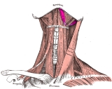
The digastric muscle (named digastric as it has two bellies) is a small muscle
located under the jaw
. so digastric muscles are muscle fibers in ligament of treitz ,omohyoid , occipitofrontalis.
It lies below the body of the mandible, and extends, in a curved form, from the mastoid process
to the symphysis menti
. It belongs to the suprahyoid muscles
group.
A broad aponeurotic layer is given off from the tendon of the digastricus on either side, to be attached to the body and greater cornu
of the hyoid bone
; this is termed the suprahyoid aponeurosis.
The two bellies of the digastric muscle have different embryological
origins, and are supplied by different cranial nerves.
of the temporal bone
and a deep groove between the mastoid process and the styloid process
called the digastric groove.
The posterior belly is supplied by the digastric branch of facial nerve
.
The digastric muscle stretches between the mastoid process of the cranium to the mandible at the chin, and part-way between, it becomes a tendon which passes through a tendinous pulley attached to the hyoid bone. It originates from the second pharyngeal arch.
, and passes downward and backward.
The anterior body is supplied by the trigeminal
via the mylohyoid nerve
, a branch of the inferior alveolar nerve
, itself a branch of the mandibular division of the trigeminal nerve
. It originates from the first pharyngeal arch.
which perforates the Stylohyoideus muscle, and is held in connection with the side of the body and the greater cornu
of the hyoid bone
by a fibrous loop, which is sometimes lined by a mucous sheath.
.
If the hyoid is being held in place (by the infrahyoid muscles), it will tend to depress the mandible (open the mouth).
The posterior belly may arise partly or entirely from the styloid process, or be connected by a slip to the middle or inferior constrictor; the anterior belly may be double or extra slips from this belly may pass to the jaw or Mylohyoideus or decussate with a similar slip on opposite side; anterior belly may be absent and posterior belly inserted into the middle of the jaw or hyoid bone.
The tendon may pass in front, more rarely behind the Stylohoideus. The Mentohyoideus muscle passes from the body of hyoid bone to chin.

into three smaller triangles.
Muscle
Muscle is a contractile tissue of animals and is derived from the mesodermal layer of embryonic germ cells. Muscle cells contain contractile filaments that move past each other and change the size of the cell. They are classified as skeletal, cardiac, or smooth muscles. Their function is to...
located under the jaw
Jaw
The jaw is any opposable articulated structure at the entrance of the mouth, typically used for grasping and manipulating food. The term jaws is also broadly applied to the whole of the structures constituting the vault of the mouth and serving to open and close it and is part of the body plan of...
. so digastric muscles are muscle fibers in ligament of treitz ,omohyoid , occipitofrontalis.
It lies below the body of the mandible, and extends, in a curved form, from the mastoid process
Mastoid process
The mastoid process is a conical prominence projecting from the undersurface of the mastoid portion of the temporal bone. It is located just behind the external acoustic meatus, and lateral to the styloid process...
to the symphysis menti
Symphysis menti
The external surface of the mandible is marked in the median line by a faint ridge, indicating the symphysis menti, mandibular symphysis, or line of junction of the two pieces of which the bone is composed at an early period of life....
. It belongs to the suprahyoid muscles
Suprahyoid muscles
The term suprahyoid refers to the region above the hyoid bone in the neck. The suprahyoid muscles include digastric, stylohyoid, geniohyoid, and mylohyoid...
group.
A broad aponeurotic layer is given off from the tendon of the digastricus on either side, to be attached to the body and greater cornu
Greater cornu
The greater cornua of the hyoid bone project backward from the lateral borders of the body; they are flattened from above downward and diminish in size from before backward; each ends in a tubercle to which is fixed the lateral hyothyroid ligament.The upper surface is rough close to its lateral...
of the hyoid bone
Hyoid bone
The hyoid bone is a horseshoe-shaped bone situated in the anterior midline of the neck between the chin and the thyroid cartilage. At rest, it lies at the level of the base of the mandible in the front and the third cervical vertebra behind.Unlike other bones, the hyoid is only distantly...
; this is termed the suprahyoid aponeurosis.
Structure
The digastricus (digastric muscle) consists of two fleshy bellies united by an intermediate rounded tendon.The two bellies of the digastric muscle have different embryological
Embryology
Embryology is a science which is about the development of an embryo from the fertilization of the ovum to the fetus stage...
origins, and are supplied by different cranial nerves.
Posterior belly
The posterior belly, longer than the anterior belly, arises on the inferior surface of the skull, from the mastoid notch on the medial surface of the mastoid processMastoid process
The mastoid process is a conical prominence projecting from the undersurface of the mastoid portion of the temporal bone. It is located just behind the external acoustic meatus, and lateral to the styloid process...
of the temporal bone
Temporal bone
The temporal bones are situated at the sides and base of the skull, and lateral to the temporal lobes of the cerebrum.The temporal bone supports that part of the face known as the temple.-Parts:The temporal bone consists of four parts:* Squama temporalis...
and a deep groove between the mastoid process and the styloid process
Styloid process (temporal)
The styloid process is a pointed piece of bone that extends down from the human skull, just below the ear.-Structure:The styloid process is a slender pointed piece of bone just below the ear...
called the digastric groove.
The posterior belly is supplied by the digastric branch of facial nerve
Digastric branch of facial nerve
The digastric branch of facial nerve arises close to the stylomastoid foramen, and divides into several filaments, which supply the posterior belly of the Digastricus; one of these filaments joins the glossopharyngeal nerve.-External links:...
.
The digastric muscle stretches between the mastoid process of the cranium to the mandible at the chin, and part-way between, it becomes a tendon which passes through a tendinous pulley attached to the hyoid bone. It originates from the second pharyngeal arch.
Anterior belly
The anterior belly arises from a depression on the inner side of the lower border of the mandible, close to the symphysisSymphysis
A symphysis is a fibrocartilaginous fusion between two bones. It is a type of cartilaginous joint, specifically a secondary cartilaginous joint.1.A symphysis is an amphiarthrosis, a slightly movable joint.2.A growing together of parts or structures...
, and passes downward and backward.
The anterior body is supplied by the trigeminal
Trigeminal nerve
The trigeminal nerve contains both sensory and motor fibres. It is responsible for sensation in the face and certain motor functions such as biting, chewing, and swallowing. Sensory information from the face and body is processed by parallel pathways in the central nervous system...
via the mylohyoid nerve
Mylohyoid nerve
The mylohyoid nerve is a nerve that innervates the mylohyoid muscle and the anterior belly of the digastric muscle.-Structure:...
, a branch of the inferior alveolar nerve
Inferior alveolar nerve
The inferior alveolar nerve is a branch of the mandibular nerve, which is itself the third branch of the trigeminal nerve .-Path:...
, itself a branch of the mandibular division of the trigeminal nerve
Mandibular nerve
The mandibular nerve is the largest of the three branches of the trigeminal nerve.-Roots:It is made up of two roots:* a large sensory root proceeding from the inferior angle of the trigeminal ganglion....
. It originates from the first pharyngeal arch.
Intermediate tendon
The two bellies end in an intermediate tendonTendon
A tendon is a tough band of fibrous connective tissue that usually connects muscle to bone and is capable of withstanding tension. Tendons are similar to ligaments and fasciae as they are all made of collagen except that ligaments join one bone to another bone, and fasciae connect muscles to other...
which perforates the Stylohyoideus muscle, and is held in connection with the side of the body and the greater cornu
Greater cornu
The greater cornua of the hyoid bone project backward from the lateral borders of the body; they are flattened from above downward and diminish in size from before backward; each ends in a tubercle to which is fixed the lateral hyothyroid ligament.The upper surface is rough close to its lateral...
of the hyoid bone
Hyoid bone
The hyoid bone is a horseshoe-shaped bone situated in the anterior midline of the neck between the chin and the thyroid cartilage. At rest, it lies at the level of the base of the mandible in the front and the third cervical vertebra behind.Unlike other bones, the hyoid is only distantly...
by a fibrous loop, which is sometimes lined by a mucous sheath.
Action
When the digastric muscle contracts, it acts to elevate the hyoid boneHyoid bone
The hyoid bone is a horseshoe-shaped bone situated in the anterior midline of the neck between the chin and the thyroid cartilage. At rest, it lies at the level of the base of the mandible in the front and the third cervical vertebra behind.Unlike other bones, the hyoid is only distantly...
.
If the hyoid is being held in place (by the infrahyoid muscles), it will tend to depress the mandible (open the mouth).
Variations
Variations are numerous.The posterior belly may arise partly or entirely from the styloid process, or be connected by a slip to the middle or inferior constrictor; the anterior belly may be double or extra slips from this belly may pass to the jaw or Mylohyoideus or decussate with a similar slip on opposite side; anterior belly may be absent and posterior belly inserted into the middle of the jaw or hyoid bone.
The tendon may pass in front, more rarely behind the Stylohoideus. The Mentohyoideus muscle passes from the body of hyoid bone to chin.
Triangles
The Digastricus divides the anterior triangle of the neckAnterior triangle of the neck
-Boundaries:The triangle is inverted with its apex inferior to its base which is under the chin.-Nerve supply:2 Bellies of Digastric* Anterior: Mylohyoid nerve* Posterior: Facial nerve-Divisions:...
into three smaller triangles.
- (1) the submandibular triangle(also called Digastric Triangle), bounded above by the lower border of the body of the mandible, and a line drawn from its angle to the Sternocleidomastoideus, below by the posterior belly of the Digastricus and the Stylohyoideus, in front by the anterior belly of the Diagastricus;
- (2) the carotid triangle, bounded above by the posterior belly of the Digastricus and Stylohyoideus, behind by the Sternocleidomastoideus, below by the Omohyoideus;
- (3) the suprahyoid or submental triangle, bounded laterally by the anterior belly of the Digastricus, medially by the middle line of the neck from the hyoid bone to the symphysis menti, and inferiorly by the body of the hyoid bone.