
Development of the urinary and reproductive organs
Encyclopedia
The development of the urinary and reproductive organs as a part of the prenatal development, concerns the urinary system
and sex organs. The latter is a part of the stages of sexual differentiation
.
The urinary and reproductive organs are developed from the intermediate mesoderm
. The permanent organs of the adult are preceded by a set of structures which are purely embryonic, and which with the exception of the ducts disappear almost entirely before the end of fetal life. These embryonic structures are on either side; the pronephros
, the mesonephros
and the metanephros of the kidney
, and the Wolffian
and Müllerian duct
s of the sex organ
. The pronephros disappears very early; the structural elements of the mesonephros mostly degenerate, but the gonad
is developed in their place, with which the Wolffian duct remains as the duct in males, and the Müllerian as that of the female. Some of the tubules of the mesonephros form part of the permanent kidney.
, immediately under the ectoderm, in the region from the fifth cervical segment to the third thoracic segment, a series of short evaginations from each segment grows dorsally and extends caudally, fusing successively from before backward to form the pronephric duct
. This continues to grow caudally until it opens into the ventral part of the cloaca
; beyond the pronephros it is termed the Wolffian duct
. Thus, the Wolffian duct is what remains of the pronephric duct after the atrophy of the pronephros.
, and in the course of each duct a glomerulus also is developed. A secondary glomerulus is formed ventral to each of these, and the complete group constitutes the pronephros. In humans, the pronephros is just rudimentary, and undergoes rapid atrophy and disappears.
, develops. They increase in number by outgrowths from the original tubules. They change from solid masses of cells to instead become hollowed in the center. One end grows toward and finally opens into the Wolffian duct, the other dilates and is invaginated by a tuft of capillary bloodvessels to form a glomerulus. The tubules collectively constitute the mesonephros
.
The mesonephros persists and form the permanent kidneys in fishes and amphibians, but in reptiles, birds, and mammals, it atrophies and for the most part disappears rapidly as the permanent kidney (metanephros) develops beginning during the sixth or seventh week, so that by the beginning of the fifth month only the ducts and a few of the tubules of the mesonephros remain.
persists, and forms for example the epididymis
, the ductus deferens, the ejaculatory duct
, seminal vesicle
and efferent ducts.
In the female, on the other hand, the Wolffian bodies and ducts atrophy, leaving behind only remnants in the adult, involving e.g. the development of the suspensory ligament of the ovary
.
 Shortly after the formation of the Wolffian ducts a second pair of ducts is developed; these are the Müllerian ducts. Each arises on the lateral aspect of the corresponding Wolffian duct as a tubular invagination of the cells lining the abdominal cavity. The orifice of the invagination remains open, and undergoes enlargement and modification to form the abdominal ostium of the fallopian tube. The ducts pass backward lateral to the Wolffian ducts, but toward the posterior end of the embryo they cross to the medial side of these ducts, and thus come to lie side by side between and behind the latter—the four ducts forming what is termed the common genital cord, to distinguish it from the genital cords of the germinal epithelium seen later in this article. The Müllerian ducts end in an epithelial elevation, the Müllerian eminence
, on the ventral part of the cloaca between the orifices of the Wolffian ducts. At a later stage the eminence opens in the middle, connecting the Müllerian ducts with the cloaca.
.
and vagina
. This fusion of the Müllerian ducts begins in the third month, and the septum formed by their fused medial walls disappears from below upward.
The parts outside this cord remain separate, and each forms the corresponding Fallopian tube
. The ostium of the fallopian tube remains from the anterior extremity of the original tubular invagination from the abdominal cavity.
About the fifth month a ring-like constriction marks the position of the cervix
of the uterus, and after the sixth month the walls of the uterus begin to thicken. For a time the vagina is represented by a solid rod of epithelial cells. A ring-like outgrowth of this epithelium occurs at the lower end of the uterus and marks the future vaginal fornix
. At about the fifth or sixth month the lumen
of the vagina is produced by the breaking down of the central cells of the epithelium. The hymen
represents the remains of the Müllerian eminence .
.
of the yolk sac
. Once they have reached the gonadal ridge they are called oogonia. Development proceeds and the oogonia become fully surrounded by a layer of connective tissue cells (pre-granulosa cells) In this way, the rudiments of the ovarian follicles are formed. The embryological origin of granulosa cells, on the other hand, remains controversial. Just as in the male, there is a gubernaculum
in the female, which pulls it downward, albeit not as much as in males. The gubernaculum later becomes the proper ovarian ligament
and the round ligament of the uterus.
. Cords of the central mass run together and form a network which becomes the rete testis
, and another network, which develops the seminiferous tubules
. Via the rete testis, the seminiferous tubules become connected with outgrowths from the mesonephros, which form the efferent ducts of the testis.
In short, the descent of the testes consists of the opening of a connection from the testis to its final location at the anterior abdominal wall, followed by the development of the gubernaculum, which subsequently pulls and translocates the testis down into the developing scrotum. Ultimately, the passageway closes behind the testis. A failure in this process causes indirect inguinal hernia
.
from the caudal end of the Wolffian duct, which, in turn, originates from intermediate mesoderm
. The ureteric bud starts close to where the Wolffian duct opens into the cloaca, and grows dorsalward and rostralward along the posterior abdominal wall, where its blind extremity expands and subsequently divides into several buds, which form the rudiments of the renal pelvis
and renal calyces; by continued growth and subdivision it gives rise to the collecting duct system
of the kidney. The other, more superficial, portion of the diverticulum, on the other hand, becomes the ureter.
s and renal tubules, in contrast, are developed from the metanephrogenic blastema instead of from the ureteric bud. The metanephrogenic blastema is moulded over the growing end of the latter, and becomes a part of the metanephros in this way. The renal tubules of the metanephros, unlike those of the pronephros and mesonephros, do not open into the Wolffian duct. Instead, the tubules rapidly elongate to form the parts of the nephron
: the proximal tubule
s, the loops of Henle and the distal convoluted tubule
s. These last join and establish communications with the collecting duct system derived from the ultimate ramifications of the ureteric bud. In the other end, the renal tubules give rise to Bowman's capsule
s and glomeruli.
. The renal tubules become arranged into renal pyramids
, and the lobulated condition of the kidneys exists for some time after birth, while traces of it may be found even in the adult. The kidney of the ox and many other animals, on the other hand, remains lobulated throughout life.
 The urinary bladder
is formed partly from the entodermal cloaca and partly from the ends of the Wolffian ducts. In other words, the allantois
takes no share in its formation.
After the separation of the rectum from the dorsal part of the cloaca, the ventral part becomes the primary urogenital sinus. The urogenital sinus, in turn, divides into the superficial definitive urogenital sinus and the deeper anterior vesico-urethral portion.
. The remainder of the vesico-urethral portion forms the body of the bladder and part of the prostatic urethra; its apex is prolonged to the umbilicus as a narrow canal, the urachus
, which later is obliterated and becomes the median umbilical ligament
of the adult.
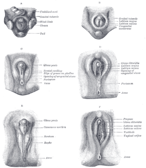 Until about the ninth week of gestational age, the external genitalia of males and females look the same, and follow a common development. This includes the development of a genital tubercle and a membrane dorsally to it, covering the developing urogenital opening
, and the development of labioscrotal folds.
Even after differentiation can be seen between the sexes, some stages are common, e.g. the disappearing of the membrane. On the other hand, sex-dependent development include further protrusion of the genital tubercle in the male to form the penis. Furthermore, the labioscrotal folds evolve into the scrotum in males, while they evolve into labia in females.
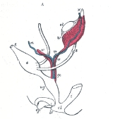
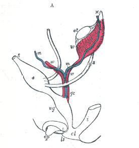
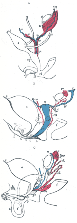 A.—Diagram of the primitive urogenital organs in the embryo previous to sexual distinction.
B.—Diagram of the female type of sexual organs.
C.—Diagram of the male type of sexual organs.
Urinary system
The urinary system is the organ system that produces, stores, and eliminates urine. In humans it includes two kidneys, two ureters, the bladder and the urethra.-Kidney:...
and sex organs. The latter is a part of the stages of sexual differentiation
Sexual differentiation
Sexual differentiation is the process of development of the differences between males and females from an undifferentiated zygote...
.
The urinary and reproductive organs are developed from the intermediate mesoderm
Intermediate mesoderm
Intermediate mesenchyme or intermediate mesoderm is a type of mesoderm that is located between the paraxial mesoderm and the lateral plate.It develops into the part of the urogenital system * forms of urogenital system...
. The permanent organs of the adult are preceded by a set of structures which are purely embryonic, and which with the exception of the ducts disappear almost entirely before the end of fetal life. These embryonic structures are on either side; the pronephros
Pronephros
Pronephros the most basic of the three excretory organs that develop in vertebrates, corresponding to the first stage of kidney development. It is succeeded by the mesonephros, which in fish and amphibians remains as the adult kidney. In amniotes the mesonephros is the embryonic kidney and a more...
, the mesonephros
Mesonephros
The mesonephros is one of three excretory organs that develop in vertebrates. It serves as the main excretory organ of aquatic vertebrates and as a temporary kidney in reptiles, birds, and mammals. The mesonephros is included in the Wolffian body after Caspar Friedrich Wolff who described it in 1759...
and the metanephros of the kidney
Kidney
The kidneys, organs with several functions, serve essential regulatory roles in most animals, including vertebrates and some invertebrates. They are essential in the urinary system and also serve homeostatic functions such as the regulation of electrolytes, maintenance of acid–base balance, and...
, and the Wolffian
Wolffian duct
The mesonephric duct is a paired organ found in mammals including humans during embryogenesis....
and Müllerian duct
Müllerian duct
Müllerian ducts are paired ducts of the embryo that run down the lateral sides of the urogenital ridge and terminate at the Müllerian eminence in the primitive urogenital sinus. In the female, they will develop to form the Fallopian tubes, uterus, cervix, and the upper two-third of the vagina; in...
s of the sex organ
Sex organ
A sex organ, or primary sexual characteristic, as narrowly defined, is any of the anatomical parts of the body which are involved in sexual reproduction and constitute the reproductive system in a complex organism; flowers are the reproductive organs of flowering plants, cones are the reproductive...
. The pronephros disappears very early; the structural elements of the mesonephros mostly degenerate, but the gonad
Gonad
The gonad is the organ that makes gametes. The gonads in males are the testes and the gonads in females are the ovaries. The product, gametes, are haploid germ cells. For example, spermatozoon and egg cells are gametes...
is developed in their place, with which the Wolffian duct remains as the duct in males, and the Müllerian as that of the female. Some of the tubules of the mesonephros form part of the permanent kidney.
Wolffian duct
In the outer part of the intermediate mesodermIntermediate mesoderm
Intermediate mesenchyme or intermediate mesoderm is a type of mesoderm that is located between the paraxial mesoderm and the lateral plate.It develops into the part of the urogenital system * forms of urogenital system...
, immediately under the ectoderm, in the region from the fifth cervical segment to the third thoracic segment, a series of short evaginations from each segment grows dorsally and extends caudally, fusing successively from before backward to form the pronephric duct
Pronephric duct
-Development:The development of the pronephric duct is a part of the development of the urinary and reproductive organs.In the outer part of the intermediate mesoderm, immediately under the ectoderm, in the region from the fifth cervical segment to the third thoracic segment, a series of short...
. This continues to grow caudally until it opens into the ventral part of the cloaca
Cloaca
In zoological anatomy, a cloaca is the posterior opening that serves as the only such opening for the intestinal, reproductive, and urinary tracts of certain animal species...
; beyond the pronephros it is termed the Wolffian duct
Wolffian duct
The mesonephric duct is a paired organ found in mammals including humans during embryogenesis....
. Thus, the Wolffian duct is what remains of the pronephric duct after the atrophy of the pronephros.
Pronephros
The original evaginations form a series of transverse tubules each of which communicates by means of a funnel-shaped ciliated opening with the abdominal cavityAbdominal cavity
The abdominal cavity is the body cavity of the human body that holds the bulk of the viscera. It is located below the thoracic cavity, and above the pelvic cavity. Its dome-shaped roof is the thoracic diaphragm , and its oblique floor is the pelvic inlet...
, and in the course of each duct a glomerulus also is developed. A secondary glomerulus is formed ventral to each of these, and the complete group constitutes the pronephros. In humans, the pronephros is just rudimentary, and undergoes rapid atrophy and disappears.
Mesonephros
On the medial side of the Wolffian duct, from the sixth cervical to the third lumbar segments, a series of tubules, the Wolffian tubulesWolffian tubules
The Wolffian tubules are precursors of the mesonephros.-Development:On the medial side of the Wolffian duct, from the sixth cervical to the third lumbar segments, a series of tubules, the Wolffian tubules, develops. They increase in number by outgrowths from the original tubules...
, develops. They increase in number by outgrowths from the original tubules. They change from solid masses of cells to instead become hollowed in the center. One end grows toward and finally opens into the Wolffian duct, the other dilates and is invaginated by a tuft of capillary bloodvessels to form a glomerulus. The tubules collectively constitute the mesonephros
Mesonephros
The mesonephros is one of three excretory organs that develop in vertebrates. It serves as the main excretory organ of aquatic vertebrates and as a temporary kidney in reptiles, birds, and mammals. The mesonephros is included in the Wolffian body after Caspar Friedrich Wolff who described it in 1759...
.
The mesonephros persists and form the permanent kidneys in fishes and amphibians, but in reptiles, birds, and mammals, it atrophies and for the most part disappears rapidly as the permanent kidney (metanephros) develops beginning during the sixth or seventh week, so that by the beginning of the fifth month only the ducts and a few of the tubules of the mesonephros remain.
Development in male
In the male the Wolffian ductWolffian duct
The mesonephric duct is a paired organ found in mammals including humans during embryogenesis....
persists, and forms for example the epididymis
Epididymis
The epididymis is part of the male reproductive system and is present in all male amniotes. It is a narrow, tightly-coiled tube connecting the efferent ducts from the rear of each testicle to its vas deferens. A similar, but probably non-homologous, structure is found in cartilaginous...
, the ductus deferens, the ejaculatory duct
Ejaculatory duct
-Anatomy:The ejaculatory ducts are paired structures in male anatomy. Each ejaculatory duct is formed by the union of the vas deferens with the duct of the seminal vesicle. They pass through the prostate, and open into the urethra at the Colliculus seminalis...
, seminal vesicle
Seminal vesicle
The seminal vesicles or vesicular glands are a pair of simple tubular glands posteroinferior to the urinary bladder of male mammals...
and efferent ducts.
In the female, on the other hand, the Wolffian bodies and ducts atrophy, leaving behind only remnants in the adult, involving e.g. the development of the suspensory ligament of the ovary
Development of the suspensory ligament of the ovary
The prenatal development of the suspensory ligament of the ovary is a part of the development of the reproductive system.The suspensory ligament originates from the mesonephros, which, in turn, originates from the Wolffian duct:...
.
The Müllerian Duct
Müllerian eminence
The Müllerian eminence is an epithelial on the ventral part of the cloaca between the orifices of the Wolffian ducts. It appears during the development of the urinary and reproductive organs.-Function:The Müllerian ducts end here...
, on the ventral part of the cloaca between the orifices of the Wolffian ducts. At a later stage the eminence opens in the middle, connecting the Müllerian ducts with the cloaca.
Atrophy in males
In the male the Müllerian ducts atrophy, but traces of their anterior ends are represented by the appendices testis (hydatids of Morgagni of the male), while their terminal fused portions form the utriculus in the floor of the prostatic urethraProstatic urethra
The prostatic urethra, the widest and most dilatable part of the urethra canal, is about 3 cm. long.It runs almost vertically through the prostate from its base to its apex, lying nearer its anterior than its posterior surface; the form of the canal is spindle-shaped, being wider in the middle...
.
Development in females
In the female the Müllerian ducts persist and undergo further development. The portions which lie in the genital cord fuse to form the uterusUterus
The uterus or womb is a major female hormone-responsive reproductive sex organ of most mammals including humans. One end, the cervix, opens into the vagina, while the other is connected to one or both fallopian tubes, depending on the species...
and vagina
Vagina
The vagina is a fibromuscular tubular tract leading from the uterus to the exterior of the body in female placental mammals and marsupials, or to the cloaca in female birds, monotremes, and some reptiles. Female insects and other invertebrates also have a vagina, which is the terminal part of the...
. This fusion of the Müllerian ducts begins in the third month, and the septum formed by their fused medial walls disappears from below upward.
The parts outside this cord remain separate, and each forms the corresponding Fallopian tube
Fallopian tube
The Fallopian tubes, also known as oviducts, uterine tubes, and salpinges are two very fine tubes lined with ciliated epithelia, leading from the ovaries of female mammals into the uterus, via the utero-tubal junction...
. The ostium of the fallopian tube remains from the anterior extremity of the original tubular invagination from the abdominal cavity.
About the fifth month a ring-like constriction marks the position of the cervix
Cervix
The cervix is the lower, narrow portion of the uterus where it joins with the top end of the vagina. It is cylindrical or conical in shape and protrudes through the upper anterior vaginal wall...
of the uterus, and after the sixth month the walls of the uterus begin to thicken. For a time the vagina is represented by a solid rod of epithelial cells. A ring-like outgrowth of this epithelium occurs at the lower end of the uterus and marks the future vaginal fornix
Vaginal fornix
The fornices of the vagina are the deepest portions of the vagina, extending into the recesses created by the vaginal portion of cervix. The word 'fornix' is Latin for 'arch'....
. At about the fifth or sixth month the lumen
Lumen (anatomy)
A lumen in biology is the inside space of a tubular structure, such as an artery or intestine...
of the vagina is produced by the breaking down of the central cells of the epithelium. The hymen
Hymen
The hymen is a membrane that surrounds or partially covers the external vaginal opening. It forms part of the vulva, or external genitalia. The size of the hymenal opening increases with age. Although an often practiced method, it is not possible to confirm with certainty that a girl or woman is a...
represents the remains of the Müllerian eminence .
Gonads
The gonads are the precursors of the testes in males and ovaries in females. They initially develop from the mesothelial layer of the peritoneumPeritoneum
The peritoneum is the serous membrane that forms the lining of the abdominal cavity or the coelom — it covers most of the intra-abdominal organs — in amniotes and some invertebrates...
.
Ovaries
The ovary is differentiated into a central part, the medulla of ovary, covered by a surface layer, the germinal epithelium. The immature ova originate from cells from the dorsal endodermEndoderm
Endoderm is one of the three primary germ cell layers in the very early embryo. The other two layers are the ectoderm and mesoderm , with the endoderm as the intermost layer...
of the yolk sac
Yolk sac
The yolk sac is a membranous sac attached to an embryo, providing early nourishment in the form of yolk in bony fishes, sharks, reptiles, birds, and primitive mammals...
. Once they have reached the gonadal ridge they are called oogonia. Development proceeds and the oogonia become fully surrounded by a layer of connective tissue cells (pre-granulosa cells) In this way, the rudiments of the ovarian follicles are formed. The embryological origin of granulosa cells, on the other hand, remains controversial. Just as in the male, there is a gubernaculum
Gubernaculum
The paired Gubernacula are embryonic structures which begin as undifferentiated mesenchyme attaching to the caudal end of the gonads .-Function during development:...
in the female, which pulls it downward, albeit not as much as in males. The gubernaculum later becomes the proper ovarian ligament
Ovarian ligament
The ovarian ligament is a fibrous ligament that connects the ovary to the lateral surface of the uterus....
and the round ligament of the uterus.
Testes
The periphery of the testes are converted into the tunica albugineaTunica albuginea
Tunica albuginea is an anatomy term that literally means "white covering."It is used to refer to three anatomical areas which include:*Tunica albuginea , the tough fibrous layer of connective tissue that surrounds the corpora cavernosa of the penis...
. Cords of the central mass run together and form a network which becomes the rete testis
Rete testis
Rete testis is an anastomosing network of delicate tubules located in the hilum of the testicle that carries sperm from the seminiferous tubules to the vasa efferentia....
, and another network, which develops the seminiferous tubules
Seminiferous tubules
Seminiferous tubules are located in the testes, and are the specific location of meiosis, and the subsequent creation of gametes, namely spermatozoa....
. Via the rete testis, the seminiferous tubules become connected with outgrowths from the mesonephros, which form the efferent ducts of the testis.
In short, the descent of the testes consists of the opening of a connection from the testis to its final location at the anterior abdominal wall, followed by the development of the gubernaculum, which subsequently pulls and translocates the testis down into the developing scrotum. Ultimately, the passageway closes behind the testis. A failure in this process causes indirect inguinal hernia
Indirect inguinal hernia
An indirect inguinal hernia is an inguinal hernia that results from the failure of embryonic closure of the deep inguinal ring after the testicle has passed through it. Like other inguinal hernias, it protrudes through the superficial inguinal ring...
.
Metanephros and definitive kidney
The metanephros is the definite, permanent, but yet immature kidney. It arises from two directions. On one hand, the precursor of the ureter buds from the Wolffian duct, while on the other hand, the precursor of the renal tubules develop from the metanephrogenic blastema. The ureteric bud subsequently grows into the latter mass, forming the parts of the nephron. Other changes include e.g. the translocation of the ureteric opening directly into the cloaca.Ureteric bud
The rudiments of the permanent kidneys make their appearance about the end of the first or the beginning of the second month. Each kidney originate as an ureteric budUreteric bud
The ureteric bud is a protrusion from the mesonephric duct during the development of the urinary and reproductive organs. It later develops into the adult kidney, except for the nephrons, which, in contrast, originate from the metanephric blastema....
from the caudal end of the Wolffian duct, which, in turn, originates from intermediate mesoderm
Intermediate mesoderm
Intermediate mesenchyme or intermediate mesoderm is a type of mesoderm that is located between the paraxial mesoderm and the lateral plate.It develops into the part of the urogenital system * forms of urogenital system...
. The ureteric bud starts close to where the Wolffian duct opens into the cloaca, and grows dorsalward and rostralward along the posterior abdominal wall, where its blind extremity expands and subsequently divides into several buds, which form the rudiments of the renal pelvis
Renal pelvis
The renal pelvis or pyelum is the funnel-like dilated proximal part of the ureter in the kidney.In humans, the renal pelvis is the point of convergence of two or three major calyces...
and renal calyces; by continued growth and subdivision it gives rise to the collecting duct system
Collecting duct system
The collecting duct system of the kidney consists of a series of tubules and ducts that connect the nephrons to the ureter. It participates in electrolyte and fluid balance through reabsorption and excretion, processes regulated by the hormones aldosterone and antidiuretic hormone.There are several...
of the kidney. The other, more superficial, portion of the diverticulum, on the other hand, becomes the ureter.
Metanephrogenic blastema
The renal corpuscleRenal corpuscle
In the kidney, a renal corpuscle is the initial blood-filtering component of a nephron. It consists of two structures: a glomerulus and a Bowman's capsule. The glomerulus is a small tuft of capillaries containing two cell types. Endothelial cells, which have large fenestrae, are not covered by...
s and renal tubules, in contrast, are developed from the metanephrogenic blastema instead of from the ureteric bud. The metanephrogenic blastema is moulded over the growing end of the latter, and becomes a part of the metanephros in this way. The renal tubules of the metanephros, unlike those of the pronephros and mesonephros, do not open into the Wolffian duct. Instead, the tubules rapidly elongate to form the parts of the nephron
Nephron
The renal tubule is the portion of the nephron containing the tubular fluid filtered through the glomerulus. After passing through the renal tubule, the filtrate continues to the collecting duct system, which is not part of the nephron....
: the proximal tubule
Proximal tubule
The proximal tubule is the portion of the duct system of the nephron of the kidney which leads from Bowman's capsule to the loop of Henle.-Structure and appearance:...
s, the loops of Henle and the distal convoluted tubule
Distal convoluted tubule
The distal convoluted tubule is a portion of kidney nephron between the loop of Henle and the collecting duct system.- Physiology :It is partly responsible for the regulation of potassium, sodium, calcium, and pH...
s. These last join and establish communications with the collecting duct system derived from the ultimate ramifications of the ureteric bud. In the other end, the renal tubules give rise to Bowman's capsule
Bowman's capsule
The Bowman's capsule is a cup-like sac at the beginning of the tubular component of a nephron in the mammalian kidney that performs the first step in the filtration of blood to form urine. A glomerulus is enclosed in the sac...
s and glomeruli.
Other changes
The mesoderm around the tubules becomes condensed to form the connective tissue of the kidney. The ureter opens at first into the hind-end of the Wolffian duct; after the sixth week it separates from the Wolffian duct, and opens independently into the part of the cloaca which ultimately becomes the urinary bladderUrinary bladder
The urinary bladder is the organ that collects urine excreted by the kidneys before disposal by urination. A hollow muscular, and distensible organ, the bladder sits on the pelvic floor...
. The renal tubules become arranged into renal pyramids
Renal pyramids
Renal pyramids are cone-shaped tissues of the kidney. The renal medulla is made up of 7 to 18 of these conical subdivisions . The broad base of each pyramid faces the renal cortex, and its apex, or papilla, points internally...
, and the lobulated condition of the kidneys exists for some time after birth, while traces of it may be found even in the adult. The kidney of the ox and many other animals, on the other hand, remains lobulated throughout life.
The Urinary Bladder
Urinary bladder
The urinary bladder is the organ that collects urine excreted by the kidneys before disposal by urination. A hollow muscular, and distensible organ, the bladder sits on the pelvic floor...
is formed partly from the entodermal cloaca and partly from the ends of the Wolffian ducts. In other words, the allantois
Allantois
Allantois is a part of a developing animal conceptus . It helps the embryo exchange gases and handle liquid waste....
takes no share in its formation.
After the separation of the rectum from the dorsal part of the cloaca, the ventral part becomes the primary urogenital sinus. The urogenital sinus, in turn, divides into the superficial definitive urogenital sinus and the deeper anterior vesico-urethral portion.
Definitive urogenital sinus
The definitive urogenital sinus consists of a caudal phallic portion and an intermediate narrow channel, the pelvic portion.Vesico-urethral portion
The vesico-urethral portion is the deepest portion, continuous with the allantois. It absorbs the ends of the Wolffian ducts and the associated ends of the renal diverticula, and these give rise to the trigone of urinary bladder and part of the prostatic urethraProstatic urethra
The prostatic urethra, the widest and most dilatable part of the urethra canal, is about 3 cm. long.It runs almost vertically through the prostate from its base to its apex, lying nearer its anterior than its posterior surface; the form of the canal is spindle-shaped, being wider in the middle...
. The remainder of the vesico-urethral portion forms the body of the bladder and part of the prostatic urethra; its apex is prolonged to the umbilicus as a narrow canal, the urachus
Urachus
The urachus is a fibrous remnant of the allantois, a canal that drains the urinary bladder of the fetus that joins and runs within the umbilical cord...
, which later is obliterated and becomes the median umbilical ligament
Median umbilical ligament
The median umbilical ligament is a structure in human anatomy. It is a shrivelled piece of tissue that represents the remnant of the embryonic urachus.It extends from the apex of the bladder to the umbilicus, on the deep surface of the anterior abdominal wall...
of the adult.
External genitalia
Urogenital opening
The urogenital opening is where waste products of the body and reproductive fluids are expelled to the environment outside of the body cavity. In some organisms, including birds and many fish, discharge from the urological, digestive, and reproductive systems empty into a common sac called the...
, and the development of labioscrotal folds.
Even after differentiation can be seen between the sexes, some stages are common, e.g. the disappearing of the membrane. On the other hand, sex-dependent development include further protrusion of the genital tubercle in the male to form the penis. Furthermore, the labioscrotal folds evolve into the scrotum in males, while they evolve into labia in females.
Diagram of internal differentiation
- 3. Ureter.
- 4. Urinary bladder.
- 5. Urachus.
- cl. Cloaca.
- cp. Elevation which becomes clitoris or penis.
- i. Lower part of the intestine.
- ls. Fold of integument from which the labia majora or scrotum are formed.
- m, m. Right and left Müllerian ducts uniting together and running with the Wolffian ducts in gc, the genital cord.
- ot. The genital ridge from which either the ovary or testis is formed.
- ug. Sinus urogenitalis.
- W. Left Wolffian body.
- w, w. Right and left Wolffian ducts.
B.—Diagram of the female type of sexual organs.
- C. Greater vestibular gland, and immediately above it the urethra.
- cc. Corpus cavernosum clitoridis.
- dG. Remains of the left Wolffian duct, such as give rise to the duct of Gärtner, represented by dotted lines; that of the right side is marked w.
- f. The abdominal opening of the left uterine tube.
- g. Round ligament, corresponding to gubernaculum.
- h. Situation of the hymen.
- i. Lower part of the intestine.
- l. Labium major.
- n. Labium minus.
- o. The left ovary.
- po. Epoophoron.
- sc. Corpus cavernosum urethrae.
- u. Uterus. The uterine tube of the right side is marked m.
- v. Vulva.
- va. Vagina.
- W. Scattered remains of Wolffian tubes near it (paroöphoron of Waldeyer).
C.—Diagram of the male type of sexual organs.
- C. Bulbo-urethral gland of one side.
- cp. Corpora cavernosa penis cut short.
- e. Caput epididymis.
- g. The gubernaculum.
- i. Lower part of the intestine.
- m. Müllerian duct, the upper part of which remains as the hydatid of Morgagni; the lower part, represented by a dotted line descending to the prostatic utricle, constitutes the occasionally existing cornu and tube of the uterus masculinus.
- pr. The prostate.
- s. Scrotum.
- sp. Corpus cavernosum urethrae.
- t. Testis in the place of its original formation.
- t’, together with the dotted lines above, indicates the direction in which the testis and epididymis descend from the abdomen into the scrotum.
- vd. Ductus deferens.
- vh. Ductus aberrans.
- vs. The vesicula seminalis.
- W. Scattered remains of the Wolffian body, constituting the organ of Giraldès, or the paradidymis of Waldeyer.
General
- Henry Gray (1821–1865). Anatomy of the Human Body. 1918. 3. The Urogenital Apparatus
- UNSW Embryology - Development of the Kidney and Reproduction Systems
- How the Body Works / Sex Development / Sexual Differentiation / Duct Differentiation - The Hospital for Sick Children (GTA - Toronto, Ontario, Canada)