

Common carotid artery
Encyclopedia
In human anatomy
, the common carotid artery is an artery
that supplies the head and neck with oxygenated blood; it divides in the neck to form the external
and internal carotid arteries
.
in the thoracic region.
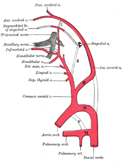
The left common carotid artery can be thought of as having two parts: a thoracic (chest) part and a cervical (neck) part. The right common carotid originates in or close to the neck, so contains a small thoracic portion. There are studies in the bioengineering literature that has looked into characterizing the geometric structure of the common carotid artery from both a qualitative and mathematical quantitative stand point.
, and travels upward through the superior mediastinum
to the level of the left sternoclavicular joint, where it is continuous with the cervical portion.
During the thoracic part of its course, the left common carotid artery is related to the following structures:
In front, it is separated from the manubrium
of the sternum by the sternohyoid and sternothyroid muscles, the anterior portions of the left pleura and lung
, the left brachiocephalic vein
, and the remains of the thymus
; behind, it lies on the trachea
, esophagus
, left recurrent laryngeal nerve
, and thoracic duct
.
To its right side below is the brachiocephalic trunk, and above, the trachea, the inferior thyroid veins, and the remains of the thymus; to its left side are the left vagus and phrenic nerve
s, left pleura, and lung. The left subclavian artery
is posterior and slightly lateral to it.
 The cervical portions of the common carotids resemble each other so closely that one description will apply to both.
Each vessel passes obliquely upward, from behind the sternoclavicular joint to the level of the upper border of the thyroid cartilage
, where it divides.
At the lower part of the neck the two common carotid arteries are separated from each other by a very narrow interval which contains the trachea; but at the upper part, the thyroid gland, the larynx
and pharynx
project forward between the two vessels.
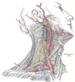
The common carotid artery is contained in a sheath known as the carotid sheath
, which is derived from the deep cervical fascia
and encloses also the internal jugular vein
and vagus nerve
, the vein lying lateral to the artery, and the nerve between the artery and vein, on a plane posterior to both. On opening the sheath, each of these three structures is seen to have a separate fibrous investment.
At approximately the level of the fourth cervical vertebra, the common carotid artery bifurcates into an internal carotid artery
(ICA) and an external carotid artery
(ECA). While both branches travel upward, the internal carotid takes a deeper (more internal) path, eventually travelling up into the skull to supply the brain via the carotid canal. The external carotid artery travels more closely to the surface, and sends off numerous branches that supply the neck and face.
 At the lower part of the neck the common carotid artery is very deeply seated, being covered by the integument, superficial fascia
, the platysma muscle, deep cervical fascia
, the sternocleidomastoid muscle
, the sternohyoid, sternothyroid, and the omohyoid; in the upper part of its course it is more superficial, being covered merely by the integument, the superficial fascia, the platysma, deep cervical fascia, and medial margin of the sternocleidomastoid.
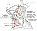
When the sternocleidomastoid muscle is drawn backward, the artery is seen to be contained in a triangular space known as the carotid triangle. This space is bounded behind by the sternocleidomastoid, above by the stylohyoid and the posterior belly of the digastric muscle
, and below by the superior belly of the omohyoid.
This part of the artery is crossed obliquely, from its medial to its lateral side, by the sternocleidomastoid branch of the superior thyroid artery
; it is also crossed by the superior
and middle thyroid vein
s (which end in the internal jugular vein
); descending in front of its sheath is the descending branch of the hypoglossal nerve
, this filament being joined by one or two branches from the cervical nerves
, which cross the vessel obliquely.
Sometimes the descending branch of the hypoglossal nerve is contained within the sheath.
The superior thyroid vein
crosses the artery near its termination, and the middle thyroid vein
a little below the level of the cricoid cartilage; the anterior jugular vein
crosses the artery just above the clavicle
, but is separated from it by the sternohyoid and sternothyroid.
Behind, the artery is separated from the transverse processes of the cervical vertebrae by the longus colli and longus capitis muscles, the sympathetic trunk
being interposed between it and the muscles. The inferior thyroid artery
crosses behind the lower part of the vessel.
Medially, it is in relation with the esophagus, trachea, and thyroid gland (which overlaps it), the inferior thyroid artery and recurrent laryngeal nerve
being interposed; higher up, with the larynx and pharynx. Lateral to the artery, inside the carotid sheath
with the common carotid, are the internal jugular vein
and vagus nerve
.
At the lower part of the neck, on the right side of the body, the right recurrent laryngeal nerve
crosses obliquely behind the artery; the right internal jugular vein
diverges from the artery. On the left side, however, the left internal jugular vein approaches and often overlaps the lower part of the artery.
Behind the angle of bifurcation of the common carotid artery is a reddish-brown oval body known as the carotid body
. It is similar in structure to the coccygeal body which is situated on the median sacral artery
.
The relations of the cervical region of the common carotid artery may be discussed in two points:
 After ligature of the common carotid, the collateral circulation can be perfectly established, by the free communication which exists between the carotid arteries of opposite sides, both without and within the cranium, and by enlargement of the branches of the subclavian artery on the side corresponding to that on which the vessel has been tied.
The chief communications outside the skull take place between the superior and inferior thyroid arteries, and the deep cervical artery
and the descending branch of the occipital artery
; the vertebral artery
takes the place of the internal carotid artery
within the cranium.
In other cases the artery on the right side may arise as a separate branch from the arch of the aorta, or in conjunction with the left carotid.
The left common carotid varies in its origin more than the right.
In the majority of abnormal cases it arises with the brachiocephalic trunk; if that artery is absent, the two carotids arise usually by a single trunk.
It is rarely joined with the left subclavian artery
, except in cases of transposition of the aortic arch
.
; more rarely, it occurs below, opposite the middle of the larynx, or the lower border of the cricoid cartilage. In at least one reported case, the artery was only 4 cm in length and divided at the root of the neck.
Very rarely, the common carotid artery ascends in the neck without any subdivision, either the external or the internal carotid being absent; and in a few cases the common carotid has itself been found to be absent, the external and internal carotids arising directly from the arch of the aorta.
This peculiarity existed on both sides in some instances, on one side in others.
or its laryngeal branch, the ascending pharyngeal artery
, the inferior thyroid artery
, or, more rarely, the vertebral artery
.
, especially in patients who are in shock and who lack a detectable pulse in the more peripheral arteries of the body.
The carotid artery should be palpated gently and while the patient is sitting or lying down. Stimulating its baroreceptors with low palpitation can provoke severe bradycardia
or even stop the heart in some sensitive persons. Also, a person's two carotid arteries should not be palpated at the same time. Doing so may limit the flow of blood to the head, possibly leading to fainting or brain ischemia
. It can be felt between the anterior border of the sternocleidomastoid muscle, above the hyoid bone and lateral to the thyroid cartilage.
Presence of a carotid pulse has been estimated to indicate a systolic blood pressure
of more than 40 mmHg, as given by the 50% percentile.
is a syndrome marked by soreness of the carotid artery near the bifurcation.
Carotid stenosis may occur in patients with atherosclerosis
.
Human anatomy
Human anatomy is primarily the scientific study of the morphology of the human body. Anatomy is subdivided into gross anatomy and microscopic anatomy. Gross anatomy is the study of anatomical structures that can be seen by the naked eye...
, the common carotid artery is an artery
Artery
Arteries are blood vessels that carry blood away from the heart. This blood is normally oxygenated, exceptions made for the pulmonary and umbilical arteries....
that supplies the head and neck with oxygenated blood; it divides in the neck to form the external
External carotid artery
In human anatomy, the external carotid artery is a major artery of the head and neck. It arises from the common carotid artery when it bifurcates into the external and internal carotid artery.-Course:...
and internal carotid arteries
Internal carotid artery
In human anatomy, the internal carotid arteries are two major arteries, one on each side of the head and neck. They arise from the common carotid arteries where these bifurcate into the internal and external carotid artery, and they supply the brain....
.
Structure
The common carotid artery is a paired structure, meaning that there are two in the body, one for each half. The left and right common carotid arteries follow the same course with the exception of their origin. The right common carotid originates in the neck from the brachiocephalic trunk. The left arises from the aortic archAortic arch
The arch of the aorta or the transverse aortic arch is the part of the aorta that begins at the level of the upper border of the second sternocostal articulation of the right side, and runs at first upward, backward, and to the left in front of the trachea; it is then directed backward on the left...
in the thoracic region.
The left common carotid artery can be thought of as having two parts: a thoracic (chest) part and a cervical (neck) part. The right common carotid originates in or close to the neck, so contains a small thoracic portion. There are studies in the bioengineering literature that has looked into characterizing the geometric structure of the common carotid artery from both a qualitative and mathematical quantitative stand point.
Thoracic part
Only the left common carotid artery has a substantial presence in the thoracic region. It originates along the aortic archAortic arch
The arch of the aorta or the transverse aortic arch is the part of the aorta that begins at the level of the upper border of the second sternocostal articulation of the right side, and runs at first upward, backward, and to the left in front of the trachea; it is then directed backward on the left...
, and travels upward through the superior mediastinum
Mediastinum
The mediastinum is a non-delineated group of structures in the thorax, surrounded by loose connective tissue. It is the central compartment of the thoracic cavity...
to the level of the left sternoclavicular joint, where it is continuous with the cervical portion.
During the thoracic part of its course, the left common carotid artery is related to the following structures:
In front, it is separated from the manubrium
Manubrium
The manubrium or manubrium sterni is the broad, upper part of the sternum. Located ventrally with a quadrangular shape, wider superiorly and narrower inferiorly, it articulates with the clavicles and the first two ribs.-Borders:The superior border is the thickest and presents at its center the...
of the sternum by the sternohyoid and sternothyroid muscles, the anterior portions of the left pleura and lung
Lung
The lung is the essential respiration organ in many air-breathing animals, including most tetrapods, a few fish and a few snails. In mammals and the more complex life forms, the two lungs are located near the backbone on either side of the heart...
, the left brachiocephalic vein
Brachiocephalic vein
The left and right brachiocephalic veins in the upper chest are formed by the union of each corresponding internal jugular vein and subclavian vein...
, and the remains of the thymus
Thymus
The thymus is a specialized organ of the immune system. The thymus produces and "educates" T-lymphocytes , which are critical cells of the adaptive immune system....
; behind, it lies on the trachea
Vertebrate trachea
In tetrapod anatomy the trachea, or windpipe, is a tube that connects the pharynx or larynx to the lungs, allowing the passage of air. It is lined with pseudostratified ciliated columnar epithelium cells with goblet cells that produce mucus...
, esophagus
Esophagus
The esophagus is an organ in vertebrates which consists of a muscular tube through which food passes from the pharynx to the stomach. During swallowing, food passes from the mouth through the pharynx into the esophagus and travels via peristalsis to the stomach...
, left recurrent laryngeal nerve
Recurrent laryngeal nerve
The recurrent laryngeal nerve is a branch of the vagus nerve that supplies motor function and sensation to the larynx . It travels within the endoneurium...
, and thoracic duct
Thoracic duct
In human anatomy, the thoracic duct of the lymphatic system is the largest lymphatic vessel in the body. It is also known as the left lymphatic duct, alimentary duct, chyliferous duct, and Van Hoorne's canal....
.
To its right side below is the brachiocephalic trunk, and above, the trachea, the inferior thyroid veins, and the remains of the thymus; to its left side are the left vagus and phrenic nerve
Phrenic nerve
The phrenic nerve originates mainly from the 4th cervical nerve, but also receives contributions from the 5th and 3rd cervical nerves in humans....
s, left pleura, and lung. The left subclavian artery
Subclavian artery
In human anatomy, the subclavian arteries are two major arteries of the upper thorax , below the clavicle . They receive blood from the top of the aorta...
is posterior and slightly lateral to it.
Cervical part
Each vessel passes obliquely upward, from behind the sternoclavicular joint to the level of the upper border of the thyroid cartilage
Thyroid cartilage
The thyroid cartilage is the largest of the nine cartilages that make up the laryngeal skeleton, the cartilage structure in and around the trachea that contains the larynx....
, where it divides.
At the lower part of the neck the two common carotid arteries are separated from each other by a very narrow interval which contains the trachea; but at the upper part, the thyroid gland, the larynx
Larynx
The larynx , commonly called the voice box, is an organ in the neck of amphibians, reptiles and mammals involved in breathing, sound production, and protecting the trachea against food aspiration. It manipulates pitch and volume...
and pharynx
Pharynx
The human pharynx is the part of the throat situated immediately posterior to the mouth and nasal cavity, and anterior to the esophagus and larynx. The human pharynx is conventionally divided into three sections: the nasopharynx , the oropharynx , and the laryngopharynx...
project forward between the two vessels.
The common carotid artery is contained in a sheath known as the carotid sheath
Carotid sheath
The carotid sheath is an anatomical term for the fibrous connective tissue that surrounds the vascular compartment of the neck. It is part of the deep cervical fascia of the neck, below the superficial cervical fascia meaning the subcutaneous adipose tissue immediately beneath the skin.The deep...
, which is derived from the deep cervical fascia
Deep cervical fascia
The deep cervical fascia lies under cover of the Platysma, and invests the neck; it also forms sheaths for the carotid vessels, and for the structures situated in front of the vertebral column...
and encloses also the internal jugular vein
Internal jugular vein
The two internal jugular veins collect the blood from the brain, the superficial parts of the face, and the neck.-Path:On both sides and at the base of the brain, the inferior petrosal sinus and the sigmoid sinus join to form the internal jugular vein...
and vagus nerve
Vagus nerve
The vagus nerve , also called pneumogastric nerve or cranial nerve X, is the tenth of twelve paired cranial nerves...
, the vein lying lateral to the artery, and the nerve between the artery and vein, on a plane posterior to both. On opening the sheath, each of these three structures is seen to have a separate fibrous investment.
At approximately the level of the fourth cervical vertebra, the common carotid artery bifurcates into an internal carotid artery
Internal carotid artery
In human anatomy, the internal carotid arteries are two major arteries, one on each side of the head and neck. They arise from the common carotid arteries where these bifurcate into the internal and external carotid artery, and they supply the brain....
(ICA) and an external carotid artery
External carotid artery
In human anatomy, the external carotid artery is a major artery of the head and neck. It arises from the common carotid artery when it bifurcates into the external and internal carotid artery.-Course:...
(ECA). While both branches travel upward, the internal carotid takes a deeper (more internal) path, eventually travelling up into the skull to supply the brain via the carotid canal. The external carotid artery travels more closely to the surface, and sends off numerous branches that supply the neck and face.
Superficial fascia
Superficial fascia is found in the subcutis in most regions of the body, blending with the reticular layer of the dermis. It is present on the face, over the upper portion of the sternocleidomastoid, at the nape of the neck, and overlying the sternum. It is mainly loose areolar connective tissue...
, the platysma muscle, deep cervical fascia
Deep cervical fascia
The deep cervical fascia lies under cover of the Platysma, and invests the neck; it also forms sheaths for the carotid vessels, and for the structures situated in front of the vertebral column...
, the sternocleidomastoid muscle
Sternocleidomastoid muscle
In human anatomy, the sternocleidomastoid muscle , also known as sternomastoid and commonly abbreviated as SCM, is a paired muscle in the superficial layers of the anterior portion of the neck...
, the sternohyoid, sternothyroid, and the omohyoid; in the upper part of its course it is more superficial, being covered merely by the integument, the superficial fascia, the platysma, deep cervical fascia, and medial margin of the sternocleidomastoid.
When the sternocleidomastoid muscle is drawn backward, the artery is seen to be contained in a triangular space known as the carotid triangle. This space is bounded behind by the sternocleidomastoid, above by the stylohyoid and the posterior belly of the digastric muscle
Digastric muscle
The digastric muscle is a small muscle located under the jaw. so digastric muscles are muscle fibers in ligament of treitz ,omohyoid , occipitofrontalis....
, and below by the superior belly of the omohyoid.
This part of the artery is crossed obliquely, from its medial to its lateral side, by the sternocleidomastoid branch of the superior thyroid artery
Superior thyroid artery
The superior thyroid artery arises from the external carotid artery just below the level of the greater cornu of the hyoid bone and ends in the thyroid gland.-Relations:...
; it is also crossed by the superior
Superior thyroid vein
The superior thyroid vein begins in the substance and on the surface of the thyroid gland, by tributaries corresponding with the branches of the superior thyroid artery, and ends in the upper part of the internal jugular vein....
and middle thyroid vein
Middle thyroid vein
The middle thyroid vein collects the blood from the lower part of the thyroid gland, and after being joined by some veins from the larynx and trachea, ends in the lower part of the internal jugular vein....
s (which end in the internal jugular vein
Internal jugular vein
The two internal jugular veins collect the blood from the brain, the superficial parts of the face, and the neck.-Path:On both sides and at the base of the brain, the inferior petrosal sinus and the sigmoid sinus join to form the internal jugular vein...
); descending in front of its sheath is the descending branch of the hypoglossal nerve
Hypoglossal nerve
The hypoglossal nerve is the twelfth cranial nerve , leading to the tongue. The nerve arises from the hypoglossal nucleus and emerges from the medulla oblongata in the preolivary sulcus separating the olive and the pyramid. It then passes through the hypoglossal canal...
, this filament being joined by one or two branches from the cervical nerves
Cervical nerves
The cervical nerves are the spinal nerves from the cervical vertebrae.Although there are seven cervical vertebrae , there are eight cervical nerves . All nerves except C8 emerge above their corresponding vertebrae, while the C8 nerve emerges below the C7 vertebra...
, which cross the vessel obliquely.
Sometimes the descending branch of the hypoglossal nerve is contained within the sheath.
The superior thyroid vein
Superior thyroid vein
The superior thyroid vein begins in the substance and on the surface of the thyroid gland, by tributaries corresponding with the branches of the superior thyroid artery, and ends in the upper part of the internal jugular vein....
crosses the artery near its termination, and the middle thyroid vein
Middle thyroid vein
The middle thyroid vein collects the blood from the lower part of the thyroid gland, and after being joined by some veins from the larynx and trachea, ends in the lower part of the internal jugular vein....
a little below the level of the cricoid cartilage; the anterior jugular vein
Anterior jugular vein
The anterior jugular vein begins near the hyoid bone by the confluence of several superficial veins from the submaxillary region.It descends between the median line and the anterior border of the Sternocleidomastoideus, and, at the lower part of the neck, passes beneath that muscle to open into the...
crosses the artery just above the clavicle
Clavicle
In human anatomy, the clavicle or collar bone is a long bone of short length that serves as a strut between the scapula and the sternum. It is the only long bone in body that lies horizontally...
, but is separated from it by the sternohyoid and sternothyroid.
Behind, the artery is separated from the transverse processes of the cervical vertebrae by the longus colli and longus capitis muscles, the sympathetic trunk
Sympathetic trunk
The sympathetic trunks are a paired bundle of nerve fibers that run from the base of the skull to the coccyx.-Structure:...
being interposed between it and the muscles. The inferior thyroid artery
Inferior thyroid artery
The inferior thyroid artery arrises from the thyrocervical trunk and passes upward, in front of the vertebral artery and Longus colli, then turns medially behind the carotid sheath and its contents, and also behind the sympathetic trunk, the middle cervical ganglion resting upon the...
crosses behind the lower part of the vessel.
Medially, it is in relation with the esophagus, trachea, and thyroid gland (which overlaps it), the inferior thyroid artery and recurrent laryngeal nerve
Recurrent laryngeal nerve
The recurrent laryngeal nerve is a branch of the vagus nerve that supplies motor function and sensation to the larynx . It travels within the endoneurium...
being interposed; higher up, with the larynx and pharynx. Lateral to the artery, inside the carotid sheath
Carotid sheath
The carotid sheath is an anatomical term for the fibrous connective tissue that surrounds the vascular compartment of the neck. It is part of the deep cervical fascia of the neck, below the superficial cervical fascia meaning the subcutaneous adipose tissue immediately beneath the skin.The deep...
with the common carotid, are the internal jugular vein
Internal jugular vein
The two internal jugular veins collect the blood from the brain, the superficial parts of the face, and the neck.-Path:On both sides and at the base of the brain, the inferior petrosal sinus and the sigmoid sinus join to form the internal jugular vein...
and vagus nerve
Vagus nerve
The vagus nerve , also called pneumogastric nerve or cranial nerve X, is the tenth of twelve paired cranial nerves...
.
At the lower part of the neck, on the right side of the body, the right recurrent laryngeal nerve
Recurrent laryngeal nerve
The recurrent laryngeal nerve is a branch of the vagus nerve that supplies motor function and sensation to the larynx . It travels within the endoneurium...
crosses obliquely behind the artery; the right internal jugular vein
Internal jugular vein
The two internal jugular veins collect the blood from the brain, the superficial parts of the face, and the neck.-Path:On both sides and at the base of the brain, the inferior petrosal sinus and the sigmoid sinus join to form the internal jugular vein...
diverges from the artery. On the left side, however, the left internal jugular vein approaches and often overlaps the lower part of the artery.
Behind the angle of bifurcation of the common carotid artery is a reddish-brown oval body known as the carotid body
Carotid body
The carotid body is a small cluster of chemoreceptors and supporting cells located near the fork of the carotid artery ....
. It is similar in structure to the coccygeal body which is situated on the median sacral artery
Median sacral artery
The median sacral artery is a small vessel, which arises from the back of the aorta, a little above its bifurcation....
.
The relations of the cervical region of the common carotid artery may be discussed in two points:
- Internal relations of organs present inside the carotid sheath
- two external relations of carotid sheath
Collateral circulation
The chief communications outside the skull take place between the superior and inferior thyroid arteries, and the deep cervical artery
Deep cervical artery
-Course:It arises, in most cases, from the costocervical trunk, and is analogous to the posterior branch of an aortic intercostal artery: occasionally it is a separate branch from the subclavian artery....
and the descending branch of the occipital artery
Occipital artery
The occipital artery arises from the external carotid artery opposite the facial artery, its path is below the posterior belly of digastric to the occipital region. This artery supplies blood to the back of the scalp and sterno-mastoid muscles...
; the vertebral artery
Vertebral artery
The vertebral arteries are major arteries of the neck. They branch from the subclavian arteries and merge to form the single midline basilar artery in a complex called the vertebrobasilar system, which supplies blood to the posterior part of the circle of Willis and thus significant portions of the...
takes the place of the internal carotid artery
Internal carotid artery
In human anatomy, the internal carotid arteries are two major arteries, one on each side of the head and neck. They arise from the common carotid arteries where these bifurcate into the internal and external carotid artery, and they supply the brain....
within the cranium.
Origin
The right common carotid may arise above the level of the upper border of the sternoclavicular joint; this variation occurs in about 12 percent of cases.In other cases the artery on the right side may arise as a separate branch from the arch of the aorta, or in conjunction with the left carotid.
The left common carotid varies in its origin more than the right.
In the majority of abnormal cases it arises with the brachiocephalic trunk; if that artery is absent, the two carotids arise usually by a single trunk.
It is rarely joined with the left subclavian artery
Subclavian artery
In human anatomy, the subclavian arteries are two major arteries of the upper thorax , below the clavicle . They receive blood from the top of the aorta...
, except in cases of transposition of the aortic arch
Aortic arch
The arch of the aorta or the transverse aortic arch is the part of the aorta that begins at the level of the upper border of the second sternocostal articulation of the right side, and runs at first upward, backward, and to the left in front of the trachea; it is then directed backward on the left...
.
Point of division
In the majority of abnormal cases, the bifurcation occurs higher than usual, the artery dividing opposite or even above the hyoid boneHyoid bone
The hyoid bone is a horseshoe-shaped bone situated in the anterior midline of the neck between the chin and the thyroid cartilage. At rest, it lies at the level of the base of the mandible in the front and the third cervical vertebra behind.Unlike other bones, the hyoid is only distantly...
; more rarely, it occurs below, opposite the middle of the larynx, or the lower border of the cricoid cartilage. In at least one reported case, the artery was only 4 cm in length and divided at the root of the neck.
Very rarely, the common carotid artery ascends in the neck without any subdivision, either the external or the internal carotid being absent; and in a few cases the common carotid has itself been found to be absent, the external and internal carotids arising directly from the arch of the aorta.
This peculiarity existed on both sides in some instances, on one side in others.
Occasional branches
The common carotid usually gives off no branch previous to its bifurcation, but it occasionally gives origin to the superior thyroid arterySuperior thyroid artery
The superior thyroid artery arises from the external carotid artery just below the level of the greater cornu of the hyoid bone and ends in the thyroid gland.-Relations:...
or its laryngeal branch, the ascending pharyngeal artery
Ascending pharyngeal artery
The ascending pharyngeal artery, the smallest branch of the external carotid, is a long, slender vessel, deeply seated in the neck, beneath the other branches of the external carotid and under the Stylopharyngeus...
, the inferior thyroid artery
Inferior thyroid artery
The inferior thyroid artery arrises from the thyrocervical trunk and passes upward, in front of the vertebral artery and Longus colli, then turns medially behind the carotid sheath and its contents, and also behind the sympathetic trunk, the middle cervical ganglion resting upon the...
, or, more rarely, the vertebral artery
Vertebral artery
The vertebral arteries are major arteries of the neck. They branch from the subclavian arteries and merge to form the single midline basilar artery in a complex called the vertebrobasilar system, which supplies blood to the posterior part of the circle of Willis and thus significant portions of the...
.
Clinical significance
The common carotid artery is often used in measuring the pulsePulse
In medicine, one's pulse represents the tactile arterial palpation of the heartbeat by trained fingertips. The pulse may be palpated in any place that allows an artery to be compressed against a bone, such as at the neck , at the wrist , behind the knee , on the inside of the elbow , and near the...
, especially in patients who are in shock and who lack a detectable pulse in the more peripheral arteries of the body.
The carotid artery should be palpated gently and while the patient is sitting or lying down. Stimulating its baroreceptors with low palpitation can provoke severe bradycardia
Bradycardia
Bradycardia , in the context of adult medicine, is the resting heart rate of under 60 beats per minute, though it is seldom symptomatic until the rate drops below 50 beat/min. It may cause cardiac arrest in some patients, because those with bradycardia may not be pumping enough oxygen to their heart...
or even stop the heart in some sensitive persons. Also, a person's two carotid arteries should not be palpated at the same time. Doing so may limit the flow of blood to the head, possibly leading to fainting or brain ischemia
Ischemia
In medicine, ischemia is a restriction in blood supply, generally due to factors in the blood vessels, with resultant damage or dysfunction of tissue. It may also be spelled ischaemia or ischæmia...
. It can be felt between the anterior border of the sternocleidomastoid muscle, above the hyoid bone and lateral to the thyroid cartilage.
Presence of a carotid pulse has been estimated to indicate a systolic blood pressure
Blood pressure
Blood pressure is the pressure exerted by circulating blood upon the walls of blood vessels, and is one of the principal vital signs. When used without further specification, "blood pressure" usually refers to the arterial pressure of the systemic circulation. During each heartbeat, BP varies...
of more than 40 mmHg, as given by the 50% percentile.
Pathophysiology
CarotidyniaCarotidynia
Carotidynia is a syndrome characterized by unilateral soreness of the carotid artery, near the bifurcation. It was first described in 1927 by Temple Fay. The most common cause of carotidynia is migraine, and then it is usually self-correcting. Common migraine treatments may help alleviate the...
is a syndrome marked by soreness of the carotid artery near the bifurcation.
Carotid stenosis may occur in patients with atherosclerosis
Atherosclerosis
Atherosclerosis is a condition in which an artery wall thickens as a result of the accumulation of fatty materials such as cholesterol...
.
See also
- Head and neck anatomyHead and neck anatomyHead and neck anatomy focuses on the structures of the head and neck of the human body, including the brain, bones, muscles, blood vessels, nerves, glands, nose, mouth, teeth, tongue, and throat...
- Carotid sheathCarotid sheathThe carotid sheath is an anatomical term for the fibrous connective tissue that surrounds the vascular compartment of the neck. It is part of the deep cervical fascia of the neck, below the superficial cervical fascia meaning the subcutaneous adipose tissue immediately beneath the skin.The deep...
- Carotid sinusCarotid sinusIn human anatomy, the carotid sinus is a localized dilation of the internal carotid artery at its origin, the common carotid artery.-Functions:...
- Carotid bodyCarotid bodyThe carotid body is a small cluster of chemoreceptors and supporting cells located near the fork of the carotid artery ....
- Carotid Doppler machineCarotid Doppler machineA carotid Doppler machine is a device which can be used to measure blood flow velocities within the cervical carotid arteries, as well as the vertebral arteries and sometimes the subclavian arteries by means of non-invasive ultrasonic scanning in which the Doppler effect is utilized...
- CarotidyniaCarotidyniaCarotidynia is a syndrome characterized by unilateral soreness of the carotid artery, near the bifurcation. It was first described in 1927 by Temple Fay. The most common cause of carotidynia is migraine, and then it is usually self-correcting. Common migraine treatments may help alleviate the...