
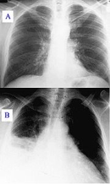
Chest X-ray
Encyclopedia
In medicine
, a chest radiograph, commonly called a chest X-ray (CXR), is a projection radiograph
of the chest
used to diagnose conditions affecting the chest, its contents, and nearby structures. Chest radiographs are among the most common films taken, being diagnostic of many conditions.
Like all methods of radiography
, chest radiography employs ionizing radiation
in the form of X-ray
s to generate images of the chest. The typical radiation
dose to an adult from a chest radiograph is around 0.06 mSv.
Chest radiographs are used to diagnose many conditions involving the chest wall, bones of the thorax
, and structures contained within the thoracic cavity
including the lung
s, heart
, and great vessels. Pneumonia
and congestive heart failure
are very commonly diagnosed by chest radiograph. Chest radiographs are used to screen for job-related lung disease in industries such as mining where workers are exposed to dust.
For some conditions of the chest, radiography is good for screening but poor for diagnosis. When a condition is suspected based on chest radiography, additional imaging of the chest can be obtained to definitively diagnose the condition or to provide evidence in favor of the diagnosis suggested by initial chest radiography.
Unless a fractured rib is suspected of being displaced, and therefore likely to cause damage to the lungs and other tissue structures, x-ray of the chest is not necessary as it will not alter patient management.
The main regions where a chest X-ray may identify problems may be summarized as ABCDEF by their first letters:
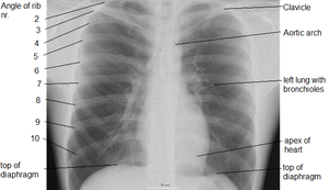 Different views of the chest can be obtained by changing the relative orientation of the body and the direction of the x-ray beams. The most common views are posteroanterior, anteroposterior, and lateral. In an posteroanterior (PA) view, the x-ray source is positioned so that x-rays enter through the posterior (back) aspect of the chest, and exit out of the anterior (front) aspect where they are detected. To obtain this view, individuals stand facing a flat surface behind which is an x-ray detector. A radiation source is positioned behind the patient at a standard distance, and x-ray beams are transmitted toward the patient.
In anteroposterior (AP) views, the positions of the x-ray source and detector are reversed: x-rays enter through the anterior aspect and exit through the posterior aspect of the chest. AP chest x-rays are harder to interpret than PA x-rays and are therefore generally reserved for situations where it is difficult for the patient to obtain a normal chest x-ray, such as when the patient cannot get out of bed. In this situation, mobile X-ray equipment is used to obtain a lying down chest x-ray (known as a "supine film"). As a result most supine films are also AP.
Lateral views of the chest are obtained in a similar fashion as the posteroanterior views, except in the lateral view, the patient stands with both arms raised and the left side of the chest pressed against a flat surface.
, initial imaging of the chest generally consists of PA and lateral views. In other countries, the PA view suffices as an initial study; the lateral view is only added if indicated from interpretation of the PA view.
There are a number of features that are helpful in suggesting the diagnosis:
If the nodules are multiple, the differential is then smaller:
The causes include:
. There needs to be at least 75ml of pleural fluid in order to blunt the costophrenic angle
on the lateral chest radiograph, and 200ml on the posteroanterior chest radiograph. On a lateral decubitus, amounts as small as 5ml of fluid are possible. Pleural effusions typically have a meniscus
visible on an erect chest radiograph, but loculated effusions (as occur with an empyema
) may have a lenticular
shape (the fluid making an obtuse angle with the chest wall).
Pleural thickening may cause blunting of the costophrenic angle, but is distinguished from pleural fluid by the fact that it occurs as a linear shadow ascending vertically and clinging to the ribs.
of the chest is usually required and sometimes a lung biopsy
. The following features should be noted:
Pleural effusion
s may occur with cancer, sarcoid, connective tissue diseases and lymphangioleiomyomatosis
. The presence of a pleural effusion argues against pneumocystis pneumonia.
Reticular (linear) pattern
Nodular pattern
Cystic
Ground glass
Consolidation
Medicine
Medicine is the science and art of healing. It encompasses a variety of health care practices evolved to maintain and restore health by the prevention and treatment of illness....
, a chest radiograph, commonly called a chest X-ray (CXR), is a projection radiograph
Projectional radiography
Projectional radiography or plain film radiography is the practice of producing two-dimensional images using x-ray radiation. Radiographic exams are typically performed by Radiologic Technologists, highly trained medical professionals who specialize in the usage of radiographic equipment, patient...
of the chest
Chest
The chest is a part of the anatomy of humans and various other animals. It is sometimes referred to as the thorax or the bosom.-Chest anatomy - Humans and other hominids:...
used to diagnose conditions affecting the chest, its contents, and nearby structures. Chest radiographs are among the most common films taken, being diagnostic of many conditions.
Like all methods of radiography
Radiography
Radiography is the use of X-rays to view a non-uniformly composed material such as the human body. By using the physical properties of the ray an image can be developed which displays areas of different density and composition....
, chest radiography employs ionizing radiation
Ionizing radiation
Ionizing radiation is radiation composed of particles that individually have sufficient energy to remove an electron from an atom or molecule. This ionization produces free radicals, which are atoms or molecules containing unpaired electrons...
in the form of X-ray
X-ray
X-radiation is a form of electromagnetic radiation. X-rays have a wavelength in the range of 0.01 to 10 nanometers, corresponding to frequencies in the range 30 petahertz to 30 exahertz and energies in the range 120 eV to 120 keV. They are shorter in wavelength than UV rays and longer than gamma...
s to generate images of the chest. The typical radiation
Radiation
In physics, radiation is a process in which energetic particles or energetic waves travel through a medium or space. There are two distinct types of radiation; ionizing and non-ionizing...
dose to an adult from a chest radiograph is around 0.06 mSv.
Problems identified
|
Chest radiographs are used to diagnose many conditions involving the chest wall, bones of the thorax
Thorax
The thorax is a division of an animal's body that lies between the head and the abdomen.-In tetrapods:...
, and structures contained within the thoracic cavity
Thoracic cavity
The thoracic cavity is the chamber of the human body that is protected by the thoracic wall ....
including the lung
Lung
The lung is the essential respiration organ in many air-breathing animals, including most tetrapods, a few fish and a few snails. In mammals and the more complex life forms, the two lungs are located near the backbone on either side of the heart...
s, heart
Heart
The heart is a myogenic muscular organ found in all animals with a circulatory system , that is responsible for pumping blood throughout the blood vessels by repeated, rhythmic contractions...
, and great vessels. Pneumonia
Pneumonia
Pneumonia is an inflammatory condition of the lung—especially affecting the microscopic air sacs —associated with fever, chest symptoms, and a lack of air space on a chest X-ray. Pneumonia is typically caused by an infection but there are a number of other causes...
and congestive heart failure
Congestive heart failure
Heart failure often called congestive heart failure is generally defined as the inability of the heart to supply sufficient blood flow to meet the needs of the body. Heart failure can cause a number of symptoms including shortness of breath, leg swelling, and exercise intolerance. The condition...
are very commonly diagnosed by chest radiograph. Chest radiographs are used to screen for job-related lung disease in industries such as mining where workers are exposed to dust.
For some conditions of the chest, radiography is good for screening but poor for diagnosis. When a condition is suspected based on chest radiography, additional imaging of the chest can be obtained to definitively diagnose the condition or to provide evidence in favor of the diagnosis suggested by initial chest radiography.
Unless a fractured rib is suspected of being displaced, and therefore likely to cause damage to the lungs and other tissue structures, x-ray of the chest is not necessary as it will not alter patient management.
The main regions where a chest X-ray may identify problems may be summarized as ABCDEF by their first letters:
- Airways, including hilar adenopathy or enlargement
- Breast shadows
- Bones, e.g. rib fractures and lytic bone lesions
- Cardiac silhoutte, detecting cardiac enlargement
- Costophrenic angles, including pleural effusions
- Diaphragm, e.g. evidence of free air
- Edges, e.g. apices for fibrosis, pneumothorax, pleural thickening or plaques
- Extrathoracic tissues
- Fields (lung parenchyma), being evidence of alveolar filling
- Failure, e.g. alveolar air space disease with prominent vascularity with or without pleural effusions
Views
In anteroposterior (AP) views, the positions of the x-ray source and detector are reversed: x-rays enter through the anterior aspect and exit through the posterior aspect of the chest. AP chest x-rays are harder to interpret than PA x-rays and are therefore generally reserved for situations where it is difficult for the patient to obtain a normal chest x-ray, such as when the patient cannot get out of bed. In this situation, mobile X-ray equipment is used to obtain a lying down chest x-ray (known as a "supine film"). As a result most supine films are also AP.
Lateral views of the chest are obtained in a similar fashion as the posteroanterior views, except in the lateral view, the patient stands with both arms raised and the left side of the chest pressed against a flat surface.
Typical views
In the United StatesUnited States
The United States of America is a federal constitutional republic comprising fifty states and a federal district...
, initial imaging of the chest generally consists of PA and lateral views. In other countries, the PA view suffices as an initial study; the lateral view is only added if indicated from interpretation of the PA view.
Additional views
- DecubitusDecubitusCommonly used in medicine, the word decubitus is used to mean "lying down". It is derived from the Latin verb “decumbere” meaning "to lie down"....
- useful for differentiating pleural effusionPleural effusionPleural effusion is excess fluid that accumulates between the two pleural layers, the fluid-filled space that surrounds the lungs. Excessive amounts of such fluid can impair breathing by limiting the expansion of the lungs during ventilation.-Pathophysiology:...
s from consolidation (e.g. pneumonia)and Loculated effusions from free fluid in the pleura. In effusions, the fluid layers out (by comparison to an up-right view, when it often accumulates in the costophrenic angleCostophrenic angleIn anatomy, the costophrenic angles are the places where the diaphragm meet the ribs .Each costophrenic angle can normally be seen as on chest x-ray as a sharply-pointed, downward indentation between each hemi-diaphragm and the adjacent chest wall . A small portion of each lung normally reaches...
s). - Lordotic view - used to visualize the apex of the lung, to pick-up abnormalities such as a Pancoast tumour.
- Expiratory view - helpful for the diagnosis of pneumothorax
- ObliqueOblique projectionOblique projection is a simple type of graphical projection used for producing pictorial, two-dimensional images of three-dimensional objects.- Overview :Oblique projection is a type of parallel projection:...
view
Nodule
A nodule is a discrete opacity in the lung which may be caused by:- Neoplasm: benign or malignant
- Granuloma: tuberculosisTuberculosisTuberculosis, MTB, or TB is a common, and in many cases lethal, infectious disease caused by various strains of mycobacteria, usually Mycobacterium tuberculosis. Tuberculosis usually attacks the lungs but can also affect other parts of the body...
- Infection: round pneumonia
- Vascular: infarct, varixEsophageal varicesIn medicine , esophageal varices are extremely dilated sub-mucosal veins in the lower esophagus...
, Wegener's granulomatosisWegener's granulomatosisWegener's granulomatosis , more recently granulomatosis with polyangiitis , is an incurable form of vasculitis that affects the nose, lungs, kidneys and other organs. Due to its end-organ damage, it is life-threatening and requires long-term immunosuppression...
, rheumatoid arthritisRheumatoid arthritisRheumatoid arthritis is a chronic, systemic inflammatory disorder that may affect many tissues and organs, but principally attacks synovial joints. The process produces an inflammatory response of the synovium secondary to hyperplasia of synovial cells, excess synovial fluid, and the development...
There are a number of features that are helpful in suggesting the diagnosis:
- rate of growth
- Doubling time of less than one month: sarcoma/infection/infarction/vascular
- Doubling time of six to 18 months: benign tumour/malignant granuloma
- Doubling time of more than 24 months: benign nodule malignancy
- calcificationCalcificationCalcification is the process in which calcium salts build up in soft tissue, causing it to harden. Calcifications may be classified on whether there is mineral balance or not, and the location of the calcification.-Causes:...
- margin
- smooth
- lobulated
- presence of a corona radiataCorona radiataIn neuroanatomy, the corona radiata is a white matter sheet that continues caudally as the internal capsule and rostrally as the centrum semiovale. This sheet of axons contains both descending and ascending axons that carry nearly all of the neural traffic from and to the cerebral cortex...
- shape
- site
If the nodules are multiple, the differential is then smaller:
- infection: tuberculosisTuberculosisTuberculosis, MTB, or TB is a common, and in many cases lethal, infectious disease caused by various strains of mycobacteria, usually Mycobacterium tuberculosis. Tuberculosis usually attacks the lungs but can also affect other parts of the body...
, fungal infection, septic emboliSeptic embolismA septic embolism is a type of embolism that is infected with bacteria, resulting in the formation of pus. These may become dangerous if dislodged from their original location. Like other emboli, a septic embolism may be fatal.... - neoplasm: e.g., metastasesMetastasisMetastasis, or metastatic disease , is the spread of a disease from one organ or part to another non-adjacent organ or part. It was previously thought that only malignant tumor cells and infections have the capacity to metastasize; however, this is being reconsidered due to new research...
, lymphomaLymphomaLymphoma is a cancer in the lymphatic cells of the immune system. Typically, lymphomas present as a solid tumor of lymphoid cells. Treatment might involve chemotherapy and in some cases radiotherapy and/or bone marrow transplantation, and can be curable depending on the histology, type, and stage...
, hamartomaHamartomaA hamartoma is a benign, focal malformation that resembles a neoplasm in the tissue of its origin. This is not a malignant tumor, and it grows at the same rate as the surrounding tissues. It is composed of tissue elements normally found at that site, but which are growing in a disorganized mass... - sarcoidosisSarcoidosisSarcoidosis , also called sarcoid, Besnier-Boeck disease or Besnier-Boeck-Schaumann disease, is a disease in which abnormal collections of chronic inflammatory cells form as nodules in multiple organs. The cause of sarcoidosis is unknown...
- alveolitisAlveolitisAlveolitis can refer to two inflammatory conditions. It can refer to inflammation of the alveoli in the lungs, or the dental alveolus in the jaw.Alveolus in the jaw is also known as dry socket...
- auto-immune disease: e.g., Wegener's granulomatosisWegener's granulomatosisWegener's granulomatosis , more recently granulomatosis with polyangiitis , is an incurable form of vasculitis that affects the nose, lungs, kidneys and other organs. Due to its end-organ damage, it is life-threatening and requires long-term immunosuppression...
, rheumatoid arthritisRheumatoid arthritisRheumatoid arthritis is a chronic, systemic inflammatory disorder that may affect many tissues and organs, but principally attacks synovial joints. The process produces an inflammatory response of the synovium secondary to hyperplasia of synovial cells, excess synovial fluid, and the development... - inhalation (e.g., pneumoconiosisPneumoconiosisPneumoconiosis is an occupational lung disease and a restrictive lung disease caused by the inhalation of dust, often in mines.-Types:Depending upon the type of dust, the disease is given different names:...
)
Cavities
A cavity is a walled hollow structure within the lungs. Diagnosis is aided by noting:- wall thickness
- wall outline
- changes in the surrounding lung
The causes include:
- cancerCancerCancer , known medically as a malignant neoplasm, is a large group of different diseases, all involving unregulated cell growth. In cancer, cells divide and grow uncontrollably, forming malignant tumors, and invade nearby parts of the body. The cancer may also spread to more distant parts of the...
(usually malignant) - infarct (usually from a pulmonary embolus)
- infection: e.g., Staphylococcus aureusStaphylococcus aureusStaphylococcus aureus is a facultative anaerobic Gram-positive coccal bacterium. It is frequently found as part of the normal skin flora on the skin and nasal passages. It is estimated that 20% of the human population are long-term carriers of S. aureus. S. aureus is the most common species of...
, tuberculosisTuberculosisTuberculosis, MTB, or TB is a common, and in many cases lethal, infectious disease caused by various strains of mycobacteria, usually Mycobacterium tuberculosis. Tuberculosis usually attacks the lungs but can also affect other parts of the body...
, Gram negative bacteria (especially Klebsiella pneumoniaeKlebsiella pneumoniaeKlebsiella pneumoniae is a Gram-negative, non-motile, encapsulated, lactose fermenting, facultative anaerobic, rod shaped bacterium found in the normal flora of the mouth, skin, and intestines....
),anaerobic bacteria, and fungal - Wegener's granulomatosisWegener's granulomatosisWegener's granulomatosis , more recently granulomatosis with polyangiitis , is an incurable form of vasculitis that affects the nose, lungs, kidneys and other organs. Due to its end-organ damage, it is life-threatening and requires long-term immunosuppression...
Pleural abnormalities
Fluid in space between the lung and the chest wall is termed a pleural effusionPleural effusion
Pleural effusion is excess fluid that accumulates between the two pleural layers, the fluid-filled space that surrounds the lungs. Excessive amounts of such fluid can impair breathing by limiting the expansion of the lungs during ventilation.-Pathophysiology:...
. There needs to be at least 75ml of pleural fluid in order to blunt the costophrenic angle
Costophrenic angle
In anatomy, the costophrenic angles are the places where the diaphragm meet the ribs .Each costophrenic angle can normally be seen as on chest x-ray as a sharply-pointed, downward indentation between each hemi-diaphragm and the adjacent chest wall . A small portion of each lung normally reaches...
on the lateral chest radiograph, and 200ml on the posteroanterior chest radiograph. On a lateral decubitus, amounts as small as 5ml of fluid are possible. Pleural effusions typically have a meniscus
Meniscus
The meniscus is the curve in the upper surface of a liquid close to the surface of the container or another object, caused by surface tension. It can be either convex or concave. A convex meniscus occurs when the molecules have a stronger attraction to each other than to the material of the...
visible on an erect chest radiograph, but loculated effusions (as occur with an empyema
Empyema
Pleural empyema is an accumulation of pus in the pleural cavity. Most pleural empyemas arise from an infection within the lung , often associated with parapneumonic effusions. There are three stages: exudative, fibrinopurulent and organizing. In the exudative stage, the pus accumulates...
) may have a lenticular
Lens (geometry)
In geometry, a lens is a biconvex shape comprising two circular arcs, joined at their endpoints. If the arcs have equal radii, it is called a symmetric lens.A concave-convex shape is called a lune...
shape (the fluid making an obtuse angle with the chest wall).
Pleural thickening may cause blunting of the costophrenic angle, but is distinguished from pleural fluid by the fact that it occurs as a linear shadow ascending vertically and clinging to the ribs.
Diffuse shadowing
The differential for diffuse shadowing is very broad and can defeat even the most experienced radiologist. It is seldom possible to reach a diagnosis on the basis of the chest radiograph alone: high-resolution CTComputed tomography
X-ray computed tomography or Computer tomography , is a medical imaging method employing tomography created by computer processing...
of the chest is usually required and sometimes a lung biopsy
Biopsy
A biopsy is a medical test involving sampling of cells or tissues for examination. It is the medical removal of tissue from a living subject to determine the presence or extent of a disease. The tissue is generally examined under a microscope by a pathologist, and can also be analyzed chemically...
. The following features should be noted:
- type of shadowing (lines, dots or rings)
- reticular (crisscrossing lines)
- companion shadowCompanion shadowCompanion shadow is a term used in describing radiographs that denotes the appearance of a smooth, homogenous, radiodensity with a well-defined margin that runs parallel with a bony landmark. Companion shadows represent soft tissue that overlies the respective bony landmark in profile...
(lines paralleling bony landmarks) - nodular (lots of small dots)
- rings or cysts
- ground glass
- consolidation (diffuse opacity with air bronchograms)
- location (where is the lesion worst?)
- upper (e.g., sarcoidSarcoidosisSarcoidosis , also called sarcoid, Besnier-Boeck disease or Besnier-Boeck-Schaumann disease, is a disease in which abnormal collections of chronic inflammatory cells form as nodules in multiple organs. The cause of sarcoidosis is unknown...
, tuberculosisTuberculosisTuberculosis, MTB, or TB is a common, and in many cases lethal, infectious disease caused by various strains of mycobacteria, usually Mycobacterium tuberculosis. Tuberculosis usually attacks the lungs but can also affect other parts of the body...
, silicosisSilicosisSilicosis, also known as Potter's rot, is a form of occupational lung disease caused by inhalation of crystalline silica dust, and is marked by inflammation and scarring in forms of nodular lesions in the upper lobes of the lungs...
/pneumoconiosisPneumoconiosisPneumoconiosis is an occupational lung disease and a restrictive lung disease caused by the inhalation of dust, often in mines.-Types:Depending upon the type of dust, the disease is given different names:...
, ankylosing spondylitisAnkylosing spondylitisAnkylosing spondylitis , previously known as Bekhterev's disease, Bekhterev syndrome, and Marie-Strümpell disease is a chronic inflammatory disease of the axial skeleton with variable involvement of peripheral joints and nonarticular structures...
, Langerhans cell histiocytosisLangerhans cell histiocytosisLangerhans cell Histiocytosis is a rare disease involving clonal proliferation of Langerhans cells, abnormal cells deriving from bone marrow and capable of migrating from skin to lymph nodes...
) - lower (e.g., cryptogenic fibrosing alveolitis, connective tissue diseaseConnective tissue diseaseA connective tissue disease is any disease that has the connective tissues of the body as a target of pathology. Connective tissue is any type of biological tissue with an extensive extracellular matrix that supports, binds together, and protects organs...
, asbestosis, drug reactions) - central (e.g., pulmonary oedema, alveolar proteinosis, lymphomaLymphomaLymphoma is a cancer in the lymphatic cells of the immune system. Typically, lymphomas present as a solid tumor of lymphoid cells. Treatment might involve chemotherapy and in some cases radiotherapy and/or bone marrow transplantation, and can be curable depending on the histology, type, and stage...
, Kaposi's sarcomaKaposi's sarcomaKaposi's sarcoma is a tumor caused by Human herpesvirus 8 , also known as Kaposi's sarcoma-associated herpesvirus . It was originally described by Moritz Kaposi , a Hungarian dermatologist practicing at the University of Vienna in 1872. It became more widely known as one of the AIDS defining...
, PCPPneumocystis pneumoniaPneumocystis pneumonia or pneumocystosis is a form of pneumonia, caused by the yeast-like fungus Pneumocystis jirovecii...
) - peripheral (e.g., cryptogenic fibrosing alveolitis, connective tissue diseaseConnective tissue diseaseA connective tissue disease is any disease that has the connective tissues of the body as a target of pathology. Connective tissue is any type of biological tissue with an extensive extracellular matrix that supports, binds together, and protects organs...
, chronic eosinophilic pneumonia, bronchiolitis obliterans organizing pneumoniaBronchiolitis obliterans organizing pneumoniaBronchiolitis obliterans organizing pneumonia is an inflammation of the bronchioles and surrounding tissue in the lungs. It is an non infectious pneumonia. BOOP is often caused by a pre-existing chronic inflammatory disease like rheumatoid arthritis. BOOP can also be a side effect of certain...
)
- upper (e.g., sarcoid
- lung volume
- increased (e.g., Langerhans cell histiocytosisLangerhans cell histiocytosisLangerhans cell Histiocytosis is a rare disease involving clonal proliferation of Langerhans cells, abnormal cells deriving from bone marrow and capable of migrating from skin to lymph nodes...
, lymphangioleiomyomatosisLymphangioleiomyomatosisLymphangioleiomyomatosis is a rare lung disease that results in a proliferation of disorderly smooth muscle growth throughout the lungs, in the bronchioles, alveolar septa, perivascular spaces, and lymphatics, resulting in the obstruction of small airways and lymphatics...
, cystic fibrosisCystic fibrosisCystic fibrosis is a recessive genetic disease affecting most critically the lungs, and also the pancreas, liver, and intestine...
, allergic bronchopulmonary aspergillosisAllergic bronchopulmonary aspergillosisIn medicine, allergic bronchopulmonary aspergillosis is a condition characterised by an exaggerated response of the immune system to the fungus Aspergillus . It occurs most often in patients with asthma or cystic fibrosis...
) - decreased (e.g., fibrotic lung disease, chronic sarcoidosis, chronic extrinsic allergic alveolitis)
- increased (e.g., Langerhans cell histiocytosis
Pleural effusion
Pleural effusion
Pleural effusion is excess fluid that accumulates between the two pleural layers, the fluid-filled space that surrounds the lungs. Excessive amounts of such fluid can impair breathing by limiting the expansion of the lungs during ventilation.-Pathophysiology:...
s may occur with cancer, sarcoid, connective tissue diseases and lymphangioleiomyomatosis
Lymphangioleiomyomatosis
Lymphangioleiomyomatosis is a rare lung disease that results in a proliferation of disorderly smooth muscle growth throughout the lungs, in the bronchioles, alveolar septa, perivascular spaces, and lymphatics, resulting in the obstruction of small airways and lymphatics...
. The presence of a pleural effusion argues against pneumocystis pneumonia.
Reticular (linear) pattern
- cryptogenic fibrosing alveolitis
- connective tissue diseaseConnective tissue diseaseA connective tissue disease is any disease that has the connective tissues of the body as a target of pathology. Connective tissue is any type of biological tissue with an extensive extracellular matrix that supports, binds together, and protects organs...
- sarcoidosisSarcoidosisSarcoidosis , also called sarcoid, Besnier-Boeck disease or Besnier-Boeck-Schaumann disease, is a disease in which abnormal collections of chronic inflammatory cells form as nodules in multiple organs. The cause of sarcoidosis is unknown...
- radiation fibrosis
- asbestosis
- lymphangitis carcinomatosis
- PCPPneumocystis pneumoniaPneumocystis pneumonia or pneumocystosis is a form of pneumonia, caused by the yeast-like fungus Pneumocystis jirovecii...
Nodular pattern
- sarcoidosisSarcoidosisSarcoidosis , also called sarcoid, Besnier-Boeck disease or Besnier-Boeck-Schaumann disease, is a disease in which abnormal collections of chronic inflammatory cells form as nodules in multiple organs. The cause of sarcoidosis is unknown...
- silicosisSilicosisSilicosis, also known as Potter's rot, is a form of occupational lung disease caused by inhalation of crystalline silica dust, and is marked by inflammation and scarring in forms of nodular lesions in the upper lobes of the lungs...
/pneumoconiosis - extrinsic allergic alveolitis
- Langerhans cell histiocytosisLangerhans cell histiocytosisLangerhans cell Histiocytosis is a rare disease involving clonal proliferation of Langerhans cells, abnormal cells deriving from bone marrow and capable of migrating from skin to lymph nodes...
- lymphangitis carcinomatosaLymphangitis carcinomatosaLymphangitis carcinomatosa is inflammation of the lymph vessels secondary to a malignancy. Breast, lung, stomach, pancreas, and prostate cancers are the most common tumors that result in lymphangitis. First described by pathologist Gabriel Andral in 1829 in a patient with uterine cancer.-References:...
- miliary tuberculosisMiliary tuberculosisMiliary tuberculosis is a form of tuberculosis that is characterized by a wide dissemination into the human body and by the tiny size of the lesions...
- metastases
Cystic
- cryptogenic fibrosing alveolitis (late stage "honeycomb lung")
- cystic bronchiectasisBronchiectasisBronchiectasis is a disease state defined by localized, irreversible dilation of part of the bronchial tree caused by destruction of the muscle and elastic tissue. It is classified as an obstructive lung disease, along with emphysema, bronchitis, asthma, and cystic fibrosis...
- Langerhans cell histiocytosisLangerhans cell histiocytosisLangerhans cell Histiocytosis is a rare disease involving clonal proliferation of Langerhans cells, abnormal cells deriving from bone marrow and capable of migrating from skin to lymph nodes...
- lymphangioleiomyomatosisLymphangioleiomyomatosisLymphangioleiomyomatosis is a rare lung disease that results in a proliferation of disorderly smooth muscle growth throughout the lungs, in the bronchioles, alveolar septa, perivascular spaces, and lymphatics, resulting in the obstruction of small airways and lymphatics...
Ground glass
- Extrinsic allergic alveolitis
- Diffuse interstitial pneumonitis
- Alveolar proteinosis
- Infant respiratory distress syndromeInfant respiratory distress syndromeInfant respiratory distress syndrome , also called neonatal respiratory distress syndrome or respiratory distress syndrome of newborn, previously called hyaline membrane disease, is a syndrome in premature infants caused by developmental insufficiency of surfactant production and structural...
(RDS)
Consolidation
- Alveolar haemorrhage
- Alveolar cell carcinoma
- vasculitisVasculitisVasculitis refers to a heterogeneous group of disorders that are characterized by inflammatory destruction of blood vessels. Both arteries and veins are affected. Lymphangitis is sometimes considered a type of vasculitis...
- chronic eosinophilic pneumonia
Signs
- The silhouette signSilhouette signIn radiology, the silhouette sign refers to loss of normal border between structures,, or an intra-thoracic radio-opacity obscuring the border of the heart or aorta due to a contiguous structure...
is especially helpful in localizing lung lesions. (e.g., loss of right heart border in right middle lobe pneumonia), - The air bronchogram sign, where branching radiolucent columns of air corresponding to bronchi is seen, usually indicates air-space (alveolar) disease, as from blood, pus, mucous, cells, protein surrounding the air bronchograms. This is seen in Respiratory distress syndromeRespiratory distress syndromeThere are two forms of respiratory distress syndrome:* ARDS, which is acute respiratory distress syndrome* Infant respiratory distress syndrome , which is a complication of premature birth, also known as hyaline membrane disease Also, respiratory distress can mean:* Shortness of breath*...
Limitations
While chest radiographs are a cheap and relatively safe method of investigating diseases of the chest, there are a number of serious chest conditions that may be associated with a normal chest radiograph and other means of assessment may be necessary to make the diagnosis. For example, a patient with an acute myocardial infarction may have a completely normal chest radiograph.External links
- Chest X-ray Atlas
- USUHS: Basic Chest X-Ray Review
- eMedicine Radiology: Chest articles
- Database of chest radiology related to emergency medicine
- Introduction to chest radiology: a tutorial for learning to read a chest x-ray
- Chest Radiology Tutorials Free Web Tutorials for Chest Anatomy and Lung Malignancies in Radiology