
Cardiac action potential
Overview
Action potential
In physiology, an action potential is a short-lasting event in which the electrical membrane potential of a cell rapidly rises and falls, following a consistent trajectory. Action potentials occur in several types of animal cells, called excitable cells, which include neurons, muscle cells, and...
in the heart
Heart
The heart is a myogenic muscular organ found in all animals with a circulatory system , that is responsible for pumping blood throughout the blood vessels by repeated, rhythmic contractions...
, necessary for the electrical conduction system of the heart
Electrical conduction system of the heart
The normal intrinsic electrical conduction of the heart allows electrical propagation to be transmitted from the Sinoatrial Node through both atria and forward to the Atrioventricular Node. Normal/baseline physiology allows further propagation from the AV node to the ventricle or Purkinje Fibers...
.
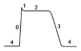
The cardiac action potential differs significantly in different portions of the heart. This differentiation of the action potentials allows the different electrical characteristics of the different portions of the heart. For instance, the specialized conduction tissue of the heart has the special property of depolarizing without any external influence.
Discussions