
CD8
Encyclopedia
CD8 is a transmembrane
glycoprotein
that serves as a co-receptor
for the T cell receptor
(TCR). Like the TCR, CD8 binds to a major histocompatibility complex
(MHC) molecule, but is specific for the class I MHC protein. There are two isoforms of the protein, alpha and beta, each encoded by a different gene. In humans, both genes are located on chromosome 2 in position 2p12.
s, but can also be found on natural killer cell
s, cortical thymocyte
s, and dendritic cell
s. It is expressed in T cell lymphoblastic lymphoma and hypo-pigmented mycosis fungoides
, but is frequently lost in other T-cell neoplasms.
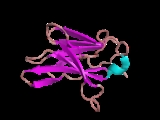
with an immunoglobulin variable (IgV)-like extracellular domain
connected to the membrane by a thin stalk, and an intracellular tail. Less-common homodimers of the CD8-α chain are also expressed on some cells. The molecular weight of CD8 is about 13,463.2 Da. Structure of the CD8 molecule was determined by Leahy, D.J., Axel, R., and Hendrickson, W.A. by X-ray Diffraction at a 2.6A resolution. The structure was determined to have an immunoglobulin-like beta-sandwich folding and 114 amino acid residues. 2% of the protein is wound into α-helices and 46% into β-sheets, with the remaining 52% of the molecules remaining in the loop portions.

of the cytotoxic T cell and the target cell bound closely together during antigen-specific activation. Cytotoxic T cells with CD8 surface protein are called CD8+ T cells. The main recognition site is a flexible loop at the a3 domain of an MHC molecule. This was discovered by doing mutational analyses. The flexible a3 domain is located between residues 223 and 229 in the genome.
Transmembrane protein
A transmembrane protein is a protein that goes from one side of a membrane through to the other side of the membrane. Many TPs function as gateways or "loading docks" to deny or permit the transport of specific substances across the biological membrane, to get into the cell, or out of the cell as...
glycoprotein
Glycoprotein
Glycoproteins are proteins that contain oligosaccharide chains covalently attached to polypeptide side-chains. The carbohydrate is attached to the protein in a cotranslational or posttranslational modification. This process is known as glycosylation. In proteins that have segments extending...
that serves as a co-receptor
Co-receptor
A co-receptor is a cell surface receptor that binds a signalling molecule in addition to a primary receptor in order to facilitate ligand recognition and initiate biological processes, such as entry of a pathogen into a host cell.-Co-receptor Properties:...
for the T cell receptor
T cell receptor
The T cell receptor or TCR is a molecule found on the surface of T lymphocytes that is responsible for recognizing antigens bound to major histocompatibility complex molecules...
(TCR). Like the TCR, CD8 binds to a major histocompatibility complex
Major histocompatibility complex
Major histocompatibility complex is a cell surface molecule encoded by a large gene family in all vertebrates. MHC molecules mediate interactions of leukocytes, also called white blood cells , which are immune cells, with other leukocytes or body cells...
(MHC) molecule, but is specific for the class I MHC protein. There are two isoforms of the protein, alpha and beta, each encoded by a different gene. In humans, both genes are located on chromosome 2 in position 2p12.
Tissue distribution
The CD8 co-receptor is predominantly expressed on the surface of cytotoxic T cellCytotoxic T cell
A cytotoxic T cell belongs to a sub-group of T lymphocytes that are capable of inducing the death of infected somatic or tumor cells; they kill cells that are infected with viruses , or are otherwise damaged or...
s, but can also be found on natural killer cell
Natural killer cell
Natural killer cells are a type of cytotoxic lymphocyte that constitute a major component of the innate immune system. NK cells play a major role in the rejection of tumors and cells infected by viruses...
s, cortical thymocyte
Thymocyte
Thymocytes are hematopoietic progenitor cells present in the thymus. Thymopoiesis is the process in the thymus by which thymocytes differentiate into mature T lymphocytes. The primary function of thymocytes is the generation of T lymphocytes . The thymus provides an inductive environment, which...
s, and dendritic cell
Dendritic cell
Dendritic cells are immune cells forming part of the mammalian immune system. Their main function is to process antigen material and present it on the surface to other cells of the immune system. That is, dendritic cells function as antigen-presenting cells...
s. It is expressed in T cell lymphoblastic lymphoma and hypo-pigmented mycosis fungoides
Mycosis fungoides
-External links:* * *...
, but is frequently lost in other T-cell neoplasms.
Structure
To function, CD8 forms a dimer, consisting of a pair of CD8 chains. The most common form of CD8 is composed of a CD8-α and CD8-β chain, both members of the immunoglobulin superfamilyImmunoglobulin superfamily
The immunoglobulin superfamily is a large group of cell surface and soluble proteins that are involved in the recognition, binding, or adhesion processes of cells. Molecules are categorized as members of this superfamily based on shared structural features with immunoglobulins ; they all possess a...
with an immunoglobulin variable (IgV)-like extracellular domain
Immunoglobulin domain
The immunoglobulin domain is a type of protein domain that consists of a 2-layer sandwich of between 7 and 9 antiparallel β-strands arranged in two β-sheets with a Greek key topology....
connected to the membrane by a thin stalk, and an intracellular tail. Less-common homodimers of the CD8-α chain are also expressed on some cells. The molecular weight of CD8 is about 13,463.2 Da. Structure of the CD8 molecule was determined by Leahy, D.J., Axel, R., and Hendrickson, W.A. by X-ray Diffraction at a 2.6A resolution. The structure was determined to have an immunoglobulin-like beta-sandwich folding and 114 amino acid residues. 2% of the protein is wound into α-helices and 46% into β-sheets, with the remaining 52% of the molecules remaining in the loop portions.
Function
The extracellular IgV-like domain of CD8-α interacts with to the α3 portion of the Class I MHC molecule. This affinity keeps the T cell receptorT cell receptor
The T cell receptor or TCR is a molecule found on the surface of T lymphocytes that is responsible for recognizing antigens bound to major histocompatibility complex molecules...
of the cytotoxic T cell and the target cell bound closely together during antigen-specific activation. Cytotoxic T cells with CD8 surface protein are called CD8+ T cells. The main recognition site is a flexible loop at the a3 domain of an MHC molecule. This was discovered by doing mutational analyses. The flexible a3 domain is located between residues 223 and 229 in the genome.
External links
- http://www.tcells.org/scientific/CD8/ T-cell Group - Cardiff University]
- Mouse CD Antigen Chart
- Human CD Antigen Chart