All Topics  
Auditory system


   Email Print
   Bookmark   Link


Auditory system


The auditory system is the sensory system
Sensory system

A sensory system is a part of the nervous system responsible for processing sense information. A sensory system consists of sensory receptors, neural pathways, and parts of the brain involved in sensory perception....
 for the sense of hearing
Hearing (sense)

Hearing is one of the traditional five senses. It is the ability to perceive sound by detecting vibrations via an organ such as the ear. The inability to hear is called deafness....
.

folds of cartilage surrounding the ear canal are called the pinna. Sound waves are reflected and attenuated when they hit the pinna, and these changes provide additional information that will help the brain determine the direction from which the sounds came.

The sound waves enter the auditory canal, a deceptively simple tube.


Discussion
Ask a question about 'Auditory system'
Start a new discussion about 'Auditory system'
Answer questions from other users
Full Discussion Forum


Encyclopedia


The auditory system is the sensory system
Sensory system

A sensory system is a part of the nervous system responsible for processing sense information. A sensory system consists of sensory receptors, neural pathways, and parts of the brain involved in sensory perception....
 for the sense of hearing
Hearing (sense)

Hearing is one of the traditional five senses. It is the ability to perceive sound by detecting vibrations via an organ such as the ear. The inability to hear is called deafness....
.

Ear

Humanear

Outer ear

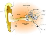
The folds of cartilage surrounding the ear canal are called the pinna. Sound waves are reflected and attenuated when they hit the pinna, and these changes provide additional information that will help the brain determine the direction from which the sounds came.

The sound waves enter the auditory canal, a deceptively simple tube. The ear canal amplifies sounds that are between 3 and 12 kHz. At the far end of the ear canal is the eardrum
Eardrum

The tympanic membrane , is a thin biological membrane that separates the external ear from the middle ear. Its function is to transmit sound from the air to the ossicles inside the middle ear....
 (or tympanic membrane), which marks the beginning of the middle ear
Middle ear

The middle ear is the portion of the ear internal to the eardrum, and external to the oval window of the cochlea. The mammalian middle ear contains three ossicles, which couple vibration of the eardrum into waves in the fluid and membranes of the inner ear....
.

Middle ear

Sound waves traveling through the ear canal will hit the tympanic membrane, or eardrum
Eardrum

The tympanic membrane , is a thin biological membrane that separates the external ear from the middle ear. Its function is to transmit sound from the air to the ossicles inside the middle ear....
. This wave information travels across the air-filled middle ear cavity via a series of delicate bones: the malleus
Malleus

The malleus or hammer is a hammer-shaped small bone or ossicles of the middle ear which connects with the incus and is attached to the inner surface of the eardrum....
 (hammer), incus
Incus

The incus or anvil is the anvil-shaped small bone or ossicles in themiddle ear. It connects the malleus to the stapes. It was first described by Alessandro Achillin of Bologna....
 (anvil) and stapes
Stapes

The stapes or stirrup is the stirrup-shaped small bone or ossicles in themiddle ear which is attached to the incus laterally and to the fenestra ovalis, the "oval window" medially....
 (stirrup). These ossicles
Ossicles

Not to be confused with ossicones.The ossicles are the three smallest bones in the human body. They are contained within the middle ear space and serve to transmit sounds from the air to the fluid-filled labyrinth ....
 act as a lever and a teletype, converting the lower-pressure eardrum sound vibrations into higher-pressure sound vibrations at another, smaller membrane called the oval (or elliptical) window
Oval window

The oval window is a membrane-covered opening which leads from the middle ear to the Vestibule of the ear of the inner ear.Vibrations that come into contact with the tympanic membrane travel through the three ossicles and into the inner ear....
. The malleus articulates with the tympanic membrane via the manubrium, where the stapes articulates with the oval window via its footplate. Higher pressure is necessary because the inner ear beyond the oval window contains liquid rather than air. The sound is not amplified uniformly across the ossicular chain. The auditory reflex of the middle ear muscles helps protect the inner ear from damage. The middle ear still contains the sound information in wave form; it is converted to nerve impulses in the cochlea.

Inner ear


The inner ear consists of the cochlea
Cochlea

The cochlea is the auditory portion of the inner ear. Its core component is the Organ of Corti, the sensory organ of hearing , which is distributed along the partition separating fluid chambers in the coiled tapered tube of the cochlea....
 and several non-auditory structures. The cochlea has three fluid-filled sections, and supports a fluid wave driven by pressure across the basilar membrane
Basilar membrane

The basilar membrane within the cochlea of the inner ear is a stiff structural element that separates two liquid-filled tubes that run along the coil of the cochlea, the scala media and the scala tympani ....
 separating two of the sections. Strikingly, one section, called the cochlear duct or scala media
Scala media

The cochlear duct is an endolymph filled cavity inside the cochlea, located in between the scala tympani and the scala vestibuli, separated by the basilar membrane and Reissner's membrane respectively....
, contains an extracellular fluid similar in composition to endolymph
Endolymph

Endolymph is the fluid contained in the membranous labyrinth of the inner ear.It is also called Scarpa's fluid, after Antonio Scarpa....
, which is usually found inside of cells. The organ of Corti is located at this duct, and transforms mechanical waves to electric signals in neurons. The other two sections are known as the scala tympani and the scala vestibuli, these are located within the bony labyrinth which is filled with fluid called perilymph. The chemical difference between the two fluids (endolymph & perilymph) is important for the function of the inner ear.

Organ of Corti
The organ of Corti forms a ribbon of sensory epithelium which runs lengthwise down the entire cochlea. The hair cells of the organ of Corti transform the fluid waves into nerve signals. The journey of a billion nerves begins with this first step; from here further processing leads to a panoply of auditory reactions and sensations.

Cochlea Crosssection

Hair cell
Hair cells are columnar cells, each with a bundle of 100-200 specialized cilia at the top, for which they are named. These cilia are the mechanosensors for hearing. Lightly resting atop the longest cilia is the tectorial membrane, which moves back and forth with each cycle of sound, tilting the cilia and allowing electric current into the hair cell.

Hair cells, like the photoreceptors of the eye, show a graded response, instead of the spikes
Action potential

An action potential is a self-regenerating wave of electrochemical activity that allows nerve cells to carry a signal over a distance. It is the primary electrical signal generated by nerve cells, and arises from changes in the permeability of the nerve cell's axonal Cell membranes to specific ions....
 typical of other neurons. These graded potentials are not bound by the “all or none” properties of an action potential.

At this point, one may ask how such a wiggle of a hair bundle triggers a difference in membrane potential. The current model is that cilia are attached to one another by “tip links”, structures which link the tips of one cilium to another. Stretching and compressing the tip links may open an ion channel and produce the receptor potential in the hair cell. Recently it has been shown that cdh23 and pchh15 are the adhesion molecules associated with these tip links. It is thought that a calcium driven motor causes a shortening of these links to regenerate tensions. This regeneration of tension allows for apprehension of prolonged auditory stimulation.

Neurons

Afferent neurons innervate cochlear inner hair cells, at synapses where the neurotransmitter glutamate communicates signals from the hair cells to the dendrites of the primary auditory neurons.

There are far fewer inner hair cells in the cochlea than afferent nerve fibers. The neural dendrites belong to neurons of the auditory nerve, which in turn joins the vestibular nerve
Vestibular nerve

The vestibular nerve is one of the two branches of the Vestibulocochlear nerve . It goes to the semicircular canals via the vestibular ganglion....
 to form the vestibulocochlear nerve
Vestibulocochlear nerve

The vestibulocochlear nerve is the eighth of twelve cranial nerves, and is responsible for transmitting sound and equilibrium information from the inner ear to the brain....
, or cranial nerve number VIII.

Efferent projections from the brain to the cochlea also play a role in the perception of sound. Efferent synapses occur on outer hair cells and on afferent (towards the brain) dendrites under inner hair cells.

Central auditory system


This sound information, now re-encoded, travels down the vestibulocochlear nerve
Vestibulocochlear nerve

The vestibulocochlear nerve is the eighth of twelve cranial nerves, and is responsible for transmitting sound and equilibrium information from the inner ear to the brain....
, through intermediate stations such as the cochlear nuclei
Cochlear nuclei

The cochlear nuclei consist of:* the dorsal cochlear nucleus, corresponding to the tuberculum acusticum on the dorso-lateral surface of the inferior peduncle; and...
 and superior olivary complex of the brainstem and the inferior colliculus
Inferior colliculus

Name = Inferior colliculus | Latin = colliculus inferior | GraySubject = 188 | GrayPage= 806 | Image = Gray711.png |...
 of the midbrain, being further processed at each waypoint. The information eventually reaches the thalamus
Thalamus

The thalamus is a pair and symmetric part of the brain. It constitutes the main part of the diencephalon....
, and from there it is relayed to the cortex. In the human brain
Human brain

The human brain is the center of the human nervous system and is a highly complex organ. It has the same general structure as the brains of other mammals, but is over five times as large as the "average brain" of a mammal with the same body size....
, the primary auditory cortex
Primary auditory cortex

The primary auditory cortex is the region of the brain that is responsible for processing of auditory system information....
 is located in the temporal lobe
Temporal lobe

The temporal lobe is a region of the cerebral cortex that is located beneath the Sylvian fissure on both the left and right hemispheres of the brain....
.

Associated anatomical structures include:

Cochlear nucleus


The cochlear nucleus is the first site of the neuronal processing of the newly converted “digital” data from the inner ear. This region is anatomically and physiologically split into two regions, the dorsal cochlear nucleus
Dorsal cochlear nucleus

The dorsal cochlear nucleus , is a cortex-like structure on the dorso-lateral surface of the brainstem. Along with the ventral cochlear nucleus, it forms the cochlear nucleus, where all auditory nerve fibers from the cochlea form their first synapses....
 (DCN), and ventral cochlear nucleus (VCN).

Trapezoid body


The Trapezoid body
Trapezoid body

The trapezoid body is part of the acoustic pathway. It is a bundle of fibers and cells in the pontine tegmentum. It consists of fibers arising from the ventral cochlear nucleus....
 is a bundle of decussating fibers in the ventral pons that carry information used for binaural computations in the brainstem.

Superior olivary complex


The superior olivary complex is located in the pons
Pons

The pons is a structure located on the brain stem. It is cranial to the medulla oblongata, caudal to the midbrain, and ventral to the cerebellum....
, and receives projections predominantly from the ventral cochlear nucleus, although the posterior cochlear nucleus projects there as well, via the ventral acoustic stria. Within the superior olivary complex lies the lateral superior olive (LSO) and the medial superior olive (MSO). The former is important in detecting interaural level differences while the latter is important in distinguishing interaural time difference.

Lateral lemniscus

The lateral lemniscus is a tract of axon
Axon

An axon or nerve fiber is a long, slender projectionof a nerve cell, or neuron, that conducts action potentialaway from the neuron's cell body or soma....
s in the brainstem that carries information about sound from the cochlear nucleus to various brainstem nuclei and ultimately the contralateral inferior colliculus
Inferior colliculus

Name = Inferior colliculus | Latin = colliculus inferior | GraySubject = 188 | GrayPage= 806 | Image = Gray711.png |...
 of the midbrain.

Inferior colliculi


The IC
Inferior colliculus

Name = Inferior colliculus | Latin = colliculus inferior | GraySubject = 188 | GrayPage= 806 | Image = Gray711.png |...
 are located just below the visual processing centers known as the superior colliculi. The central nucleus of the IC is a nearly obligatory relay in the ascending auditory system, and most likely acts to integrate information (specifically regarding sound source localization from the superior olivary complex and dorsal cochlear nucleus
Dorsal cochlear nucleus

The dorsal cochlear nucleus , is a cortex-like structure on the dorso-lateral surface of the brainstem. Along with the ventral cochlear nucleus, it forms the cochlear nucleus, where all auditory nerve fibers from the cochlea form their first synapses....
) before sending it to the thalamus and cortex.

Medial geniculate nucleus


The medial geniculate nucleus
Medial geniculate nucleus

The Medial Geniculate Nucleus or Medial Geniculate Body is part of the auditory thalamus and represents the thalamic relay between the inferior colliculus and the auditory cortex ....
 is part of the thalamic relay system.

Primary auditory cortex


The primary auditory cortex
Primary auditory cortex

The primary auditory cortex is the region of the brain that is responsible for processing of auditory system information....
 is the first region of cerebral cortex
Cerebral cortex

The cerebral cortex is a structure within the brain that plays a key role in memory, attention, perceptual awareness, thought, language, and consciousness....
 to receive auditory input.

Perception of sound is associated with the right posterior superior temporal gyrus
Superior temporal gyrus

A gyrus is a bump or ridge on the surface of the brain. The superior temporal gyrus is one of three gyri in the temporal lobe of the human brain....
 (STG). The superior temporal gyrus contains several important structures of the brain, including Brodmann areas 41 and 42, marking the location of the primary auditory cortex
Primary auditory cortex

The primary auditory cortex is the region of the brain that is responsible for processing of auditory system information....
, the cortical region responsible for the sensation of basic characteristics of sound such as pitch and rhythm.

The auditory association area is located within the temporal lobe
Temporal lobe

The temporal lobe is a region of the cerebral cortex that is located beneath the Sylvian fissure on both the left and right hemispheres of the brain....
 of the brain, in an area called the Wernicke's area
Wernicke's area

Wernicke's area is a part of the human brain that forms part of the Cerebral cortex , on the posterior section of the superior temporal gyrus, encircling the auditory cortex, on the Sylvian fissure ....
, or area 22. This area, near the lateral cerebral sulcus, is an important region for the processing of acoustic signals so that they can be distinguished as speech, music, or noise.

Bibliography


Kandel, et al Principles of Neuroscience. Fourth ed. pp 591-624. Copyright 2000, by McGraw-Hill Co.

See also

  • Music and the brain
    Music and the brain

    Sounds and noises are only separated by the experience of the listener. In the domain of the mind subjectivity reigns, and yet attempts are still made to chip away at individual variations to quantify the actions of the brain....
  • Noise health effects
    Noise health effects

    Noise health effects are the health consequences of elevated sound levels. Elevated workplace or other noise can cause hearing impairment, hypertension, ischemic heart disease, annoyance, sleep disturbance, and decreased school performance....
  • Sound
    Sound

    Sound is vibration transmitted through a solid, liquid, or gas, composed of frequencies within the range of hearing and of a threshold of hearing to be heard, or the sensation stimulated in organs of hearing by such vibrations....
  • Tinnitus
    Tinnitus

    Tinnitus is the perception of sound within the human ear in the absence of corresponding external sound.Tinnitus can be perceived in one or both ears or in the head....
  • Auditory Brainstem Response
    Auditory Brainstem Response

    Auditory brainstem response is an electrical signal evoked from the brainstem of a human or other mammal by the presentation of a sound such as a click....
     (ABR) and ABR audiometry test for newborn hearing


External links

  • Washington University Neuroscience Tutorial -