
.gif)
Atlas (anatomy)
Encyclopedia
In anatomy
, the atlas (C1) is the most superior (first) cervical vertebra of the spine.
It is named for the Atlas
of Greek mythology
, because it supports the globe of the head.
The atlas is the topmost vertebra, and – along with the Axis
– forms the joint connecting the skull
and spine. The atlas and axis are specialized to allow a greater range of motion than normal vertebrae. They are responsible for the nodding and rotation movements of the head.
The atlanto-occipital joint allows the head to nod up and down on the vertebral column.
The dens acts as a pivot that allows the atlas and attached head to rotate on the axis, side to side.
The Atlas' chief peculiarity is that it has no body, it is ring-like, and consists of an anterior and a posterior arch and two lateral masses.
The Atlas and Axis are important neurologically because the brain stem extends down to the Axis.
s and the anterior longitudinal ligament
; posteriorly it is concave, and marked by a smooth, oval or circular facet (fovea dentis), for articulation with the odontoid process (dens) of the axis.
The upper and lower borders respectively give attachment to the anterior atlantooccipital membrane
and the anterior atlantoaxial ligament
; the former connects it with the occipital bone
above, and the latter with the axis below.
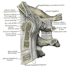 The posterior arch forms about two-fifths of the circumference of the ring: it ends behind in the posterior tubercle, which is the rudiment of a spinous process and gives origin to the Recti capitis posteriores minores and the ligamentum nuchae. The diminutive size of this process prevents any interference with the movements between the atlas and the skull.
The posterior part of the arch presents above and behind a rounded edge for the attachment of the posterior atlantooccipital membrane
, while immediately behind each superior articular process is a groove (sulcus arteriae vertebralis), sometimes converted into a foramen by a delicate bony spiculum which arches backward from the posterior end of the superior articular process.
This groove represents the superior vertebral notch, and serves for the transmission of the vertebral artery
, which, after ascending through the foramen in the transverse process, winds around the lateral mass in a direction backward and medially; it also transmits the suboccipital nerve
(first spinal nerve). In a common anatomic variant the vertebral artery
passes through an arcuate foramen
.
On the under surface of the posterior arch, behind the articular facets, are two shallow grooves, the inferior vertebral notches. The lower border gives attachment to the posterior atlantoaxial ligament
, which connects it with the axis.
Each carries two articular facets, a superior and an inferior.
into two unequal parts:
This part of the vertebral canal is of considerable size, much greater than is required for the accommodation of the spinal cord.
The transverse processes are large; they project laterally and downward from the lateral masses, and serve for the attachment of muscle
s which assist in rotating the head. They are long, and their anterior and posterior tubercles are fused into one mass; the foramen transversarium is directed from below, upward and backward.
 The atlas is usually ossified
from three centers.
Of these, one appears in each lateral mass about the seventh week of fetal life, and extends backward; at birth, these portions of bone are separated from one another behind by a narrow interval filled with cartilage
.
Between the third and fourth years they unite either directly or through the medium of a separate center developed in the cartilage.
At birth, the anterior arch consists of cartilage; in this a separate center appears about the end of the first year after birth, and joins the lateral masses from the sixth to the eighth year.
The lines of union extend across the anterior portions of the superior articular facets.
Occasionally there is no separate center, the anterior arch being formed by the forward extension and ultimate junction of the two lateral masses; sometimes this arch is ossified from two centers, one on either side of the middle line.
.
The pharyngeal and retropharyngeal inflammations may cause decalcification of atlas vertebra. This may lead to loosening of attachments of transverse ligament which may eventually yield. This allows the dens
of axis to move and exert pressure on spinal cord causing sudden death.
Anatomy
Anatomy is a branch of biology and medicine that is the consideration of the structure of living things. It is a general term that includes human anatomy, animal anatomy , and plant anatomy...
, the atlas (C1) is the most superior (first) cervical vertebra of the spine.
It is named for the Atlas
Atlas (mythology)
In Greek mythology, Atlas was the primordial Titan who supported the heavens. Although associated with various places, he became commonly identified with the Atlas Mountains in north-west Africa...
of Greek mythology
Greek mythology
Greek mythology is the body of myths and legends belonging to the ancient Greeks, concerning their gods and heroes, the nature of the world, and the origins and significance of their own cult and ritual practices. They were a part of religion in ancient Greece...
, because it supports the globe of the head.
The atlas is the topmost vertebra, and – along with the Axis
Axis (anatomy)
In anatomy, the second cervical vertebra of the spine is named the axis or epistropheus.It forms the pivot upon which the first cervical vertebra , which carries the head, rotates....
– forms the joint connecting the skull
Human skull
The human skull is a bony structure, skeleton, that is in the human head and which supports the structures of the face and forms a cavity for the brain.In humans, the adult skull is normally made up of 22 bones...
and spine. The atlas and axis are specialized to allow a greater range of motion than normal vertebrae. They are responsible for the nodding and rotation movements of the head.
The atlanto-occipital joint allows the head to nod up and down on the vertebral column.
The dens acts as a pivot that allows the atlas and attached head to rotate on the axis, side to side.
The Atlas' chief peculiarity is that it has no body, it is ring-like, and consists of an anterior and a posterior arch and two lateral masses.
The Atlas and Axis are important neurologically because the brain stem extends down to the Axis.
Anterior arch
The anterior arch forms about one-fifth of the ring: its anterior surface is convex, and presents at its center the anterior tubercle for the attachment of the Longus colli muscleMuscle
Muscle is a contractile tissue of animals and is derived from the mesodermal layer of embryonic germ cells. Muscle cells contain contractile filaments that move past each other and change the size of the cell. They are classified as skeletal, cardiac, or smooth muscles. Their function is to...
s and the anterior longitudinal ligament
Anterior longitudinal ligament
The anterior longitudinal ligament is a ligament that runs down the anterior surface of the spine. It traverses all of the vertebral bodies and intervertebral discs....
; posteriorly it is concave, and marked by a smooth, oval or circular facet (fovea dentis), for articulation with the odontoid process (dens) of the axis.
The upper and lower borders respectively give attachment to the anterior atlantooccipital membrane
Anterior atlantoöccipital membrane
The anterior atlantoöccipital membrane is broad and composed of densely woven fibers, which pass between the anterior margin of the foramen magnum above, and the upper border of the anterior arch of the atlas below....
and the anterior atlantoaxial ligament
Anterior atlantoaxial ligament
The anterior atlantoaxial ligament is a strong membrane, fixed, above, to the lower border of the anterior arch of the atlas; below, to the front of the body of the axis....
; the former connects it with the occipital bone
Occipital bone
The occipital bone, a saucer-shaped membrane bone situated at the back and lower part of the cranium, is trapezoidal in shape and curved on itself...
above, and the latter with the axis below.
Posterior arch
The posterior part of the arch presents above and behind a rounded edge for the attachment of the posterior atlantooccipital membrane
Posterior atlantoöccipital membrane
The posterior atlantoöccipital membrane , broad but thin, is connected above, to the posterior margin of the foramen magnum; below, to the upper border of the posterior arch of the atlas....
, while immediately behind each superior articular process is a groove (sulcus arteriae vertebralis), sometimes converted into a foramen by a delicate bony spiculum which arches backward from the posterior end of the superior articular process.
This groove represents the superior vertebral notch, and serves for the transmission of the vertebral artery
Vertebral artery
The vertebral arteries are major arteries of the neck. They branch from the subclavian arteries and merge to form the single midline basilar artery in a complex called the vertebrobasilar system, which supplies blood to the posterior part of the circle of Willis and thus significant portions of the...
, which, after ascending through the foramen in the transverse process, winds around the lateral mass in a direction backward and medially; it also transmits the suboccipital nerve
Suboccipital nerve
The first cervical nerve, the suboccipital nerve exits the spinal cord between the skull and the first cervical vertebra, the atlas.It supplies muscles around the suboccipital triangle including the rectus capitis posterior major, obliquus capitis superior, and obliquus capitis inferior. The...
(first spinal nerve). In a common anatomic variant the vertebral artery
Vertebral artery
The vertebral arteries are major arteries of the neck. They branch from the subclavian arteries and merge to form the single midline basilar artery in a complex called the vertebrobasilar system, which supplies blood to the posterior part of the circle of Willis and thus significant portions of the...
passes through an arcuate foramen
Arcuate foramen
In human anatomy, arcuate foramen, also known as ponticulus posticus , refers to a bony bridge on the atlas that covers the groove for the vertebral artery. It is a common anatomic variant and estimated to occur in approximately 3-15% of the population...
.
On the under surface of the posterior arch, behind the articular facets, are two shallow grooves, the inferior vertebral notches. The lower border gives attachment to the posterior atlantoaxial ligament
Posterior atlantoaxial ligament
The posterior atlantoaxial ligament is a broad, thin membrane attached, above, to the lower border of the posterior arch of the atlas; below, to the upper edges of the laminæ of the axis....
, which connects it with the axis.
Lateral masses
The lateral masses are the most bulky and solid parts of the atlas, in order to support the weight of the head.Each carries two articular facets, a superior and an inferior.
- The superior facets are of large size, oval, concave, and approach each other in front, but diverge behind: they are directed upward, medially, and a little backward, each forming a cup for the corresponding condyle of the occipital bone, and are admirably adapted to the nodding movements of the head. Not infrequently they are partially subdivided by indentations which encroach upon their margins.
- The inferior articular facets are circular in form, flattened or slightly convex and directed downward and medially, articulating with the axis, and permitting the rotatory movements of the head.
Vertebral foramen
Just below the medial margin of each superior facet is a small tubercle, for the attachment of the transverse atlantal ligament which stretches across the ring of the atlas and divides the vertebral foramenVertebral foramen
In a typical vertebra, the vertebral foramen is the foramen formed by the anterior segment , and the posterior part, the vertebral arch....
into two unequal parts:
- the anterior or smaller receiving the odontoid process of the axis
- the posterior transmitting the spinal cordSpinal cordThe spinal cord is a long, thin, tubular bundle of nervous tissue and support cells that extends from the brain . The brain and spinal cord together make up the central nervous system...
(medulla spinalis) and its membranes
This part of the vertebral canal is of considerable size, much greater than is required for the accommodation of the spinal cord.
The transverse processes are large; they project laterally and downward from the lateral masses, and serve for the attachment of muscle
Muscle
Muscle is a contractile tissue of animals and is derived from the mesodermal layer of embryonic germ cells. Muscle cells contain contractile filaments that move past each other and change the size of the cell. They are classified as skeletal, cardiac, or smooth muscles. Their function is to...
s which assist in rotating the head. They are long, and their anterior and posterior tubercles are fused into one mass; the foramen transversarium is directed from below, upward and backward.
Development
Ossification
Ossification is the process of laying down new bone material by cells called osteoblasts. It is synonymous with bone tissue formation...
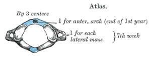
from three centers.
Of these, one appears in each lateral mass about the seventh week of fetal life, and extends backward; at birth, these portions of bone are separated from one another behind by a narrow interval filled with cartilage
Cartilage
Cartilage is a flexible connective tissue found in many areas in the bodies of humans and other animals, including the joints between bones, the rib cage, the ear, the nose, the elbow, the knee, the ankle, the bronchial tubes and the intervertebral discs...
.
Between the third and fourth years they unite either directly or through the medium of a separate center developed in the cartilage.
At birth, the anterior arch consists of cartilage; in this a separate center appears about the end of the first year after birth, and joins the lateral masses from the sixth to the eighth year.
The lines of union extend across the anterior portions of the superior articular facets.
Occasionally there is no separate center, the anterior arch being formed by the forward extension and ultimate junction of the two lateral masses; sometimes this arch is ossified from two centers, one on either side of the middle line.
Injuries
A break in the first vertebra is referred to as a Jefferson fractureJefferson fracture
A Jefferson fracture is a bone fracture of the anterior and posterior arches of the C1 vertebra, though it may also appear as a three or two part fracture...
.
The pharyngeal and retropharyngeal inflammations may cause decalcification of atlas vertebra. This may lead to loosening of attachments of transverse ligament which may eventually yield. This allows the dens
Dens
Dens may refer to:* Den *Dens , also known as odontoid process or odontoid peg*Tooth...
of axis to move and exert pressure on spinal cord causing sudden death.
External links
- Netter, Frank. Atlas of Human Anatomy, "High Cervical Spine: C1-C2"