

Angiogram
Encyclopedia
Angiography or arteriography is a medical imaging
technique used to visualize the inside, or lumen
, of blood vessels and organs of the body, with particular interest in the arteries
, vein
s and the heart chamber
s. This is traditionally done by injecting a radio-opaque contrast agent
into the blood vessel and imaging using X-ray
based techniques such as fluoroscopy
.
The word itself comes from the Greek
words angeion, "vessel", and graphein, "to write or record". The film or image of the blood vessel
s is called an angiograph, or more commonly, an angiogram. Though the word itself can describe both an arteriogram and a venogram, in its everyday usage, the terms angiogram and arteriogram are often used synonymously, whereas the term venogram is used more precisely.
The term angiography is strictly defined as based on projectional radiography
; however, the term has been applied to newer vascular imaging techniques such as CT angiography
and MR angiography
. The term isotope angiography has also been used, although this more correctly is referred to as isotope perfusion scanning.
physician and neurologist Egas Moniz
at the University of Lisbon to provide contrasted x-ray cerebral angiography
in order to diagnose several kinds of nervous diseases, such as tumors, artery disease and arteriovenous malformations. He is usually recognized as one of the pioneers in this field. Moniz performed the first cerebral angiogram in Lisbon in 1927, and Reynaldo Cid dos Santos performed the first aortogram in the same city in 1929. With the introduction of the Seldinger technique
in 1953, the procedure became markedly safer as no sharp introductory devices needed to remain inside the vascular lumen.
 Depending on the type of angiogram, access to the blood vessels is gained most commonly through the femoral artery
, to look at the left side of the heart and the arterial system
or the jugular or femoral vein
, to look at the right side of the heart and the venous system. Using a system of guide wire
s and catheters, a type of contrast agent (which shows up by absorbing the x-rays), is added to the blood to make it visible on the x-ray images.
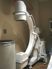
The X-ray
images taken may either be still images, displayed on a image intensifier
or film, or motion images. For all structures except the heart, the images are usually taken using a technique called digital subtraction angiography
(DSA). Images in this case are usually taken at 2 - 3 frames per second, which allows the radiologist to evaluate the flow of the blood through a vessel or vessels. This technique "subtracts" the bones and other organs so only the vessels filled with contrast agent can be seen. The heart images are taken at 15-30 frames per second, not using a subtraction technique. Because DSA requires the patient to remain motionless, it cannot be used on the heart. Both these techniques enable the radiologist or cardiologist to see stenosis
(blockages or narrowings) inside the vessel which may be inhibiting the flow of blood and causing pain.
is used to administer the x-ray contrast agent at the desired area to be visualized. The catheter is threaded into an artery in the forearm
, and the tip is advanced through the arterial system into the major coronary artery. X-ray
images of the transient radiocontrast
distribution within the blood flowing within the coronary arteries allows visualization of the size of the artery openings
. Presence or absence of atherosclerosis
or atheroma
within the walls of the arteries
cannot be clearly determined. See coronary catheterization
for more detail..
in order to visualise the arterial and venous supply to the brain. Intervention work such as coil-embolisation of aneurysm
s and AVM
gluing can also be performed.
in patients with leg claudication or cramps, caused by reduced blood flow down the legs and to the feet; in patients with renal stenosis (which commonly causes high blood pressure) and can be used in the head to find and repair stroke. These are all done routinely through the femoral artery, but can also be performed through the brachial or axillary (arm) artery. Any stenoses found may be treated by the use of atherectomy
.
l vascular disorders, such as diabetic retinopathy
and macular degeneration
.
, blood clots (which can cause heart attack or stroke), hypotension
and pericardial effusion
. Minor complications can include bleeding
or bruising at the site where the contrast is injected, blood vessel damage on the route to the heart from the catheter (rare) and allergic reaction to the contrast.
, an allergic reaction to the anaesthetic other medication or the contrast medium, blockage or damage to one of the access veins in the leg, or thrombosis
and embolism
formation. Bleeding
or bruising at the site where the contrast is injected are minor complications, delayed bleeding can also occur but is rare.
(1973) featured a scene in which the protagonist, a young girl, is subjected to carotid angiography. The scene was portrayed accurately by medical personnel—rather than actors—who regularly performed the procedure in real life. Although the movie as a whole is often cited as one of the most frightening motion pictures of all time, this angiography scene was the scene most likely to upset audiences, according to the director, William Friedkin
.
Medical imaging
Medical imaging is the technique and process used to create images of the human body for clinical purposes or medical science...
technique used to visualize the inside, or lumen
Lumen (anatomy)
A lumen in biology is the inside space of a tubular structure, such as an artery or intestine...
, of blood vessels and organs of the body, with particular interest in the arteries
Artery
Arteries are blood vessels that carry blood away from the heart. This blood is normally oxygenated, exceptions made for the pulmonary and umbilical arteries....
, vein
Vein
In the circulatory system, veins are blood vessels that carry blood towards the heart. Most veins carry deoxygenated blood from the tissues back to the heart; exceptions are the pulmonary and umbilical veins, both of which carry oxygenated blood to the heart...
s and the heart chamber
Heart chamber
aHeart chamber is a general term used to refer to any chambers of the mammalian heart. The heart consists of four chambers: the right and left atrium and the right and left ventricle. The top chambers are connected to the bottom chambers by valves and are separated by the coronary sulcus...
s. This is traditionally done by injecting a radio-opaque contrast agent
Radiocontrast
Radiocontrast agents are a type of medical contrast medium used to improve the visibility of internal bodily structures in an X-ray based imaging techniques such as computed tomography or radiography...
into the blood vessel and imaging using X-ray
X-ray
X-radiation is a form of electromagnetic radiation. X-rays have a wavelength in the range of 0.01 to 10 nanometers, corresponding to frequencies in the range 30 petahertz to 30 exahertz and energies in the range 120 eV to 120 keV. They are shorter in wavelength than UV rays and longer than gamma...
based techniques such as fluoroscopy
Fluoroscopy
Fluoroscopy is an imaging technique commonly used by physicians to obtain real-time moving images of the internal structures of a patient through the use of a fluoroscope. In its simplest form, a fluoroscope consists of an X-ray source and fluorescent screen between which a patient is placed...
.
The word itself comes from the Greek
Greek language
Greek is an independent branch of the Indo-European family of languages. Native to the southern Balkans, it has the longest documented history of any Indo-European language, spanning 34 centuries of written records. Its writing system has been the Greek alphabet for the majority of its history;...
words angeion, "vessel", and graphein, "to write or record". The film or image of the blood vessel
Blood vessel
The blood vessels are the part of the circulatory system that transports blood throughout the body. There are three major types of blood vessels: the arteries, which carry the blood away from the heart; the capillaries, which enable the actual exchange of water and chemicals between the blood and...
s is called an angiograph, or more commonly, an angiogram. Though the word itself can describe both an arteriogram and a venogram, in its everyday usage, the terms angiogram and arteriogram are often used synonymously, whereas the term venogram is used more precisely.
The term angiography is strictly defined as based on projectional radiography
Projectional radiography
Projectional radiography or plain film radiography is the practice of producing two-dimensional images using x-ray radiation. Radiographic exams are typically performed by Radiologic Technologists, highly trained medical professionals who specialize in the usage of radiographic equipment, patient...
; however, the term has been applied to newer vascular imaging techniques such as CT angiography
Computed tomography angiography
Computed tomography angiography is a computed tomography technique used to visualize arterial and venous vessels throughout the body. This ranges from arteries serving the brain to those bringing blood to the lungs, kidneys, arms and legs.-Technique:...
and MR angiography
Magnetic Resonance Angiography
Magnetic resonance angiography is a group of techniques based on Magnetic Resonance Imaging to image blood vessels. Magnetic resonance angiography is used to generate images of the arteries in order to evaluate them for stenosis , occlusion or aneurysms...
. The term isotope angiography has also been used, although this more correctly is referred to as isotope perfusion scanning.
History
The technique was first developed in 1927 by the PortuguesePortugal
Portugal , officially the Portuguese Republic is a country situated in southwestern Europe on the Iberian Peninsula. Portugal is the westernmost country of Europe, and is bordered by the Atlantic Ocean to the West and South and by Spain to the North and East. The Atlantic archipelagos of the...
physician and neurologist Egas Moniz
Egas Moniz
António Caetano de Abreu Freire Egas Moniz , known as Egas Moniz , was a Portuguese neurologist and the developer of cerebral angiography...
at the University of Lisbon to provide contrasted x-ray cerebral angiography
Cerebral angiography
Cerebral angiography is a form of angiography which provides images of blood vessels in and around the brain, thereby allowing detection of abnormalities such as arteriovenous malformations and aneurysms....
in order to diagnose several kinds of nervous diseases, such as tumors, artery disease and arteriovenous malformations. He is usually recognized as one of the pioneers in this field. Moniz performed the first cerebral angiogram in Lisbon in 1927, and Reynaldo Cid dos Santos performed the first aortogram in the same city in 1929. With the introduction of the Seldinger technique
Seldinger technique
The Seldinger technique is a medical procedure to obtain safe access to blood vessels and other hollow organs. It is named after Dr. Sven-Ivar Seldinger , a Swedish radiologist from Mora, Dalarna County, who introduced the procedure in 1953....
in 1953, the procedure became markedly safer as no sharp introductory devices needed to remain inside the vascular lumen.
Technique
Femoral artery
The femoral artery is a general term comprising a few large arteries in the thigh. They begin at the inguinal ligament and end just above the knee at adductor canal or Hunter's canal traversing the extent of the femur bone....
, to look at the left side of the heart and the arterial system
Artery
Arteries are blood vessels that carry blood away from the heart. This blood is normally oxygenated, exceptions made for the pulmonary and umbilical arteries....
or the jugular or femoral vein
Femoral vein
In the human body, the femoral vein is a blood vessel that accompanies the femoral artery in the femoral sheath. It begins at the adductor canal and is a continuation of the popliteal vein...
, to look at the right side of the heart and the venous system. Using a system of guide wire
Guide wire
A guide wire may refer to:*A step in the Seldinger technique*A misspelling of guy-wire*The character called Guide Wire from the anime series Rave Master...
s and catheters, a type of contrast agent (which shows up by absorbing the x-rays), is added to the blood to make it visible on the x-ray images.
The X-ray
X-ray
X-radiation is a form of electromagnetic radiation. X-rays have a wavelength in the range of 0.01 to 10 nanometers, corresponding to frequencies in the range 30 petahertz to 30 exahertz and energies in the range 120 eV to 120 keV. They are shorter in wavelength than UV rays and longer than gamma...
images taken may either be still images, displayed on a image intensifier
Image intensifier
An image intensifier tube is a vacuum tube device for increasing the intensity of available light in an optical system to allow use under low light conditions such as at night, to facilitate visual imaging of low-light processes such as fluorescence of materials to X-rays or gamma rays, or for...
or film, or motion images. For all structures except the heart, the images are usually taken using a technique called digital subtraction angiography
Digital subtraction angiography
Digital subtraction angiography is a type of fluoroscopy technique used in interventional radiology to clearly visualize blood vessels in a bony or dense soft tissue environment. Images are produced using contrast medium by subtracting a 'pre-contrast image' or the mask from later images, once...
(DSA). Images in this case are usually taken at 2 - 3 frames per second, which allows the radiologist to evaluate the flow of the blood through a vessel or vessels. This technique "subtracts" the bones and other organs so only the vessels filled with contrast agent can be seen. The heart images are taken at 15-30 frames per second, not using a subtraction technique. Because DSA requires the patient to remain motionless, it cannot be used on the heart. Both these techniques enable the radiologist or cardiologist to see stenosis
Stenosis
A stenosis is an abnormal narrowing in a blood vessel or other tubular organ or structure.It is also sometimes called a stricture ....
(blockages or narrowings) inside the vessel which may be inhibiting the flow of blood and causing pain.
Coronary angiography
One of most common angiograms performed is to visualize the blood in the coronary arteries. A long, thin, flexible tube called a catheterCatheter
In medicine, a catheter is a tube that can be inserted into a body cavity, duct, or vessel. Catheters thereby allow drainage, administration of fluids or gases, or access by surgical instruments. The process of inserting a catheter is catheterization...
is used to administer the x-ray contrast agent at the desired area to be visualized. The catheter is threaded into an artery in the forearm
Forearm
-See also:*Forearm flexors*Forearm muscles...
, and the tip is advanced through the arterial system into the major coronary artery. X-ray
X-ray
X-radiation is a form of electromagnetic radiation. X-rays have a wavelength in the range of 0.01 to 10 nanometers, corresponding to frequencies in the range 30 petahertz to 30 exahertz and energies in the range 120 eV to 120 keV. They are shorter in wavelength than UV rays and longer than gamma...
images of the transient radiocontrast
Radiocontrast
Radiocontrast agents are a type of medical contrast medium used to improve the visibility of internal bodily structures in an X-ray based imaging techniques such as computed tomography or radiography...
distribution within the blood flowing within the coronary arteries allows visualization of the size of the artery openings
Lumen (anatomy)
A lumen in biology is the inside space of a tubular structure, such as an artery or intestine...
. Presence or absence of atherosclerosis
Atherosclerosis
Atherosclerosis is a condition in which an artery wall thickens as a result of the accumulation of fatty materials such as cholesterol...
or atheroma
Atheroma
In pathology, an atheroma is an accumulation and swelling in artery walls that is made up of macrophage cells, or debris, that contain lipids , calcium and a variable amount of fibrous connective tissue...
within the walls of the arteries
Artery
Arteries are blood vessels that carry blood away from the heart. This blood is normally oxygenated, exceptions made for the pulmonary and umbilical arteries....
cannot be clearly determined. See coronary catheterization
Coronary catheterization
A coronary catheterization is a minimally invasive procedure to access the coronary circulation and blood filled chambers of the heart using a catheter. It is performed for both diagnostic and interventional purposes....
for more detail..
Neuro-vascular angiography
Another increasingly common angiographic procedure is neuro-vascular digital subtraction angiographyDigital subtraction angiography
Digital subtraction angiography is a type of fluoroscopy technique used in interventional radiology to clearly visualize blood vessels in a bony or dense soft tissue environment. Images are produced using contrast medium by subtracting a 'pre-contrast image' or the mask from later images, once...
in order to visualise the arterial and venous supply to the brain. Intervention work such as coil-embolisation of aneurysm
Aneurysm
An aneurysm or aneurism is a localized, blood-filled balloon-like bulge in the wall of a blood vessel. Aneurysms can commonly occur in arteries at the base of the brain and an aortic aneurysm occurs in the main artery carrying blood from the left ventricle of the heart...
s and AVM
Arteriovenous malformation
Arteriovenous malformation or AVM is an abnormal connection between veins and arteries, usually congenital. This pathology is widely known because of its occurrence in the central nervous system, but can appear in any location. An arteriovenous malformation is a vascular anomaly. It is a...
gluing can also be performed.
Peripheral angiography
Angiography is also commonly performed to identify vessel narrowingStenosis
A stenosis is an abnormal narrowing in a blood vessel or other tubular organ or structure.It is also sometimes called a stricture ....
in patients with leg claudication or cramps, caused by reduced blood flow down the legs and to the feet; in patients with renal stenosis (which commonly causes high blood pressure) and can be used in the head to find and repair stroke. These are all done routinely through the femoral artery, but can also be performed through the brachial or axillary (arm) artery. Any stenoses found may be treated by the use of atherectomy
Atherectomy
Atherectomy is a minimally invasive surgical method of removing, mainly, atherosclerosis from a large blood vessel within the body. Today, it is generally used to effectively treat peripheral arterial disease of the lower extremities...
.
Other
Other angiographic uses include the diagnosis of retinaRetina
The vertebrate retina is a light-sensitive tissue lining the inner surface of the eye. The optics of the eye create an image of the visual world on the retina, which serves much the same function as the film in a camera. Light striking the retina initiates a cascade of chemical and electrical...
l vascular disorders, such as diabetic retinopathy
Diabetic retinopathy
Diabetic retinopathy is retinopathy caused by complications of diabetes mellitus, which can eventually lead to blindness....
and macular degeneration
Macular degeneration
Age-related macular degeneration is a medical condition which usually affects older adults and results in a loss of vision in the center of the visual field because of damage to the retina. It occurs in “dry” and “wet” forms. It is a major cause of blindness and visual impairment in older adults...
.
Coronary angiography
Coronary angiographies are common and major complications are rare. These include Cardiac arrhythmias, kidney damageRenal failure
Renal failure or kidney failure describes a medical condition in which the kidneys fail to adequately filter toxins and waste products from the blood...
, blood clots (which can cause heart attack or stroke), hypotension
Hypotension
In physiology and medicine, hypotension is abnormally low blood pressure, especially in the arteries of the systemic circulation. It is best understood as a physiologic state, rather than a disease. It is often associated with shock, though not necessarily indicative of it. Hypotension is the...
and pericardial effusion
Pericardial effusion
Pericardial effusion is an abnormal accumulation of fluid in the pericardial cavity. Because of the limited amount of space in the pericardial cavity, fluid accumulation will lead to an increased intrapericardial pressure and this can negatively affect heart function...
. Minor complications can include bleeding
Bleeding
Bleeding, technically known as hemorrhaging or haemorrhaging is the loss of blood or blood escape from the circulatory system...
or bruising at the site where the contrast is injected, blood vessel damage on the route to the heart from the catheter (rare) and allergic reaction to the contrast.
Cerebral angiography
Major complications in Cerebral Angiography are also rare but include strokeStroke
A stroke, previously known medically as a cerebrovascular accident , is the rapidly developing loss of brain function due to disturbance in the blood supply to the brain. This can be due to ischemia caused by blockage , or a hemorrhage...
, an allergic reaction to the anaesthetic other medication or the contrast medium, blockage or damage to one of the access veins in the leg, or thrombosis
Thrombosis
Thrombosis is the formation of a blood clot inside a blood vessel, obstructing the flow of blood through the circulatory system. When a blood vessel is injured, the body uses platelets and fibrin to form a blood clot to prevent blood loss...
and embolism
Embolism
In medicine, an embolism is the event of lodging of an embolus into a narrow capillary vessel of an arterial bed which causes a blockage in a distant part of the body.Embolization is...
formation. Bleeding
Bleeding
Bleeding, technically known as hemorrhaging or haemorrhaging is the loss of blood or blood escape from the circulatory system...
or bruising at the site where the contrast is injected are minor complications, delayed bleeding can also occur but is rare.
In popular culture
The horror movie The ExorcistThe Exorcist (film)
The Exorcist is a 1973 American horror film directed by William Friedkin, adapted from the 1971 novel of the same name by William Peter Blatty and based on the exorcism case of Robbie Mannheim, dealing with the demonic possession of a young girl and her mother’s desperate attempts to win back her...
(1973) featured a scene in which the protagonist, a young girl, is subjected to carotid angiography. The scene was portrayed accurately by medical personnel—rather than actors—who regularly performed the procedure in real life. Although the movie as a whole is often cited as one of the most frightening motion pictures of all time, this angiography scene was the scene most likely to upset audiences, according to the director, William Friedkin
William Friedkin
William Friedkin is an American film director, producer and screenwriter best known for directing The French Connection in 1971 and The Exorcist in 1973; for the former, he won the Academy Award for Best Director...
.
See also
- Cardiac catheterizationCardiac catheterizationCardiac catheterization is the insertion of a catheter into a chamber or vessel of the heart. This is done for both investigational and interventional purposes...
- Computed Tomography AngiographyComputed tomography angiographyComputed tomography angiography is a computed tomography technique used to visualize arterial and venous vessels throughout the body. This ranges from arteries serving the brain to those bringing blood to the lungs, kidneys, arms and legs.-Technique:...
- Contrast MediumContrast mediumA medical contrast medium is a substance used to enhance the contrast of structures or fluids within the body in medical imaging...
- Echocardiogram
- ElectrocardiogramElectrocardiogramElectrocardiography is a transthoracic interpretation of the electrical activity of the heart over a period of time, as detected by electrodes attached to the outer surface of the skin and recorded by a device external to the body...
- Fluorescein angiographyFluorescein angiographyIntravenous Fluorescein angiography or fluorescent angiography is a technique for examining the circulation of the retina using the dye tracing method...
- Image intensifierImage intensifierAn image intensifier tube is a vacuum tube device for increasing the intensity of available light in an optical system to allow use under low light conditions such as at night, to facilitate visual imaging of low-light processes such as fluorescence of materials to X-rays or gamma rays, or for...
- Interventional RadiologyInterventional radiologyInterventional radiology is a specialty of radiology, in which image-guided procedures are used to diagnose and treat a multitude of diseases across all body systems...
- Intravascular ultrasoundIntravascular ultrasoundIntravascular ultrasound is a medical imaging methodology using a specially designed catheter with a miniaturized ultrasound probe attached to the distal end of the catheter. The proximal end of the catheter is attached to computerized ultrasound equipment...
- Intravenous digital subtraction angiographyIntravenous digital subtraction angiographyIntravenous digital subtraction angiography is a form of angiography which was first developed in the late 1970s.IV-DSA uses a computer technique which compares an x-ray image of a region of the body before and after radiopaque iodine based dye has been injected intravenously into the body...
- Magnetic Resonance AngiographyMagnetic Resonance AngiographyMagnetic resonance angiography is a group of techniques based on Magnetic Resonance Imaging to image blood vessels. Magnetic resonance angiography is used to generate images of the arteries in order to evaluate them for stenosis , occlusion or aneurysms...
- Peripheral artery occlusive diseasePeripheral artery occlusive diseasePeripheral vascular disease , commonly referred to as peripheral arterial disease or peripheral artery occlusive disease , refers to the obstruction of large arteries not within the coronary, aortic arch vasculature, or brain. PVD can result from atherosclerosis, inflammatory processes leading to...
External links
- RadiologyInfo - The radiology information resource for patients: Angiography procedures
- Cardiac Catheterization from Angioplasty.Org
- Angiography Equipment from Siemens Medical
- Cardiovascular and Interventional Radiological Society of Europe
- http://emedicine.medscape.com/article/1603072-overview Coronary CT angiography by Eugene Lin